The role
of
maternal
anti-A and
anti-B antibody
titers
in
predicting
ABO
hemolytic
disease
of the
newborn
Nartono Kadri
Abstrak
Telahdilakukanpenelitianterhadap26SpersalinanibugolongandarahOdenganbayinyagolongandarahAatauB. Kemampuan ibu golongan darah O memproduksi antibodi anti-A dan anti-B tidakbedabermakna (p < 0,05). Ditemukan 94 bayi (35,1Vo) mengalami ikterus potensial dan 174 bayi (64,9Vo) ikterus tidak potensial. Untuk memprediksi kelahiran bayi ikterus potensial diperoleh titer
antibodi anti-A dan anti-B ibu > 1/256 sebagai cut-off point dengan sensitiftas 83.33Vo dan spesifisitas 78,2 lVo, nilai prediksi
positif
T9,2TVodannilaiprediksinegatif lT,5TVo.Padatiterantibodianti-Adananti-Bibu>l256,risikodiLahirkannyabayi ikteruspotensial secara bermakna menjadi 18 kali lebihbesar dibandingkan titer < I/256 (RO 18,64 ;
IK
95Vo = 6.18 - 56.18).Abstract
A study has been conducted on 268 blood group O delivering mothers and their related blood group A and B newborn infants. It
reveaLed that the capability of mothers 1o produce anti-A and anti-B antibodies did not
dffir
significantly from each other (p > 0.05) .Ninety four (35.1Vo) ofthe newborns developed potentionaljaundice while the others did not. To predict the birth ofa baby with potential jaundice, a maternal anti-A or anti-B antibody titer of > l/256 was considered to be the cut-offpoint with a sensitivity and specificity
of
S3.33VoandTS.2lVorespectivelyandapositivepredictivevalueofTg.2TVoandanegativepredictivevalueof 17.57Va. Ifthematemal
anti-Aoranti-Bantibody titerwas>l/256theriskforgivingbirthofababywithpotentialjaundicerosesignificantlylStimescompared
to those with titers
of
< 1/256 (OR 18,64;
CI 95Vo = 6.18-56.18).Keywords: ABO incompatibility, anti-A/B antibody titers, hemolytic disease of the newborn
ABO hemolytic
diseaseof
thenewborn (ABO-HDN)
is at
present
the most frequently hemolytic disease
found in
newborns
after
anti-Rh0
(anti-D)
preventive
measures ag.aints Rhesushemolytic disease
have beenfound
out.'''
ABO-HDN is
more
frequently found in
blood
group
A
or
B
newborn infants born frorn blood
group
O
mothers
andthe
occurence
significantly
of-fers
from ABO incompatibility
casesin blood group
A
or B mothers.''' This
group
representsthe pregnancy
at
risk
for
ABO blood group incompatibility
for
the occurenceofneonataljaundice.
This is
actually
causedby
the fact thatanti-A
andanti-B
antibodies
ofblood
group O mothers is
in general of IgG
classorigin
which
can pass
transplacentally while in the
caseof
blood
groupA
or B mothersit
is ofIgM origin
which
cannot
pass
acrossthe
placenta.3'a
Blood
group
O
mothers havenaturally
already anti-A
andanti-B
antibodiesin their
blood.
Thefetal red cells immunization
acrossDepartment of Child Health, Faculty of Medicine
U niv e r s i ty of I nd o ne s i a./ D r. C ip t o M an gunkus umo H o sp ita l, Jakarta, Indonesia
the placenta
in
OA or
OB
incompatible
pregnanciesmay cause
animmune
responsein
the
mother
which
may
increase those
levels
of
anti-A and anti-B
an-tibody
titers.
Besidesfetal
red bloodcell
immuniza-tion,
the level
of
the maternal anti-A
and anti-B
antibody titers
depend also
on
the
environment.
Theincidence
of ABO-HDN
in
slum areas
is found to be
higher than
in
favorable healthy
living
areas.s This
isthought to be
as a consequenceofthe
fact that
in
slum
areas manyE.coli
bacteria and
Ascaris
lumbricoides
worms
arestill
prevailing while
in
those organism
a substanceresembling
A
or B redblood cell component
is
found.o'/
Continous
and
recurrent
exposure
may cause a secondaryimmune response thus resulting
in
the
presenceof
anti-A
and
anti-B
antibodies
in
themother. [n pregnant
mothers
with
high titers
of anti-A
or anti-B
antibodies, the
risk fbr
the newborn tocon-tract ABO-HDN
becomesthus also higher.8'e
The aim
of this
studyis to
find
out the
cutt-of point
of
the maternal
anti-A
and
anti-B antibody titers which
80 Kadri
MATERIALS
AND
METIIODS
The
subjectsof this
study
consistedof
blood group
Odelivering mothers
with
their
A
or
B
blood
group
newborn infants.
During
pregnancy and labor
themothers had
not
received
blood transfusion so far
while
thenewborn infants were
all live
births
which
abirthweight
of >
1500gram
or
gestation
period
of
>32
weeks,
all
without mayor congenital
malforma-tions.
Sampleswere patients
from Dr.Cipto
Mangun-kusumo
General
Hospital, the
Budi
Kemuliaan
Maternity hospital and also
from
four community
health
centers scatteredall
over Jakarta onApril
1995to
January
1997. Maternal
peripheral
blood
samplesright before delivery were
obtained
and cord
blood
samples
of
the newborn infants after birth.
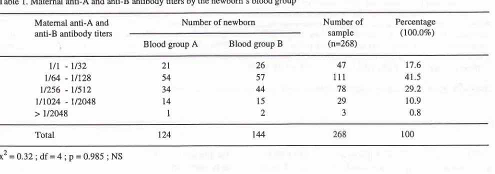
Seroim-Table
l.
Maternal anti-A and anti-B antibody titers by the newborn's blood groupMed J Indones
analysis were performed.
To find
out
the
anti-A
andanti-B
antibody
titers
aspredictor
ofthe
occurrenceof
potential jaundice,
acalculation
was made on thesen-sitivity
and
specificity at the cut-off points
of
1164,Ill28,
11256,tl5l2
and
ll1024 titers
andplotted
on thereceiver operating characterstic
(ROC)
curve.
RESULTS
Two
hundred
sixty
eight
blood
samplesof
themother
and
of its
respective
newborn infants
had been
col-lected, consisting
of blood
group O mothers
andcord
blood
group
A
or B of
the newborn.
The distribution
of anti-A
andanti-B antibody titers
in
the serumof
themother by the newborn's blood group
is
shown in
Table
1.Matemal anti-A and
antiB
antibody titersNumber of newborn Number of
sample (n=268)
Percentage
(lOO.OVo)
Blood group
A
Blood group But
-lt32
u64
-
Ut28u256 -
U5t2Ut024
- 1t2048 > u20482t 54 34
t4
126 57 44
l5
2
47
lll
78 29
17.6 41.5 29.2 10.9 0.8
100
IM
124 Total
x2 = 0.32 ; df =
4;
p = 0.985 ; NSmunologic antiglobulin
tests(Coombs'
test) wereper-formed
in
themothers
and
newbornsblood
as soon aspossible. The maternal
anti-A
and
anti-B
antibody
titers were determined
and thecord blood
wasdivided
into
two
groups, namely
thepotential
jaundice
(posi-tive Coombs'
test)
and
the non potential
jaundice
(negative Coombs' test). Design
of
the
sudy
wereperformed into two
steps,the
first
stepwas
across-sectional study
to
obtain the basic data,
while
the second step was acase-control
study (nestedcase-con-trol
study) between
thepotential
jaundice
and the nonpotential
jaundice
groups.
To
distinguish
between thetwo
groups as
a
case
group and control group,
amatching was
doneof
three variables
influencing
theoccurence
of
neonatal
jaundice namely the
gestationperiod, newborn
blood
group
andthe
sexof
the
new-born.
Data analysis was done
with
Chi-square
test.Fisher's
exact test
with
significance
limit
of p <
0.05 andby calculating relative
risk
(Odds ratio) with95Vo
confidence
interval. Model
of
bivariat
andmultivariat
There was no
significant
difference found
between the numberof
newborn
with
A
andB
blood group
in
anymaternal
titer group (p
>
0.05), meaning that
thecapability
of
blood group O mothers
in
producing
anti-A or anti-B
antibodies
wasthe
same.Results
of
antiglobulin test
of
the newborn's
cord
blood
were
divided
into
the potential
jaundice
group
(positive Coombs' test)
and the nonpotential jaundice
group (negative
Coombs'
test).
Newborn with
poten-tial
jaundice have the potential
to develop
neonatal
jaundice. Table 2
shows
the distribution of
newborn
blood group by
theresults
of
theantiglobulin
tests.As
many
as 94 out
of
the 268 studied
samples
(35.1%)showed potential jaundice,
thosenewborn
hada
tendency
to
develop neonatal
jaundice. The
prob-ability
of
becoming potential jaundice
in
the blood
[image:2.595.54.549.301.475.2]Table 2. Distribution
of
antigiobulin test by newbom,s blood groupAntiglobulin test Number ofnewborn
Blood group
A
Blood group BNumber
of
sample (n=268)
Percentage (l00.OVo)
Positive
(potential jaundice)
Negative
(non potential jaundice)
94
174 48
96 46
78
35.
l
64.9
100
TM r24
Total 268
x2
=0.27;df
=I
; p = 0.606 ;NSTable
3
reveals result
of
sensitifity and specificity
tests
on
the
various
cut-off points
of
maternal blood
group
O anti-A
andanti-B
antibody
titers which
wasthen also
plotted
as acurve
on thereceiving operation
characteristic (ROC)
chart.percentage
with
apositive predictive
value
of
79.27Vo andnegative
predictive
value
of
l7.5j%o.
With
thosesensitivity
and
specificity
values the
cut-off point
of
maternal
anti-A
and
anti-B antibodies
to
be l/256.
Plotting
thosesensitivity
andspecificity
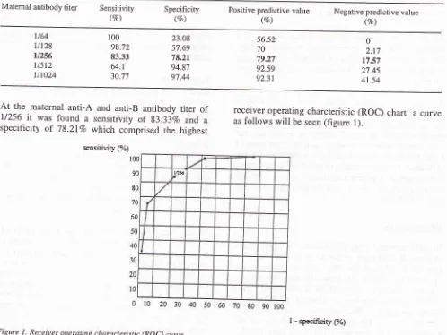
values on theTable 3. Sensitivity and spesificity at various cut-off points of matemal anti-A and anti-B antibody titers
Matemal antibody titer Sensitivity
(vo) Specificity(%) Positive predictive value(vo) Negative predictive value(vo)
l/64
llt28
u256
u5t2
tlt024
100 98.72 83.33
64.1
30.77
23.08 57.69 78.21 94.87 97.44
56.52 70 79.27 92.59 92.31
0 2.t7 17.57 27.45 41.54
At
the maternal
anti-A
and anti-B antibody
titer of
U256 1t was found
a
sensitivity
of
g3.33Vo and
aspecificity
of
78.2IVo which comprised the
highesr
sensitivity (%)
100
90
80
70
60
receiver operating
charcteristic (ROC)
chart
acurve
[image:3.595.45.537.85.318.2]as
follows
will
be seen(figure
l).
Figure
I.
Receiver operating characteistic (ROC) curve [image:3.595.38.536.349.723.2]82 Kadri
One
of
the factor influencing the
severity
of
ABO-HDN
is
the strength and quantity
of
the
mother's
antibody
cross transplacentally.l0
After
matching
thepotential
jaundice
and the nonpotential jaundice
groups
in
the
nestedcase-control methode
(gestationperiod, blood group
and sex), a
total
of
78
samples eachrespectively
was takenfor
casegroup
andcontrol
group.
Table
4
shows the
bivariat
analysis, the
influence of
maternal
anti-A
and
anti-B
antibody
on the
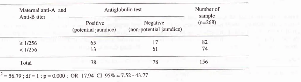
[image:4.595.55.546.239.374.2]an-tiglobulin
test
at thecut-off point of
11256.Table 4. Influence ofmaternal anti-A and anti-B antibody titer on potentialjaundice
Med J Indones
the quantity and
quality of
the antibodies,
severalother factors may also contribute
to the severity
of
ABO-HDN
among others theantibody
subclass,varia-tion of
antigen
subtypeof
thefetal
redblood cells,
the secretor systemof
the
fetal
tissue
andbody fluid,
theantibody
quantity
and speedtransfer transplacentally,
the
efficiencv
of
thefetal
mononuclear
cells
and
thematurity of
tÉefetus.l '2'to'tt'12 Afrcr
32weeks ofpreg-nancy
the
transplacental
transfer
of
IgG
antibody
increases
speedly
and at the endof
mature
pregnancythe
amount
of
IgG
molecules
in
the
placental
Matemal
anti-A
and Anti-B titerAntiglobulin test Number
of
sample(n=268) Positive
(potential jaundice)
Negative (non-potential j aundice)
> t/256 < 11256
65 13
17 61
82 74
156 78
78
Total
x2
=
56.'19; df =I
; P = 0.000;
OR
17.94Cl
95Vo =7.52-
43;:-':.By
the
bivariat
analysis
it
revealed that
atthe
cut-off
point
of
11256of the
maternal
anti-A
and
anti-B
an-tibody
titer
the
probability of
the
birth of
anewborn
with
potential
jaundice
was
significantly
greater thantiters
of <
11256.The
multivariat
analysismodel
was alsoperformed
on severalother variables
which
on the
bivariat
analysishad a significant role on the
occurence
of
potential
jaundice namely placental
inflammation,
the
way
thebaby
wasborn
and thefacilities
avaible
at the placeof
birth. On
the
multivariat
analysis
it
revealed that
theinfluence of maternal anti-A
oranti-B antibody titer
onthe occurence
of
potentialjaundice
baby
wasfound
atthe
cut-off point titer
of
2
11256with OR
18.65;
CI
95Vo=6.18-56.18.
DISCUSSION
In ABO incompatibility
pregnancies, maternal
anti-A
and
anti-B
antibodies
in
the
form
of
IgG can
crosstransplacentally
and thusmay
causedestruction of
thenewborn'
s redblood
cells. One of the factorscontribut-ing
the severity
of
ABO-HDN
is
the strength
andquantity
of
maternal
anti-A
andanti-B
antibody
crosstransplacentally.ru The higher the
antibody transfer
transplacentally
the higher
the probability
of
thedostruction
of
thenewborn's
red
blood
cells.
Besidestrophoblast
increases
so that at
birth
the
newborn
infant's
number
of IgG
molecules
is higher
than
the 13.14motner
s.To
predict
the occurenceof
potencialjaundice in
ABO
incompatible
pregnancies
it
is important to know
thecut-off point
of
the maternal anti-A and anti-B
an-tibody
titers
somuch
sothat
monitoring
action
of
theborn baby
can be
intensified. The
already
sensitizedred blood cells
of
the newborn can more easily
bedestructed
not only on the
fact
of its
condition
of
incompatibility itself but
also becauseof
other
causessuch as nosocomial infections, acidosis as
well
asdehydration. An
increaseof
thebilirubin
content
fol-lowing
exessive destruction
of
red blood cells is
al-ready
known
to be able tohinder
thebaby's growth
anddeveiopment
in
later
y"u.r.
l5'16'17On this study
with a sensitivity
and
a specificity of
83.33Vo
and
78.2lEo respectively
with
a positive
predictive
value
of
79,27Vo and anegative predictive
value
of
17.57Vo, acut-off
pointof
11256of
themater-nal anti-A
andanti-B
antibody titer
wasfound.
V/ith
those values,
by
thebivariat
analysisit
wasfound that
the risk
of
becoming
potencial
jaundice was at
thematernal
anti-A
andanti-B
antibody
titer of
>
11256work-ing
synergically as
well
as antagonistically
on
thenewborn's potential jaundice.
After
having performed
a
multivariat
analysis
on
other variables
contributing
in
the occurenceof
thenewborn's
potentional j aundiceat
that certain titer
the
ratio
odds was
OR
18.64;
CI
95Vo = 6.18 - 56.18. The obtained data revealed that the
titer
of
>
11256
of
the maternal anti-A and
anti-B
antibody the
risk of
thenewborn
infant with
potential
jaundice significantly
became
l8
times higher
com-pared
to
thosewith
low
antibody titers
(<
11256).This
study
wasconducted on the
middle to low
sosioeconomic
class
comprising the mayor
of
our
whole
population. For
clinical
application
it
can be suggested thatin
ablood group O
delivering mother with
a nonO newborn
infant,
maternal
anti-A
and
anti-B
an-tibody
titer
2
11256 on theantiglobulin
testis
amarker
and
warrant the health worker involved
to
intensify
monitoring
of
the newborn
infant. This is
dueto
thefact
that therisk for
potential jaundice
of
thenewborn
becomes
l8
times higher.
Acknowledgements
The
author
would
like to
express
his
sincere
andheartfelt gratitude
to all
themidwives of
thehospitals
and
health centers
involved for helping
in
collecting
the samples, to
referral laboratory of
theDKI
Jaya RedCross
Blood
Transfusion Service
for
the
seroim-munologic examinations,
toDrs Sudjadi
for
analyzing
the data and
to Dr.
Titi
SSularyo
for
her cooperation
in
preparing
themanuscirpt.
REFERENCES
l.
Oski FA, Naiman JL. Hematologic problems in the newborn. 3rd ed. Philadelphia: Saunders; 1982. p.283-346.2. Nathan
DG, Oski
FA.
Hematologyof
the infancy andchildhood. 4th ed. Philadelphia: Saunders; 1993. p. 17-l 15,
495-674.
3. Abelson
NM,
Rawson AJ. Studiesof
blood groupan-tibodies. V. Fractionation of examples
of
anti-B, anti-A,B,anti-M, anti-P, anti-Jkâ, anti-Lea, anti-D, anti-CD, anti-K, anti-Fya, anti-s and anti-Good. Transfu sion 196l :l :l | 6-23.
4. Rawson AJ, Abelson NM, Kochwa, et al. In: Bryant NJ, Ed.
An
introduction to immunohematology; 2nd ed. Philadel-phia: Saunders;1982. p. 197-200.5. Huntley
CC,
Lyerly AD,
Litrlejohn MP, er
al.
ABO hemolytic diseasein
PuertoRico
andNorth
Carolina. Pediatrics 197 6; 9 5 :87 5 -83.6. Oliver-Gonzales
J.
Functional antigens in helminths. J In-fect Dis 1946;7 9 :232-7 .7. Huntley CC, Lyerly AD, Patterson
MV.
Isohemagglutinins in parasitic infections. JAMA 1969; 208:l145-8.8. Zipursky
A.
Isoimumne hemolytic diseases.In:
Oski FA, Naiman JL.Eds. Hematologic
problems in the newborn. 3rd ed. Philadelphia: Saunders; 1982.p.283 -3 46.9. Mollison
PL.
Blood transfusionin
clinical medicine. In: Nathan DG, Oski FA, Eds. Hematology of the infancy andchildhood. 3rd ed. Philadelphia: Saunders; 1993. p.66-9. 10. Garraty
GG.
The serological investigationof
hemolyticdisease of the newbom. In: Sibanga CTHS, Das PC, Forfar
JO, Eds. Pediatric and blood transfusion. The Hague: Mar-tinus Nijhoff Publ; 1982. p. 4-21.
11. Bryant NJ. An introduction to immunohematology. 2nd ed.
Philadelphia: Saunders; I 982. p. 28-207, 264-326.
12. Ukita M, Tkahashi A, Nunotani T, et
al.
IgG subclassesof
anti-A and anti-B antibodies bound to the cord red cells in ABO incompatible pregnancies. Vox Sang 1989;
56:l8l-6.
13. Akin JW, Conover WB, DePriestPD.
Increasing quantityof matemal immunoglobulin G in trophoblastic tissue before
the onset oflabour. Am J Obstet Gynecol 199O1,162:1154-7.
14. Kohler PF, Farr RS. Elevation
of
cord over maternal IgG immunoglobulin: Evidencefor
an active placental IgG transport. Nature 1966; 1070-1.15. Hendarto SK, Sutomenggolo TS, Ismael S, Monintja HE. Longterm follow-up study in infants with intermediate levels
of neonatal hyperbilirubinemia. Pediatr Indon 1986;
26:83-92.
16. Nakamura H, Takada S, Shimabuku
RG.
Auditory nerve and brainstem responsesin
newbom infantswith
hyper-bilirubinemia. Pediatrics 1985; 75:703-8.17. Kemper KJ, Forsyth BW, McCarthy
PL.
Persistentpercep-tions of vunerability following neonatal jaundice. Am J Dis