Research Report
The role of human papillomavirus in
advanced laryngeal squamous cell carcinoma
Fauziah Fardizza, Bambang Hermani, Susyana Tamin Otorhinolaryngology Head and Neck Surgery Department
Faculty of Medicine Universitas Indonesia/ Dr. Cipto Mangunkusumo Hospital Jakarta
ABSTRACT
Background: Many studies had been conducted regarding the association of human papillomavirus (HPV) with laryngeal cancer. HPV was assumed to be one of the etiology of squamous cell carcinoma (SCC) besides smoking and alcohol consumption. Neck lymph node metastasis which is found in advanced laryngeal cancer could decrease the 5-year survival rate up to 50%. Purpose: This study aimed to investigate the role of HPV infections in the oncogenesis of the advanced laryngeal SCC and to evaluate the role of HPV in neck metastasis. Methods: Cross-sectional, double blind study with planned data collection. Data were taken from Formalin Fixed Paraffin Embedded (FFPE) of laryngeal cancer specimen after laryngectomy. Samples were analyzed by nested Polymerase Chain Reaction (PCR) and continuous flow-through hybridization for genotyping. Expression of Epidermal Growth Factor Receptor (EGFR), and Vascular Endothelial Growth Factor (VEGF) as metastasis biomarker were evaluated by immunohistochemistry. Results: Overall HPV proportion in laryngeal cancer was 28.7%. A total of 9% laryngeal cancer patients were infected with high risk HPV type and HPV 16 was found in 5 out of 7 samples. Mantel-Haenszel multivariate analysis found that HPV infection did not play a role in neck metastasis even though there were positive evidences of metastasis biomarker. On the contrary, in the absent of HPV, high expression metastasis biomarkers increased the risk of neck nodes metastasis: in EGFR 3.38 and VEGF 5.14 fold. Conclusion: HPV was found to be an oncogenic factor of laryngeal SCC, and HPV 16 was the most frequently observed type of HPV. HPV had protective function towards lymph node metastasis.
Keywords: Human papillomavirus, advanced laryngeal squamous cell carcinoma, expression of epidermal growth factor receptor, vascular endothelial growth factor, formalin fixed paraffin embedded
ABSTRAK
onkogenik KSS Laring, dan HPV 16 merupakan tipe yang paling sering ditemukan. HPV mempunyai fungsi protektif terhadap kejadian metastasis KGB lokoregional.
Kata kunci: Human papillomavirus, advanced laryngeal squamous cell carcinoma, expression of epidermal growth factor receptor, vascular endothelial growth factor, formalin fixed paraffin embedded
Author correspondence: DR. dr. Fauziah Fardizza, Sp. T.H.T.K.L.(K). Otorhinolaryngology Head and Neck Surgery Department Faculty of Medicine Universitas Indonesia/ Dr. Cipto Mangunkusumo Hospital, Jakarta. E-mail: [email protected]
INTRODUCTION
Head and neck malignancy accounts
for 5% of all malignancies and 3% of those
results in death. Laryngeal carcinoma is placed as the third most common malignancy after nasopharyngeal and paranasal sinus carcinoma.1 World Health Organization (WHO) reported, cited by Hermani,2 that 1.5 of 100,000 population died because of malignant tumor. In Indonesia, the incidence of laryngeal carcinoma is estimated to reach 1% of all malignancies in the otolaryngology head and neck area. The American Cancer Society reported in 2006, that America had
12,000 new cases with 4,740 mortality cases
caused by laryngeal carcinoma.
Siddiqui3 reported that Spain is also one of the countries with a high incidence of laryngeal carcinoma, with number of 20 out of 100,000 population. Countries with a high incidence of laryngeal carcinoma are Italy, France, and Poland. In India, data from the Pathology Anatomy Department, Patna Medical College Hospital, during 2002–2006,
reported 5,479 tumor cases: 768 cases in the head and neck region, 455 cases were diagnosed as benign tumors, and 214 cases
of malignant tumors with 18.7% of those cases were diagnosed with squamous cell carcinoma (SCC) of the larynx, placing above of nasopharyngeal cancer. From the data of the Pathology Anatomy Department, Faculty of Medicine Universitas of Indonesia/Cipto Mangunkusumo Hospital (FMUI-CMH), in
the period of 2000–2005, there were 3,344
cases of malignant tumors of the head and neck region with nasopharyngeal carcinoma
as the most common case with 948 cases
(28.3%), while laryngeal carcinoma with 225 cases (6.73%). Histologically, SCC comprises
95–98% of all laryngeal carcinoma cases.4 Otolaryngology Head and Neck Department
FMUI-CMH reported 310 laryngeal carcinoma cases in 2006–2014.5
Human papillomavirus (HPV) is the most common virus researched which is correlated with laryngeal carcinoma. Human papillomavirus is one of the factors that have the role as an oncovirus in developing countries. The prevalence of this virus involvement varies between 0–85%. Because of the wide variation of prevalence, it is important to know the prevalence of this oncovirus in Indonesia. Tumor with HPV involvement has a better prognosis and better response to therapy compared to tumor without HPV infection.6
Commonly, human papillomavirus infection is detected in malignancy cases, at high risk type, and among the high risk type, HPV 16 is the most common type of HPV found. In a molecular level, head and neck carcinoma with HPV has a different characteristic with malignancies without HPV. Malignancies with a positive HPV has a low expression of cyclin D,
Retinoblastoma protein (pRb) and p53 wild
type, also an increase of p16 that is not found in malignancies with negative HPV. This condition causes a difference in the molecular behavior that affects prognosis.7
tumors adhesion, damage of the protecting
barriers that confine the tumor, new growth
of blood vessels, lymphatic vessels, and others. These changes can be detected by using biomarkers. Accurate biomolecular analysis development is expected to portray the status of a patient’s tumor with or without
metastasis. This biomarker finding can be
used to determine the potential of metastasis to locoregional lymph nodes at the same time during the biopsy of the primary tumor. This
finding is expected to be used to determine
the potential of micrometastasis so that an integrated management such as neck dissection could be executed coinciding with
the definitive surgery. Molecular targeted
therapy could also be done in patients with late stadium SCC or those with metastasis.8-10
Angiogenesis role that is stated by the high expression of vascular endothelial
growth factor (VEGF) and epidermal growth factor receptor (EGFR) at the time of
diagnosis show a risk of metastasis to neck lymph nodes. Detection of these biomarker can predict metastasis to the neck lymph nodes, enabling determination of the right choice of therapy.11
According to this background, the author is interested in conducting a research regarding the analysis of laringeal SCC, how
the biomarkers expression of EGFR, MMP-9, and VEGF in laryngeal carcinoma with or
without HPV infection, and also to observe the role of the biomarker in determining the potential of metastasis by correlating it with locoregional lymph node involvement.
METHODS
This research used a cross sectional design, double blinded with a diagnostic research approach with expression of biomarkers EGFR, MMP-9, TIMP-1,
E-cadherin and collagen type IV, also the
infection of Human papillomavirus and its subtype in laryngeal SCC to determine the
diagnosis of locoregional lymph node that has potential for metastasis.
This research was conducted in Laryngopharyngeal Division, Department Otolaryngology HNS FMUI-CMH. HPV
examination used the GenoFlow Human
Papillomavirus Array Test-Kit, DiagCor with a technology of flow through hybridization. The assessment of the protein expressions
EGFR, MMP-9, and VEGF was done by
immunohistochemistry. All examinations for HPV and protein expressions were performed in the Department of Pathology Anatomy FMUI–CMH.
T h e r e s e a r c h p o p u l a t i o n w e r e laryngeal SCC patients that have undergone laryngectomy with or without neck dissection in the Department of Otolaryngology HNS FMUI-CMH with a paraffin block of the surgical tissue kept in the Department of Pathology Anatomy FMUI-CMH that were matched the inclusion criteria of no history of previous chemotherapy and/or radiation before surgery and no chronic illnesses such as TBC or hepatitis. The exclusion criteria was when the data of the laryngeal SCC patient’s medical record were incomplete
and/or the paraffin block of the patient could
not be used for laboratory analysis. Research subjects were included by random sampling
from the CMH medical record of 2006–2014.
The number of subjects were counted from the minimal research subject equation to answer the purpose of this research as large as 87 cases.
This research had been initially begun by identifying patients with the diagnosis of laryngeal SCC in the Department of O t o l a r y n g o l o g y H N S F M U I - C M H through tumor medical records in the Laryngopharyngeal Division (from 2006–
2014). Classification of the tumor stadium had
been done according to UICC 2002. Then, the data recorded into a special research status. Human papillomavirus subtype examination
Papillomavirus Array Test-Kit, DiagCor,
(GenoFlow; DiagCor Bioscience, Hong
Kong) with a technology of flow through hybridization. DNA extraction was conducted with DNeasy and Steinau et al12 modification from the paraffin block of laryngeal SCC. The
HPV genotyping results were read from the cellulose membrane in the form of dot blots
in a probe with a specific marker.
Immunohistochemistry (IHC) tissue had
been processed from the paraffin block that has been re-evaluated using Hematoxylin Eosin (HE) staining by the Department of Pathology Anatomy FMUI-CMH. The HE staining and IHC for all expressions of EGFR, TIMP-1, MMP-9, VEGF, E-cadherin and collagen type
IV of laryngeal SCC tissues were conducted in the Department of Pathology Anatomy FMUI-CMH. Immunohistochemistry staining
used specific antibodies for each protein.
The examination results were evaluated using the binocular light microscope Leica,
with a 400x enlargement. Image captured
was taken from each corner and evaluated using ImageJ software (Wayne Rasband, Bethesda, Maryland). The final score for each
research subject were the mean value of five
examinations.
Paraffin blocks from laryngeal SCC that had undergone laryngectomy from
the year 2006–2014 with or without neck
dissection were included in the study until the minimal number of subjects was reached. Anonymosity was executed in the collection of the data without attaching the name when transferring data from the medical record. The collected data was included the demographic characteristic, gender, age, cigarette smoking consumption, alcohol consumption, and clinical characteristics such
as clinical symptoms of hoarseness, difficulty
in swallowing, size of the tumor, tracheotomy b e f o r e s u rg e r y, t o t a l l a r y n g e c t o m y, pathological anatomy differentiation, therapy modality, the time distance to radiation postsurgery, and the existence of locoregional
lymph node enlargement according to UICC criteria. The existence and type of
HPV examination with GenoFlow Human
Papillomavirus Array Test-Kit, DiagCor was examined by using formalinized laryngeal SCC tissue in the form of parafin block.
Identification of HPV subtype was conducted
using the technology of flow through hybridization in the Molecular Pathology
Division and the protein expressions of EGFR MMP-9, and VEGF were assessed using
the immunohistochemistry examination in the Immunology Division, Department of Pathology Anatomy FMUI-CMH.
All numerical data were tested for normality values with Shapiro Wilk test. Quantitative data analysis that have an abnormal distribution was presented in median and range. Statistical correlation testing between 2 qualitative variables was
done using Chi Square or Fisher Exact test
if it does not comply with the parameteric conditions. Multivariate analysis was done by using binary logistic analysis. Variables that have a correlation with the locoregional lymph node status (p<0.25) from the bivariate analysis were included in the binary logistic regression. This was performed to investigate which independent variable had the strongest correlation with the dependent variable and then included in a multivariate logistic regression model.
Mantel Haenszel multivariate analysis
was done for biomarker expreession EGFR, MMP-9, VEGF and also HPV against locoregional lymph node metastatasis subject.
RESULTS
The characteristic of subjects found that male were more dominant than female with a ratio 16:1, with the biggest age group was the
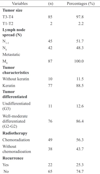
Ninety seven point eight percent of subjects were advanced laryngeal carcinoma. Lymph node spread were found in 51.7%, there was no distant metastasis. Keratin tumor was 88.5%, well-moderate differentiated
tumor G1-G2 was 86.4%. Chemoradiation was 56.3%, and recurrence was 25%.
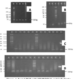
DNA HPV were found in 25 cases of Laryngeal Carcinoma with nested PCR using
MY09/MY11 and GP5+/GP6+ (Figure 3 and 4). Positive nested PCR continue with
flow-through hybridization (DiagCor) for genotyping. Sample result from DiagCor genotyping seen in Figure 5.
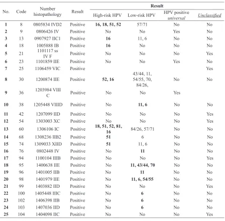
We found 7 cases of high-risk HPV and 8
cases of the low-risk type, as well as 3 cases of universal (unknown genotype) (Table 3).
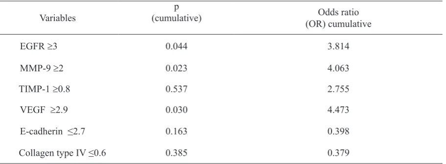
The Mantel-Haenszel test listed three kinds of positive biomarkers expression towards HPV infections that were able to be used to determine the potential of locoregional
lymph node metastasis, such as EGFR, MMP-9, and VEGF high expressions with p value
<0.05. This study had proven, when there was
no HPV infection, high expression of EGFR may pose the risk of N 3.814 times higher than low expression of EGFR. High MMP-9 expression was found to be 4.063 times likely
than low expression of MMP-9, as well as the
expression of high VEGF increased the risk to 4.473 times (Table 4).
Multivariate statistics indicated the HPV infection in SCC laryngeal cancer, despite
positive metastasis biomarkers (EGFR ≥3, MMP-9 and VGEF ≥2.9), patients were no longer at risk of locoregional lymph node metastasis. This study showed that HPV was statistically protective to locoregional lymph node.
Table 1. Demographic profile of patients
Variables (n) Percentages Young adult (18-44) 5 5.8 Smoking
Table 2. Size of tumor, lymph node spreading, metastasis, characteristic and differentiated of tumor
Variables (n) Percentages (%)
Figure 1. Immunohistochemistry EGFR expression staining of laryngeal carcinoma
Immunostaining positive on cell membrane and cytoplasm; (A) EGFR high positive expression, (B) EGFR positive ex -pression, (C) EGFR low positive ex-pression, (D) Negative control of EGFR expression (400x enlargement).
Figure 2. Immunohistochemistry VEGF expression stainning of layngeal carcinoma
Immunostainning positive on cell membrane and cyto-plasm; (A) VEGF high positive expression, (B) VEGF positive expression, (C) VEGF low positive expression, (D) Negative control of VEGF Expression (400 x enlargement).
Figure 3. DNA positivity and type of HPV in laryngeal carcinoma
Figure 4. Nested PCR with MY09/MY11 and Gp5+/Gp6+.
Figure 5. Genotyping DiagCor results
(A) Sample 39, showed genotyping DNA HPV with high risk, type 16, with coordinate; (3;C);
(B) Sample no.82, showed genotyping DNA HPV with low risk, type 11 and 54/55, with coordinate (1;B) and type 54/55 (2;A); (C) Sample 58, showed genotyping DNA HPV universal (1;F);
(D) Probe mapping for genotyping DNA HPV.
Table 3. HPV type profile using DiagCor
No. Code Number
histopathology Result
Result
High-risk HPV Low-risk HPV HPV positive
universal Unclassified
1 8 0805834 IVD2 Positive 16, 18, 51, 52 57/71 No No
2 9 0806426 IV Positive No No Yes No
3 13 0907927 IIC1 Positive 16 11, 6 No No
4 18 1005888 IB Positive 16 No No No
5 21 1101117 ss
IV F Positive No No No Yes
6 23 1101859 IIE Positive No No Yes No
7 25 1106459 VIC Positive Yes
8 30 1200874 IIE Positive 52, 16
43/44, 11, 54/55, 70,
84/26, No No
9 36 1203984 VIII
C Positive No No Yes
10 38 1205448 VIIID Positive No 11, 6 No No
11 42 1207099 IID Positive No No No Yes
12 54 1303003 XC Positive No No No Yes
13 60 1306106 IC Positive 18, 51, 52, 81,
16 84/26, 57/71 No No
14 68 1308236 IIB2 Positive 51 6 No No
15 74 1309033 XIID Positive 51 11, 6 No No
16 76 0802448 IV Positive No 11 No No
17 94 1100104 IIIB Positive No No No Yes
18 95 1400638 IIE Positive No 11, 43/44, 70 No No
19 96 1401005 IIB Positive No 11 No No
20 98 1401979 IIE Positive No 11, 6, 54/55 No No
21 99 1403882 IID Positive No No No Yes
22 100 1405448 IIIC Positive No 6 No No
23 102 1406398 IIB Positive No 6 No No
24 103 1407036 IID Positive No 6 No No
25 104 1404098 IIC Positive No No No Yes
Descriptions:
DISCUSSION
This research found HPV DNA as the etiology of SCC in laryngeal cancer, besides smoking and alcohol consumption as risk factors. Literatures have established that high risk HPV had been proven to be the etiology of malignant tumors. This research found the proportion of HPV DNA infections in laryngeal SCC patients were up to 28.7%.
Fouret et al, cited by Torente,13 found that the most common type of HPV infection in his research was HPV 16, reported 12 out of
19 (63.9%) in cervical carcinoma, all cases in laryngeal cancer, and 49 out of 55 (89%)
cases in head and neck cancer. One research
tried to compare between 93 laryngeal cancer and 49 cases normal laryngeal mucosa, 22
laryngeal nodules as control group. HPV DNA was detected by PCR, which they found
in 33 (35.5%) patients, 4 out of 49 (8.2%) were normal mucosa. Gungor et al14 used paraffin block in 99 patients and found 7.3
% was HPV-positive, which HPV type 6 and 11 were found to be the most common type, the only high risk type found was HPV 16. This research, utilized the same techniques, in 87 patients and acquired more than 28.7%
which was higher than Gungor’s, 28% were
high risk HPV.
Baumann et al15 detected HPV DNA in 6 out of 38 patients of early stage SCC laryngeal
cancer (T1 and Tis) and found subtypes 16,
26, 31, 39, and 52 HPV. HPV-positive SCC
laryngeal cancer patients mostly found to be young adults and rarely had smoking habits. This research found 7 cases (100%) of HPV-positive in adults and elderly age group, which was on the contrary to the literature. HPV-positive were also found more in well
and moderate differentiated tumors (G1/G2),
these were 6 out of 7 cases, whilst the only 1 was found in the undifferentiated tumor
(G3), otherwise, no HPV was found. This
was true according to the study conducted by Morshed et al.16 Morshed et al16 studied 40 patients of SCC laryngeal carcinoma and 33
non-neoplastic laryngeal lesions as control group. Human papilloma virus was found in
6 cases of SCC laryngeal cancer; 5 out of 6 positive-HPV showed G2 tumor differentiation
(moderately differentiated) and 1 (16,6%) cases
of G3 (poorly differentiated), whereas no HPV
infection tumor was considered as G1 (
well-differentiated). Four out of 6 positive-HPV was supraglotic, 1 case of tumor located in the glotic, and 1 subglotic case. Five out of 6
(83.4%) cases of positive-HPV were T3-T4, and one case was T
2. Three out of 6 HPV-positive tumors clinically showed no neck lymph node metastasis (N0), three of the positive-HPV Table 4. HPV influence to the expression of the metastasis with locoregional lymph node metastasis
biomarker
Variables
p
(cumulative) Odds ratio
(OR) cumulative
EGFR ≥3 0.044 3.814
MMP-9 ≥2 0.023 4.063
TIMP-1 ≥0.8 0.537 2.755
VEGF ≥2.9 0.030 4.473
E-cadherin ≤2.7 0.163 0.398
patients had N1 or N2 status. No HPV was found in control group. Morshed et al16 mentioned 15% of HPV infection may suggest the etiology of SCC laryngeal carcinoma, even though they
could not find the connection between the HPV
incidence and the stage of infection.
Bivariate analysis was implemented to measure the correlation of HPV infection and locoregional lymph node metastasis.
Three out of 7 positive-HPV (42.9%) were positive-N status and 4 (57.1%) cases were
negative-N status. This indicates there was
no statistical correlation (p=0.232, OR=0.36
and CI 95% 0.07-1.78).
The correlation of HPV infection and recurrency was found in 2 out of 7 cases of positive-HPV (28.6%), whereas the rest of the
five (71.4%) cases were not recurrent. There
were 17 recurrences in cases of
negative-HPV (24.3%) and 53 (75.7%) cases were not
recurrent, showing no statistically correlation.
Bozdayi et al17 studied 89 subjects of PCR assay and found HPV genome in 65 cases due
to inadequate tissue to amplify the G6PDH. Positive-HPV was found in 27 (41.5%)
cases. Positive-HPV was also found to be correlated to neck lymph node metastasis. The odds ratio analysis indicated positive-HPV as an important factor in neck lymph node metastasis or HPV to be the predictor for neck lymph node metastasis.
Mendhelson et al18 in his research studied the neck lymph node metastasis, HPV and p16. They stated that HPV and p16 may predict the poorly differentiated tumor. Tumor with positive-HPV and p16 increased the risk for neck lymph node metastasis (p=0.01, OR
23.9, CI 95% 2.2-265.1). Tumor with HPV and p16 positive showed superior predictive value towards lymph node metastasis compared to standardized histopathology examination
using HE. A multidiscipline team managing
the head and neck tumor with HPV and p16 must consider neck lymph node management carefully. This study stated a different result
than that of Bozdayi et al17 and Mendhelson et al18.In this study, HPV infection also indicates a tendency to a different kind of SCC laryngeal carcinoma without HPV infection, having better nature, even though not yet proven statistically.
Mantel-Haenszel test analysis showed
3 kinds of positive biomarkers expression in
the role of diagnosis of potential locoregional
lymph node metastasis, which were EGFR, MMP-9, and VEGF. This analysis also stated
that HPV infection statistically had a role in protecting locoregional lymph node metastasis. Further research is needed to prove this correlation. In SCC laryngeal carcinoma with HPV infection, with the presence of either high or low expression of the biomarkers
(EGFR, MMP-9 and VEGF) statistically had
no relationship with the N status having the
same CI 95% of all 3 biomarkers, 0.14-1.17. On the contrary, without HPV infection, the locoregional lymph node metastasis was
3.814 times likely to occur in high expression of EGFR than the low expression. And so for
MMP-9 expression, the risk is increased to
4.063 times in high expressed MMP-9, as well as high expression of VEGF, implying the risk 4.473 times higher. In cases of laryngeal
SCC with or without HPV infection, there
were differences in the profile expression of EGFR, MMP-9, TIMP-1, VEGF, E-cadherin
and collagen type IV, between patients with positive and negative N status.
Human papillomavirus was found in 28.7 % of laryngeal SCC. HPV high–risk type was found in 9.1% cases and low-risk type
in 10.4% cases. In laryngeal SCC without
HPV infection, there was a difference in the
expression of the biomarker profiles EGFR,
MMP-9, and VEGF between the positive and negative N status, but in cases with HPV infection, the difference in the expression of
the biomarker profile could not determine
REFERENCES
1. Marur S, Arlene, Forastiere. Head and neck cancer: Changing epidemiology, diagnostic and treatment. Mayo Clin Proc. 2008; 83:489‒501.
2. Hermani B. Perkembangan terapi bedah tumor ganas laring dalam upaya meningkatkan kualitas hidup [Pengukuhan guru besar]. Jakarta: Universitas Indonesia; 2007. 3. Siddiqui S, Chandra R, Aziz A, Suman S.
Epidemiology and histopatological spectrum of head and neck cancer in Bihar, a state of Eastern India Asian Pasific. J Cancer Prev. 2012;13:3949‒53.
4. Data from Pathology Anatomy Department of Faculty of Medicine Universitas Indonesia, Dr. Cipto Mangunkusumo Hospital. 2000‒2005.
5. Fardizza F. Data of laryngeal cancer patients of Otorhinolaryngology Head and Neck Surgery Department, Faculty of Medicine Universitas Indonesia, Dr. Cipto Mangunkusumo Hospital. 2006‒2011. 6. Oliveira D, Bacchi M, Macarenco R,
Tagliarini J, Cordeiro R, Bacchi C. Human papillomavirus and Epstein‒Barr Virus infection, p53 expression, and cellular proliferation in laryngeal carcinoma. Am J Clin Pathol. 2006;126:284‒93.
7. Koskinen WJ. Prognostic markers in head and neck carcinoma [disertasion]. Helsinki: Helsinki University Sentral Hospital; 2006. 8. Cortesina TG. Molecular metastases
markers in head and neck squamous cell carcinoma: Review of the literature. Acta Otorhinolayngol Ital. 2006; 26:317‒25. 9. Perez-Ordonez B, Beauchemin M, Jordan
R. Molecular biology of squamous cell carcinoma of head and neck. J Clin Pathol. 2006;59:445‒53.
10. Sno PPGB, Leemans CR, Tiwari R. Management of cervical lymph nodes in patients with head and neck cancer. Eur Arch Otorhinolaryngology. 1992; 249:187‒94.
11. Nakata Y, Uzawa N, Takashi K, Sumino J, Michikawa, Sato H, et al. EGFR gene copy number alteration is better prognostic indicator than protein overexpression in oral tongue squamous cell carcinoma. European J of Cancer. 2011; 47:2364‒72.
12. Steinau M, Patel SS, Unger ER. Technical advance: Efficient DNA extraction for HPV genotyping in formalin‒fixed, paraffin embedded tissues. J Mol Diagn. 2011;13(4):377‒81.
13. Torente MC, Ojeda JM. Exploring the relation between human papilloma virus and larynx cancer. Acta Oto‒Laryngologica. 2007;127:900‒6.
14. Gungor A, Cincik H, Baloglu H, Cekin E, Dogru S, Dursun E. Human papillomavirus prevalence in laryngeal squamous cell carcinoma. The Journal of Laryngology & Otology. 2007; 121:772–4.
15. Baumann J, Cohen S, Evjen A, Law JH, Vadivelu S, Attia A, et al. Human papillomavirus in early laryngeal carcinoma. Laryngoscope. 2009; 119:1531‒7.
16. Morshed K, Korobowicz E, Szymański M, Skomra D, Gołąbek W. Immunohistochemical demonstration of multiple HPV types in laryngeal squamous cell carcinoma. Eur Arch Otorhinolaryngol. 2005; 262(11):917‒20. 17. Bozdayi G, Kemaloglu Y, Ekinci O,
Dogan B, Ilhan M, Aydil U, et al. Role of human papillomavirus in the clinical and histopathologic features of laryngeal and hypopharyngeal cancers. J Otolaryngol Head Neck Surg. 2009; 38(1):119‒25.