Effects of initial and long-term lipid-lowering therapy on vascular
wall characteristics
Froukje L. Ubels
a,*, Jaap H.J. Muntinga
b, Jasper J. van Doormaal
a,c,
Wepco D. Reitsma
a, Andries J. Smit
aaDepartment of Internal Medicine,Uni6ersity Hospital,P.O.Box30.001,NL-9700RB Groningen,The Netherlands bDepartment of Medical Physiology,Uni6ersity of Groningen,Bloemsingel10,NL-9712KZ Groningen,The Netherlands cDepartment of Cardiology,Out-patient clinic for Atherosclerosis/Lipid Disorders,Uni6ersity Hospital,P.O.Box30.001,
NL-9700RB Groningen,The Netherlands
Received 13 September 1999; received in revised form 28 February 2000; accepted 3 March 2000
Abstract
Several studies have demonstrated the beneficial effects of 3 hydroxy-3-methylglutaryl-coenzyme A (HMG-CoA) reductase inhibitors on vascular properties, but little is known about treatment intensification. We compared patients in whom statins were started (INITIAL,n=30) for hypercholesterolaemia (\6.5 mmol l−1) with a matched patient group of long-time statin users, with similar baseline characteristics for lipids, intima-media thickness (IMT), and pulse wave velocity, in whom treatment with statins was intensified (LONG-TERM,n=54). At baseline and after 1 year, lipid profile, IMT of the carotid and femoral arteries, aortic distensibility using pulse-wave velocity and various properties of the peripheral vascular bed using a recently developed bio-impedance method were measured. After 1 year the relative changes in lipid profile were significantly better in the INITIAL compared with the LONG-TERM-group. The relative changes in IMT of the mean internal carotid and common femoral arteries significantly differed between the INITIAL and LONG-TERM-group (−8 and +11%, −11 and +22%, respectively). After 1 year, in both groups, most other vascular wall characteristics were unaltered compared with baseline. In conclusion, the beneficial structural alterations of the vascular wall were greater after starting than after intensifying already existing lipid-lowering treatment. This suggests that other effects of HMG-CoA reductase inhibitors than lipid-lowering alone must be involved in vascular changes. © 2001 Elsevier Science Ireland Ltd. All rights reserved.
Keywords:Hypercholesterolaemia; Lipid-lowering drugs; 3 Hydroxy-3-methylglutaryl-coenzyme reductase inhibitor; Clinical trial; Intima-media thickness; Vascular wall
www.elsevier.com/locate/atherosclerosis
1. Introduction
Several clinical trials have demonstrated favourable effects of lipid-lowering therapy on cardiovascular mor-bidity and mortality, and on coronary atherosclerosis measured by quantitative coronary angiography [1,2]. Lipid-lowering therapy also showed beneficial effects on both structural and functional properties of the vascular wall, using non-invasive vascular measure-ments. Some placebo-controlled trials assessed the ef-fect of lipid-lowering therapy during 2 – 4 years on the intima-media thickness (IMT) of the carotid and
femoral arteries, measured by high resolution B-mode ultrasound [3], on the endothelial function of the brachial artery and on the arterial distensibility of the carotid artery [2,4 – 7]. The observed vascular changes appeared to be usually related to the degree of lipid-lowering, even when additional effects of lipid-lowering drugs independent of lipid reduction were assumed to be operative [8 – 10]. Thus, lipid target values have now become a major issue and have initiated several ongo-ing studies comparongo-ing the effects of startongo-ing with low versus high doses of statins. However, the use of these 3 hydroxy-3-methylglutaryl-coenzyme A (HMG-CoA) reductase inhibitors has become widespread in clinical practice, the dose usually being lower than that used in the high dose studies described above. Therefore, posi-* Corresponding author. Tel.: +31-50-3616161; fax: +
31-50-3619069.
E-mail address:[email protected] (F.L. Ubels).
tive results of high dose studies may result in treatment intensification in many patients.
The aim of this study was to compare retrospectively changes in vascular wall characteristics after intensify-ing treatment, with those after startintensify-ing lipid-lowerintensify-ing drugs. If the effect of statins on the vessel wall would be independent of the degree of lipid-lowering, intensifying treatment might be less effective than expected. We compared starting and intensifying treatment in previ-ously untreated and in conventionally treated hyperlipi-daemic patients, respectively, on various vascular characteristics.
2. Methods
2.1. Study population and design
The study population consisted of participants of the Dutch project On Cardiovascular Tracing Of Risk fac-tors, Bristol-Myers Squibb (DOCTOR)-study, a study which evaluated the effects of reducing cardiovascular risk factors on changes of clinical and biochemical cardiovascular risk parameters. In our Out-patient Clinic for Atherosclerosis/Lipid Disorders, in the years 1995 – 1996, 84 patients were recruited with untreated or treated moderate to severe hypercholesterolaemia. Moderate to severe hypercholesterolaemia was defined as a fasting plasma total cholesterol between 6.5 and 12.0 mmol l−1, resistant to diet or lipid-lowering
ther-apy. Inclusion was independent of a history of cardio-vascular events or use of cardiocardio-vascular drugs. Patients with a medical history of unstable angina pectoris, poorly controlled congestive heart failure, diabetes mel-litus, impaired renal or hepatic function, rheumatoid arthritis and drugs or alcohol abuse were excluded. The use of both simvastatin or pravastatin as HMG-CoA reductase inhibitor was allowed. The dose was titrated with the aim to reduce total cholesterol B5.0 mmol l−1
and/or low-density lipoprotein (LDL) – cholesterol
B3.5 mmol l−1
. To reach this treatment goal, combi-nation therapy was given with other lipid-lowering drugs such as fibrates, bile acid resins or niacin if necessary. At baseline and after 1, 3, 6 and 12 months, fasting lipid profile and blood glucose was determined and the lipid-lowering medication adjusted if needed. Non-invasive vascular measurements were made at baseline and after 1 year.
2.2. Patients characteristics
Participants were divided retrospectively into two groups according to their treatment status at inclusion in the study. One group consisted of previously un-treated patients in which treatment with lipid-lowering drugs was initiated for the first time (INITIAL) and
another group consisted of patients using lipid-lowering therapy in which lipid-lowering treatment was inten-sified (LONG-TERM). Body mass index (BMI) was calculated by dividing the body weight by the square of the length (kg m−2). The existence or absence of
car-diovascular disease was recorded using patient histories. Coronary heart disease (CHD), cerebrovascular acci-dents (CVA), peripheral vascular disease (PVD) and a family history of cardiovascular diseases were defined according to current clinical practice. Smoking was classified as ‘never’, ‘formerly’ for patients who stopped smoking before participation in the study for at least 3 years, and ‘currently’ for patients who continued smoking.
2.3. Biochemical parameters
After an overnight fast, blood samples were taken to measure glucose, cholesterol, high-density lipoprotein (HDL) – cholesterol, triglyceride and lipoprotein(a) us-ing standard laboratory methods. LDL – cholesterol was calculated by the Friedewald formula [11] and the cholesterol/HDL – cholesterol ratio by dividing total cholesterol by HDL – cholesterol.
2.4. Non-in6asi6e6ascular parameters
The parameters were obtained by two observers (vas-cular technicians) blinded to the assignment of the patients to the INITIAL or LONG-TERM-group.
Blood pressure was measured after 5 min of rest in sitting position, using a calibrated automatic oscillo-metric manometer (mmHg).
The IMT was measured using high resolution B-mode ultrasound (Acuson XP128 duplex scanner) [12]. In supine position, the far wall of different segments of the carotid arteries (common carotid artery (CCA) and internal carotid artery (ICA) and the carotid bulb (CB)) and the right common femoral artery (CFA) and su-perficial femoral artery (SFA) were scanned and as-sessed. The IMT was defined as the distance between the intima and media double line pattern, expressed in millimetres and reported as mean value of the right and left side.
Aortic distensibility (Dao) was determined with
pulse-wave velocity measurements as extensively described elsewhere [13], using a computerised program, devel-oped in our department [14,15]. Two Doppler probes (5 MHz each) were placed on conducting gel upon the proximal part of the right subclavian artery and the right common femoral artery just below the inguinal ligament. Simultaneous registration of the Doppler pulses was made three times during 10 s with in the meantime withdrawal of the Doppler probes from the skin. Dao (in MPa
−1
upstrokes of the maximum flow velocity waveforms recorded by each Doppler transducer and the distance between the top of the manubrium sterni and the right common femoral artery as a measure of the aortic length.
Various properties of the upper arm vascular bed were investigated using an electrical bioimpedance method as described previously [16]. In brief, two po-tential measuring electrodes were applied around the left upper arm, with a specially designed blood pressure cuff around them. Likewise, two current conducting electrodes were placed around the proximal and distal part of the left arm. A continuous blood pressure registration at the left upper arm was made using a Finapres device (Ohmeda 2300) at the right hand, and correcting the recorded values for left – right and hydro-static blood pressure differences. During a measure-ment, the cuff was rapidly inflated to 20 mmHg above the maximal systolic blood pressure, subsequently the cuff pressure was kept constant during 2 min, after which the cuff was slowly deflated in 4 min. From the concomitant changes in electrical impedance, measured using a Minnesota impedance cardiograph (model 304A; Instrumentation for Medicine), and the transmu-ral pressure values, calculated by subtracting the cuff
pressure from the mean arterial blood pressure, various physiological parameters are estimated. Parameters used in this study were the arterial bloodvolume (V), compliance (C) and distensibility (D). Their values at ambient blood pressure level are represented byVa, Ca
and Da, respectively. The values at zero transmural
pressure (Va(0), Ca(0) and Da(0)) are used as
representa-tives of the largest arteries, and at higher transmural pressures, for example 80 mmHg (Va(80), Ca(80) and
Da(80)), as representatives of the arterial bed as a whole.
The initial venous blood volume and the extravascular volume are denoted byVvandVi, respectively. The cuff
pressure at which the veins start to refill during cuff deflation is denoted byPthr. The difference between the
reciprocal values of the impedances before and after cuff pressure changes (Ydiff) is used as a measure of the
myogenic response (reverse stress relaxation or delayed compliance) of the arm veins and expressed as a venous blood volume (Vdiff). The mean arterial pressure and
mean heart rate during the measurement are described by Pmn and MHR, respectively. The repeatability of
this method was comparable with established methods and within the physiologically acceptable range [17].
2.5. Statistical analysis
Statistical tests were performed with SPSS version 8.0. Results are given in mean9S.D. and interquartile ranges for the duration of existing lipid-lowering ther-apy. Differences in baseline clinical characteristics be-tween both groups of patients were determined by ANOVA. Paired t-tests were used to compare the biochemical and vascular parameters before and after 1 year in each of the defined groups of patients. Absolute and relative changes after 1 year were compared with both groups using multiple-comparisons. Univariate re-gression analysis was performed to determine different correlations of the vascular wall characteristics at base-line and after 1 year. Relative changes in biochemical and vascular parameters were defined as the percent-ages of the differences with regard to the initial values divided by the initial values, multiplied by 100. Differ-ences were considered statistically significant at P -val-ues B0.05.
3. Results
3.1. Initial parameters
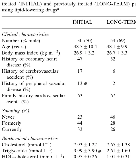
Baseline clinical and biochemical characteristics of patients in the INITIAL and LONG-TERM-group are mentioned in Table 1. The proportion of patients treated for secondary prevention of cardiovascular dis-eases was comparable in the INITIAL and LONG-TERM-group with 60 and 54%, respectively. In the Table 1
Baseline clinical and biochemical characteristics of previously un-treated (INITIAL) and previously un-treated (LONG-TERM) patients using lipid-lowering drugsa
History of coronary heart 47 52 disease (%)
History of cerebrovascular 17 6 accident (%)
13c 2
History of peripheral vascular disease (%)
Family history cardiovascular 63 67 events (%)
Cholesterol (mmol l−1) 7.6791.38
Triglyceride (mmol l−1) 3.9993.90c 2.6191.60
HDL–cholesterol (mmol l−1) 0.9590.26 1.0190.31
LDL–cholesterol (mmol l−1) 5.2891.47 5.4991.36
8.9696.10 9.1693.98
Cholesterol/HDL ratio
Lipoprotein(a) (mg l−1) 2319285 3149326
4.490.6 Glucose (mmol l−1) 4.590.6
aValues are expressed as mean9S.D.; HDL, high-density
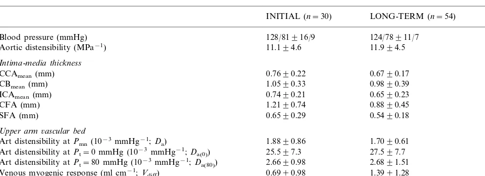
Table 2
Baseline vascular characteristics of previously untreated (INITIAL) and previously treated (LONG-TERM) patients using lipid-lowering drugsa
LONG-TERM (n=54) P-value INITIAL (n=30)
128/81916/9
Blood pressure (mmHg) 124/78911/7 n.s.
11.994.5 n.s.
11.194.6 Aortic distensibility (MPa−1)
Intima-media thickness
0.6790.17
0.7690.22 n.s.
CCAmean(mm)
0.9890.39 n.s.
CBmean(mm) 1.0590.33
0.6590.23
0.7490.21 n.s.
ICAmean(mm)
1.2190.74
CFA (mm) 0.8890.45 n.s.
SFA (mm) 0.6590.29 0.5490.18 n.s.
Upper arm6ascular bed
Art distensibility atPmn(10−3mmHg−1;Da) 1.8890.86 1.7090.61 n.s.
27.597.7
25.597.3 n.s.
Art distensibility atPt=0 mmHg (10−
3mmHg−1;D a(0))
2.6891.51
Art distensibility atPt=80 mmHg (10−3mmHg−1;Da(80)) 2.6690.98 n.s.
1.3991.28
0.6990.98 0.018
Venous myogenic response (ml cm−1;V diff)
Extravascular volume (ml cm−1;Vi) 1609104 2499168 0.012
aValues are expressed as mean9S.D.; CCA, common carotid artery; CB, carotid bifurcation; ICA, internal carotid artery; CFA, common
femoral artery; SFA, superficial femoral artery; mean, mean value of both sides; Art, arterial;Pmn, mean arterial pressure;Pt, arterial transmural
pressure; n.s., not significant.
INITIAL-group, more patients had a history of periph-eral vascular disease (PB0.05). At baseline, in the LONG-TERM-group, 23 (43%) participants were treated with pravastatin with a mean dose of 37 mg, and 28 (52%) with simvastatin in a mean dose of 44 mg, respectively. Of these patients 16 (30%) were additionally using combination therapy with one of the statins and three patients (6%) used monotherapy with other lipid-lowering drugs. Mean duration of lipid-lipid-lowering treat-ment was 18 (1.25 – 30) months. Promptly after inclusion, one patient in the LONG-TERM-group dropped out of the study because of a severe myocardial infarction.
Baseline triglyceride values were higher in the INI-TIAL-group. There were no other differences in lipid profile at baseline. The lipid profile of the patients in the LONG-TERM group at the start of the first lipid-lower-ing therapy was not significantly different from that at the moment of inclusion in the present study.
Table 2 shows the non-invasively determined vascular parameters. No significant differences were found in the mean IMT of the carotid and femoral artery in the INITIAL compared with the LONG-TERM-group, al-though the IMT of the CCA and CFA tended to be higher in the INITIAL-group. The myogenic response of the peripheral veins, and the extravascular volume were lower in the INITIAL compared with the LONG-TERM-group (P=0.018 and 0.012, respectively). At baseline, no further differences in vascular wall charac-teristics between both groups were found.
Among the observed baseline correlations, the choles-terol/HDL – cholesterol ratio was positively correlated with the IMT of the mean CCA (r=0.36,PB0.01), but also with theCa(80) (r=0.49, PB0.001), in the overall
group.
3.2. Follow-up parameters
After 1 year of treatment, in the INITIAL-group ten (33%) patients were treated with pravastatin and 20 (67%) with simvastatin in mean doses of 36 and 37 mg, respectively. Eight (27%) patients using simvastatin were additionally treated with other lipid-lowering drugs. In the LONG-TERM-group, 15 (28%) participants were treated with pravastatin and 35 (65%) with simvastatin, in mean doses of 38 and 44 mg, respectively. Eight patients using pravastatin and 17 simvastatin (in total 46%) were additionally treated with other lipid-lowering drugs. Monotherapy with other lipid-lowering drugs than HMG-CoA reductase inhibitors was prescribed in three (6%) patients in the LONG-TERM-group. After 1 year, two patients dropped out in the INITIAL and five in the LONG-TERM-group, because of moving (n=1) and non-compliance (n=1) in the INITIAL and psycho-social problems (n=1), non-compliance (n=3) and misunderstanding (n=1) in the LONG-TERM-group. After initiating or intensifying (1 year) lipid-lowering therapy, the lipid profile significantly improved in both groups, except for the triglycerides in the LONG-TERM-group. In the INITIAL-group the total cholesterol fell with 2.18 mmol l−1to a mean value of 5.63 mmol l−1,
in the LONG-TERM-group with 1.55 to 6.12 mmol l−1
(not significant (n.s.) between both groups). The triglyc-erides fell with 0.99 to 2.85 mmol l−1
in the INITIAL-group, and with 0.23 to 2.21 mmol l−1 in the
LONG-TERM-group (n.s. between both groups). The HDL-cholesterol rose with 0.25 to 1.22 mmol l−1in the
INITIAL-group, and with 0.12 to 1.19 mmol l−1
the INITIAL-group, and with 1.51 to 4.1 mmol l−1 in
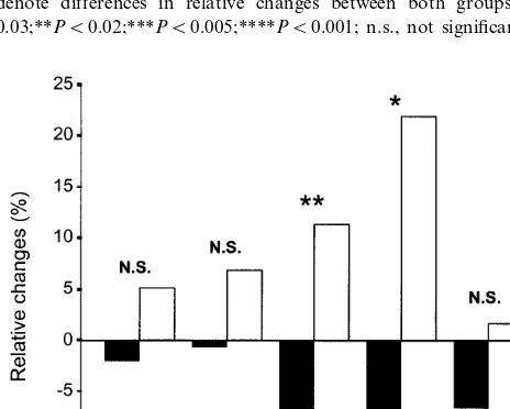
the LONG-TERM-group (n.s. between both groups). The cholesterol/HDL – cholesterol ratio fell with 3.9 to 4.6 in the INITIAL-group, and with 2.1 to 5.5 in the LONG-TERM-group (P=0.008 between both groups). Baseline lipid values of the dropouts in both groups were excluded. The relative changes in lipid profile were significantly more pronounced in the INITIAL
com-pared with the LONG-TERM group as represented in Fig. 1, except for the triglyceride levels.
The relative changes in IMT of the mean ICA and CFA as well, were significantly different between both groups, with a decrease in the INITIAL and an increase in the LONG-TERM-group (Fig. 2). The absolute change in the mean ICA IMT was −0.08 mm in the INITIAL-group, and +0.047 mm in the LONG-TERM-group (P=0.011). For the mean CFA the change was −0.25 mm in the INITIAL-group, and +0.12 mm in the LONG-TERM-group (P=0.005). The IMT of the other arterial segments showed the same tendency, although no statistical significance was reached. The mean heart rate increased in the INI-TIAL-group from 64.1910.6 to 69.7911.8 min−1
(P=0.011) but remained unaltered in the LONG-TERM-group (64.599.4 and 66.199.4 beats min−1).
The extravascular volume tended to increase in the INITIAL, and to decrease in the LONG-TERM-group, with a nearly statistical difference in relative change between both groups (P=0.059). Other IMT segments, mean bloodpressure, Dao and the upper arm vascular
properties (arterial distensibility and compliance) re-mained largely unaltered compared with baseline in both groups.
In the LONG-TERM-group a relation was found between the decrease in cholesterol/HDL – cholesterol ratio and the fall in IMT of the CCA (r=0.29, P= 0.055). Remarkably, no significant correlations were found between the relative changes in lipid profile and IMT or other vascular measurements.
4. Discussion
The present study shows that initiating lipid-lowering therapy is markedly more beneficial on the lipid profile and the IMT of the carotid and femoral arteries than intensified, long-term pharmacological lipid-lowering therapy in patients with comparable biochemical and vascular characteristics at baseline. Although the lipid profile improved in the LONG-TERM-group, several IMT segments even increased.
One year follow-up was sufficient to detect changes in the CCA IMT between both study groups. In several studies of lipid-lowering treatment, there was a ten-dency for changes in IMT after 6 months of active treatment with a significant reduction in IMT after 1 year [3]. So, the increase in IMT over 1 year during successful treatment in the LONG-TERM-group was unexpected, but may represent retarded progression. The increase was even comparable to that observed in the placebo arms of the several lipid intervention-IMT-studies and can therefore be seen as a reflection of an age-related increase in IMT [18]. Thus, remarkably, during 1 year of relatively successful additional contin-Fig. 1. Relative changes (%) in lipid profile after 1 year. Black and
white bars, INITIAL and LONG-TERM-group, respectively (see text). CHOL, total cholesterol; TG, triglyceride; HDL, high-density lipoprotein cholesterol; LDL, low-density lipoprotein cholesterol; CHOL/HDL, cholesterol/HDL-cholesterol ratio. Significance levels denote differences in relative changes between both groups,*PB 0.03;**PB0.02;***PB0.005;****PB0.001; n.s., not significant.
ued lowering of lipids, the observed IMT increases. Assuming a causal relationship between the changes in IMT and lipid levels, this is unexpected. Another point in favour of an initial vascular effect of starting statins independent of lipid-lowering is the lack of correlations between changes in lipid profile and vascular wall char-acteristics. Others have also found this [9]. This does suggest an initial effect of statins, independently of the lipid-lowering itself. Such non-lipid effects of statins may consist of, for example, the modification of en-dothelial function and insulin resistance, increase in plaque stability and antithrombotic properties [8 – 10,19]. Furthermore, the LDL particle size may be of importance, as has been shown for cardiovascular events in various regression studies [20,21].
In most regression studies a single non-invasive vas-cular measurement was used. As Muramatsu et al. [5], we investigated several vascular characteristics. Three complementary techniques were used to investigate these characteristics, reflecting functional changes mainly. However, the aortic distensibility and arterial distensibility and compliance of all vessels of the upper arm bed failed to show differences between both groups. In contrast to others [5], we found an increase in mean heart rate in the INITIAL-group after 1 year of treatment. Although speculative, this could be a result of an increased sympathetic activity to compen-sate the therapy-induced decrease in IMT in order to maintain the same vascular diameter. Because of the lack of a relationship between changes in IMT and heart rate, other factors must have been involved also. Both groups showed moderately severe hypercholes-terolaemia, with higher cholesterol levels at inclusion in 1995 – 1997 than in most other vascular studies of lipid-lowering treatment in secondary or even primary pre-vention settings. Thus, because both groups included mostly patients with previous cardiovascular disease manifestations in particular, the LONG-TERM-group was considerably undertreated at the moment of inclu-sion [22]. Considering the number of patients already on the highest registered dose of pravastatin or simvas-tatin (40 mg daily in the Netherlands) at inclusion, and the higher number of patients treated with combination therapy also, it was expected that it would be more difficult to reach the therapeutic goals in the LONG-TERM than in the INITIAL-group. Therefore, the originally aimed intensification of the treatment in the LONG-TERM-group, resulting in a quite modest dif-ference in lipid changes between both groups, was not realised by increasing the doses of statins in the first place, but by transition to another statin in the same dosage [23], frequently by the use of combination ther-apy and possibly by increased treatment compliance and alterations in time of ingestion of the medication. The significantly higher triglyceride levels at baseline, together with the significantly more pronounced
in-crease in HDL – cholesterol and dein-crease in LDL – cholesterol after 1 year of treatment in the INITIAL-group, could have been the result of a more atherogenic lipoprotein profile pattern B in the INI-TIAL-group compared with the LONG-TERM-group [24]. In that case however, the other observed differ-ences in treatment effects have to be the result of the additional lipid-lowering treatment and/or life-style modifications, since statins have been shown to influ-ence the lipid levels in a similar way in patients with different LDL-subclasses [24,25]. Since the LDL-parti-cle size, HDL-subfractions and other non-routine parameters of the atherogenic lipid profile were not determined, these questions could not be answered in the present study. Comparisons between two subsets of the INITIAL and LONG-TERM group (consisting of 27 and 50 patients, respectively) with similar triglyce-ride levels, showed similar effects of lipid-lowering ther-apy as in the original patient groups.
The IMT in both groups was relatively low at base-line. In most other lipid intervention-IMT-studies the mean CCA IMT was \0.8 mm, in many studies even
\1.0 mm [3]. Most of these studies were performed in a secondary prevention setting, but our study also concerned mostly patients with pre-existing cardiovas-cular disease manifestations. Maybe the lower age of our patients than in most other regression studies is responsible. The 1 year change in the mean CCA IMT in the INITIAL-group was relatively large compared with that in other studies, and even larger for other vessel segments, especially the femoral artery. For other segments than the CCA little data are available on changes during treatment. Only in the Regression Growth Evaluation Statin Study (REGRESS) femoral arteries were followed and also showed more marked changes compared with carotid vessels [26].
A confounding factor in this study might have been the use of cardiovascular drugs. Because these drugs remained unaltered during the study period and pa-tient’s baseline characteristics were comparable for both groups, this only can have had minimal influence on the results.
In conclusion, the beneficial effects of initiating lipid-lowering therapy on the IMT of the carotid and femoral arteries were greater than that of continuing already existing lipid-lowering treatment. As discussed above, the lipid-lowering effects of HMG-CoA reduc-tase inhibitors could not have only been responsible for that, but other factors must have been involved also.
Acknowledgements
References
[1] Waters D, Pedersen TR. Review of cholesterol-lowering therapy: coronary angiographic and events trials. Am J Med 1996;101(suppl. 4A):34S – 9S.
[2] Vogel RA, Corretti MC, Gellman J. Cholesterol, cholesterol lowering, and endothelial function. Prog Cardiovasc Dis 1998;41:117 – 36.
[3] Ubels FL, Terpstra WF, Smit AJ. Carotid intima-media thick-ness: influence of drug treatment and clinical implications. Neth J Med 1999;55:188 – 95.
[4] Vogel RA, Corretti MC, Plotnick GD. Changes in flow-medi-ated brachial artery vasoactivity with lowering of desirable cholesterol levels in healthy middle-aged men. Am J Cardiol 1996;77:37 – 40.
[5] Muramatsu J, Kobayashi A, Hasegawa N, Yokouchi S. Hemo-dynamic changes associated with reduction in total cholesterol by treatment with the HMG-CoA reductase inhibitor pravas-tatin. Atherosclerosis 1997;130:179 – 82.
[6] Simons LA, Sullivan D, Simons J, Celermajer DS. Effects of atorvastatin monotherapy and simvastatin plus cholestyramine on arterial endothelial function in patients with severe primary hypercholesterolaemia. Atherosclerosis 1998;137:197 – 203. [7] Schobel HP, Schmieder RE. Vasodilatory capacity of forearm
resistance vessels is augmented in hypercholesterolemic patients after treatment with fluvastatin. Angiology 1998;49:743 – 8. [8] Vaughan CJ, Murphy MB, Buckley BM. Statins do more than
just lower cholesterol. Lancet 1996;348:1079 – 82.
[9] Williams JK, Sukhova GK, Herrington DM, Libby P. Pravas-tatin has cholesterol-lowering independent effects on the artery wall of atherosclerotic monkeys. J Am Coll Cardiol 1998;31:684 – 91.
[10] Nordmann A, Martina B, Keller U, Battegay E. Lipid-lowering therapy: new pathophysiological aspects and clinical implica-tions. Schweiz Med Wochenschr 1998;128:665 – 70.
[11] Friedewald WT, Levy RI, Fredrickson DS. Estimation of the concentration of low-density lipoprotein cholesterol in plasma, without use of the preparative ultracentrifuge. Clin Chem 1972;18:499 – 502.
[12] Pignoli P, Tremoli E, Poli A, Oreste P, Paoletti R. Intimal plus medial thickness of the arterial wall: a direct measurement with ultrasound imaging. Circulation 1986;74:1399 – 406.
[13] Laogun AA, Gosling RG. In vivo arterial compliance in man. Clin Phys Physiol Meas 1982;3:201 – 12.
[14] Lubbers J, van Someren RR, Smit AJ, Journee HL. An instru-ment for the measureinstru-ment of pulse-wave velocity in man. Eur J Ultrasound 1994;1:S32.
[15] Journee HL, Buitenweg JR, van Someren RR, Smit AJ, Lubbers J. A real-time double spectral Doppler system with parallel non-missing sample storage over a long period for a 486/33 PC. Eur J Ultrasound 1994;1:S33.
[16] Muntinga JHJ, Visser KR. Estimation of blood pressure-related parameters by electrical impedance measurement. J Appl Physiol 1992;73(5):1946 – 57.
[17] Muntinga JHJ, Gels ME, Terpstra WF, Visser KR. Investigation of the arterial and venous upper arm vascular bed. Med Eng Phys 1995;17:264 – 72.
[18] Howard G, Sharrett AR, Heiss G, et al. Carotid artery intimal-medial thickness distribution in general populations as evaluated by B-mode ultrasound. Stroke 1993;24:1297 – 304.
[19] Rosenson RS, Tangney CC. Antiatherothrombotic properties of statins. J Am Med Assoc 1998;279:1643 – 50.
[20] Miller BD, Alderman EL, Haskell WL, Fair JM, Krauss RM. Predominance of dense low-density lipoprotein particles predicts angiographic benefit of therapy in the Stanford Coronary Risk Intervention Project. Circulation 1996;94:2146 – 53.
[21] Hodis HN, Mack WJ, Dunn M, et al. Intermediate-density lipoproteins and progression of the carotid arterial wall intima-media thickness. Circulation 1997;95:2022 – 6.
[22] Knopp RH. Drug treatment of lipid disorders. New Engl J Med 1999;341:498 – 511.
[23] Jones P, Kafonek S, Laurora I, Hunninghake D. Comparative dose efficacy study of atorvastatin versus simvastatin, pravas-tatin, lovastatin and fluvastatin in patients with hypercholes-terolemia (The CURVES Study). Am J Cardiol 1998;81:582 – 7. [24] Superko HR. Did grandma give you heart disease? The new battle against coronary artery disease. Am J Cardiol 1998;82:34Q – 46Q.
[25] Superko HR, Krauss RM, DiRicco C. Effect of fluvastatin on low-density lipoprotein peak particle diameter. Am J Cardiol 1997;80:78 – 81.
[26] De Groot ED, Jukema JW, Montauban van Swijndregt AD, et al. B-Mode ultrasound assessment of pravastatin treatment effect on carotid and femoral artery walls and its correlations with coronary arteriographic findings: a report of the Regression Growth Evaluation Statin Study (REGRESS). J Am Coll Car-diol 1998;31:1561 – 7.