Original Articles
The Efficacy of Fish Scales as Bone Graft Alternative Materials
Abdul Gani Soulissa1, Irene Nathania1
1Department of Periodontology, Faculty of Dentistry, Trisakti University –Indonesia
‘Corresponding Author: Abdul Gani Soulissa, Faculty of Dentistry, Trisakti University – Indonesia. Email: [email protected]
Received date:July 19, 2017.Accepted date:December 4, 2017.Published date:January 25, 2018.
Copyright:©2018 Soulissa AG, Nathania I. This is an open access article distributed under the terms of the Creative Commons Attribution License, which permits unrestricted use, distribution, and reproduction in any medium provided the original author and sources are credited.
ABSTRACT
Background: Bone graft application is a therapy that could be used to repair bone and minimize bone resorption. However, current bone graft materials carry risks for the recipient. Studies on alloplast, which can be found in fish bones and scales have been developed in the past few years. Objectives: This study aimed to determine the efficacy of hydroxyapatite powder from white barramundi (Lates calcarifer) fish scales as bone graft material in the mandibular defect regeneration of rats.Methods:This experimental laboratory study utilized 24 male Sprague-Dawley rats aged 16 weeks as test subjects, where 12 were used for control groups and the other 12 were used as the experimental group. All were given bone defects of 3 mm on the right mandible, split into 6 groups of 4 (3 experimental and 3 control groups), and the groups were observed for 2, 4, and 6 weeks respectively. The surgery results were assessed by radiography and histopathologic analysis. Result:Radiography results showed that the highest bone growth was found in the 6 weeks treatment group with 100% growth, followed by the 4 weeks treatment group with 88.89% growth, and last was the 2 weeks treatment group with 66.67% growth. The Mann-Whitney test showed that there is a significant difference between pre- and post-intervention (p >0.05). Histopathologic analysis showed the presence of osteocytes and osteoblasts in the 6 weeks treatment group.Conclusion: It can be concluded that hydroxyapatite powder from white barramundi fish scales can be used as a bone graft alternative material.
Keywords : bone graft, hydroxyapatite, white barramundi fish scales
Background
Bone grafting is the term used for transplanting bone from one skeletal area to another.1,2A bone graft is a treatment for damaged bone offered to periodontitis patients. It is also a treatment to minimize alveolar bone and mandible resorption. The purpose of bone grafting is to cure, strengthen, and repair bone function.1,2 The expected final results of bone grafting are new attachments that can be observed clinically and new bone regeneration. The main functions of grafts are osteogenesis, osteoinduction, osteoconduction, and to
provide mechanical support for the recipient's bone structure.1 In dentistry, bone graft materials are divided into four categories based on the source, namely: autograft, allograft, xenograft, and alloplast.1,2,3,4Each material has their own advantages, detailed below.
of carrying contagious diseases.4
Allograft is a type of bone graft derived from two individuals of the same species, which carries the risk of rejection.5Most of the bones come from a bone bank, where bones are collected from cadavers, washed with absolute alcohol, and then frozen.6
The last bone graft material is alloplast, which is a synthetic material that contains hydroxyapatite (HA) and other minerals. The alloplast is developed into scaffolds to be used as the pillars of bone formation. Ideally, a scaffold is capable of bio-degradation, osteo induction, and osteo conduction. However, alloplast materials tend to not have all those abilities optimally; they generally only have one of the three listedabilities.5
In Indonesia, periodontal disease has the second highest prevalence after dental caries. The prevalence of periodontal disease in Indonesia across all age groups is 96.58%.10 This high number might carry a high demand for bone grafts. However, all existing bone graft materials have several disadvantages for the recipients. Those disadvantages include invasive surgery, a high risk of contamination from the donor, cultural and religious issues regarding the donor sources, and the high cost. Due to these disadvantages, researchers are eager to find an alternative material that is made from synthetic material. This alternative material is expected to have all the advantages necessary for it to minimize alveolar bone internal resorption. The waste should contain hydroxyapatite (HA) to best support the bone growth process.
Moreover, in Indonesia, there is a huge amount of waste from fish production because it is the largest producer of fish with over 10 million tons of production every year.12,13 In 2011, as reported by Indonesia's Directorate General of Aquaculture Fishery, fish production increased to 12 million tons.13Each year, fish waste amounts to almost 30% of total fish production.11
Various innovations in using fish waste, including fish scales have been researched in Indonesia over the last few years. Other studies have shown that fish scales can be used as artificial bone.13 Ikoma and Tanaka have proven that artificial bone from fish scales could speed up the growth process with higher bone density, thus can potentially be developed as a bone graft alternative material.13
Hydroxyapatite (HA), also known as molecular formula Ca10(PO4)6(OH)2, is one of the inorganic compounds similar to human hard tissues such as bones, teeth, and dentine. Some research results suggest that synthetic HA could be used as a substitute for bone grafts (allograft and xenograft) due to its excellent bio-compatibility to human bones and teeth. Hydroxyapatite has been widely used as a biomedical implant and for bone regeneration because it has bioactive and biodegradable abilities.7 Fish scales from white barramundi fish are an appropriate alternative material because their composition consists of collagen and hydroxyapatite (HA). The hydroxyapatite contents can be separated from the collagen, therefore producing genuine hydroxyapatite (HA 100%). This genuine hydroxyapatite can be used as bone graft alternative material with osteo conductive characteristics, which is ideal for dentistry use.14In this study, hydroxyapatite from white barramundi fish scales were tested on a rat mandibular defect model. This study aims to determine the efficacy of hydroxyapatite powder from white barramundi (Lates calcarifer) fish scales in the healing of rat mandibular defects radio graphically and through histopathologic analysis.
Methods and Materials
Bone Graft HA Sample
at 60°C to remove water from the powder and HA 100% was sterilized by an irradiation process and then packed into dark sterile tubes, each weighing 7 mg total. Hydroxyapatite was synthesized through a wet method using the reaction between calcium hydroxide and phosphoric acid. Hydroxyapatite nano-size observations were performed by using a scanning electron microscope (SEM).
Animal Model
This study was conducted with the approval of the Bogor Agriculture Institute (IPB) Animal Care and Use Committee. Six groups of 16-weeks-old male Sprague-Dawley rats were used in this study (three experimental and three control groups), with each group consisting of four animals with weights between 150–220 g. All animals were kept and cared for at the Faculty of Veterinary (FKH), Bogor Agriculture Institute, Bogor, Indonesia.
Before surgery, the animals were looked after for a week to aid in the adaptation process and were then scaled. The rats were anesthetized by administration of ketamine (40 mg/kg body weight) and xylazine (5 mg/kg body weight) intraperitoneally. Whiskers and hair on the right mandibular site were shaved, cleaned, and sterilized with povidone iodine solution. The surgical site was located on the right mandible—near the angulus—and was sterilized with an alcohol swab. The incision was performed with surgical knife #3 and blade #15 to give a mucoperiosteal flap with a length of 2 cm. A 3-mm-diameter bone defect was given with a dental carbide round bur with constant irrigation of sterile saline solution to avoid thermal burning (Fig. 1). This study only used the right mandible as the bone defect site.The authors were aware that use of the split mouth design could reduce biological variation. However, the left mandible was not used as a control because the distance between the wound on the right side was too close to the left mandible. The bone implant material, genuine HA (100%) nano powder, was placed in the experimental groups until the defects had been completely covered.
Mucosae were closed using 6-0 a traumatic
polypropylene and the outer skins were closed using 4-0 a
traumatic polypropylene sutures. The radiographic photos taken of each rat shows defect location and shape. After each surgery was performed, the surgical site was sterilized again with an alcohol swab and covered with a dressing pad.
Radiographic and Histopathologic Analysis
Four animals per group were scaled and had another radiographic photo of the defect taken two weeks post-surgery. The other four animals per group were harvested four weeks post-surgery, and the last four animals per group were harvested six weeks post-surgery. These different healing periods were selected with the expectation that steps in the bone healing process between groups would be seen radiographically and histopathologically. The rats were harvested via euthanasia, where they were overdosed with intra-peritoneal anesthesia, which was performed at the Faculty of Veterinary, Bogor Agriculture Institute, Bogor, Indonesia.
For the radiographic analysis, the sizes of bone defects were measured in each photograph. The radiographic examination was performed using a lateral x-ray image to compare the size of bone defects at the baseline, after two weeks, after four weeks, and after six weeks between the HA group and control group. Bone defect diameters that appeared in lateral x-ray images were measured digitally.For the histopathologic analysis, the rats' heads were separated. The defect site was cut and fixated with 3% glutaraldehyde in 0.1 mol/L sodium cacodylate buffer for 48 hours. Histological examination was done to detect the presence of bone formation cells. The specimens were analyzed at the Histopathologic Diagnose Laboratory of Central Veterinary Research, Bogor, Indonesia.
Statistical Analysis
Results
Rats weighing between 150–220 g were chosen for the experiment. After surgery, the rats were kept in twelve cages (two rats per cage) according to their group (control or experimental). Before surgery and
after euthanasia, each animal was scaled to collect weight data. Observations indicated that their weight increased significantly.
Figure 1. Radiograph of the defect on the right mandible (day 1)
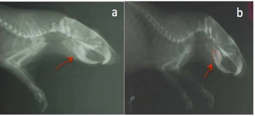
Figure 3. Four weeks after surgery: (a) defect with HA powder; (b) control
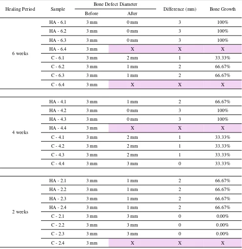
Table 1. Defect size on right mandible before (day 1) and after (healing period) *Highlighted column indicates that the rats were missing during care
Healing Period Sample Bone Defect Diameter Difference (mm) Bone Growth
Before After
6 weeks
HA - 6.1 3 mm 0 mm 3 100%
HA - 6.2 3 mm 0 mm 3 100%
HA - 6.3 3 mm 0 mm 3 100%
HA - 6.4 3 mm X X X
C - 6.1 3 mm 2 mm 1 33.33%
C - 6.2 3 mm 1 mm 2 66.67%
C - 6.3 3 mm 1 mm 2 66.67%
C - 6.4 3 mm X X X
4 weeks
HA - 4.1 3 mm 1 mm 2 66.67%
HA - 4.2 3 mm 0 mm 3 100%
HA - 4.3 3 mm 0 mm 3 100%
HA - 4.4 3 mm X X X
C - 4.1 3 mm 2 mm 1 33.33%
C - 4.2 3 mm 2 mm 1 33.33%
C - 4.3 3 mm 2 mm 1 33.33%
C - 4.4 3 mm 3 mm 0 33.33%
2 weeks
HA - 2.1 3 mm 1 mm 2 66.67%
HA - 2.2 3 mm 1 mm 2 66.67%
HA - 2.3 3 mm 1 mm 2 66.67%
HA - 2.4 3 mm 1 mm 2 66.67%
C - 2.1 3 mm 3 mm 0 0.00%
C - 2.2 3 mm 3 mm 0 0.00%
C - 2.3 3 mm 3 mm 0 0.00%
Table 1 shows the diameter of the defects before and after intervention. The Mann-Whitney test showed that there is a significant difference between before and after intervention (p= 0.026). Table 1 also shows bone growth as percentage. After 2 weeks, the implant group showed more than 50% bone growth (Fig. 2). After 4 and 6 weeks, the implant group showed perfect bone growth (100%). However, in the control group, even after 4 and 6 weeks of healing, a little shrinkage alongside an expansion of the defect area was present in several
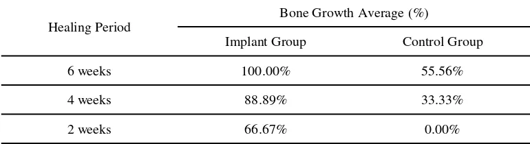
animals (Fig. 3, Fig. 4). It was observed that, 6 weeks after the surgery, bone growth in the control group was only 66.67%. The bone growth difference between the implant and control group was over 50% (Table 2).
The result of the histopathologic analysis on the rats' right mandible showed that the implant group presents the availability of hemosiderin (after two weeks), osteocytes and osteoblasts (after four and six weeks), including lymphocytes and neutrophils in every implant group rat.
Table 2.Differences of bone growth in percentage
Healing Period
Bone Growth Average (%)
Implant Group Control Group
6 weeks 100.00% 55.56%
4 weeks 88.89% 33.33%
2 weeks 66.67% 0.00%
Discussion
Hydroxyapatite is a complex calcium phosphate mineral with the chemical formula Ca10(PO4)6(OH)2, which constructs vertebrae bone structure. In the last few years, HA has been developed and produced as a bone graft material. Human bone consists of 65% HA; this
makes HA biocompatible to the human body.
Hydroxyapatite also has osteo conductive features that allows it to work as a scaffold in new bone growth.6
Based on the source, hydroxyapatite can be divided into two types: biologic hydroxyapatite, which is found in the bones or teeth of animals such as fish, and synthetic hydroxyapatite. In the field of dentistry, synthetic hydroxyapatite is the type mostly used. However, since the synthetic type requires a complex production process and is very expensive,15the HA powder used in this study was derived from discarded white barramundi fish scales that underwent mild processing. The HA powder was
packed into 7 mg dark tubes and sterilized with gamma irradiation.16 Each animal in the implant group had HA powder applied to its defect at a dose of one tube per defect/animal.
Researchers believed that HA powder could promote the bone growth process on mandibular defects. Histological examination showed that bone formation was verified on the defect area, given the presence of osteocytes as well as intense osteoblastic activity, along with intense osteogenic activity—especially at weeks four and six. This is in line with the radiographic examination results, which showed a reduction in the size of the mandibular right bone defect; it had fully recovered after six weeks.The bone growth process was supported by the results of the histopathological analysis. The histopathological analysis indicated that osteocytes, osteoblasts, collagen, neutrophil, lymphocytes, and hemosiderin were present in the implant group rats. The presence of osteocytes and osteoblasts showed that the HA powder had induced bone growth cells.18
Based on these results, HA powder from white barramundi fish scales can be considered as an alloplast alternative material. According to Fleckenstein et al., calvaria defects given to Sprague-Dawley rats that were treated with hydroxyapatite tricalcium phosphate (HA-TCP) powder showed significant bone growth because of its rigid scaffold that the rats could tolerate.19 The results of a study by Reis et al. also showed bone growth on a rabbit's ulna when given a hydroxyapatite-polyhydroxy-butyrate composite. Furthermore, direct contact between the bone graft and bone indicated osteo integration 45 days after surgery.20Osteointegration was also observed in a study by James Mah et al., who used alloplast materials available in the market on rat calvarias. However, in their study, the available materials did not show significant bone growth when compared to the control group.7Therefore, hydroxyapatite (HA) powder from white barramundi fish scales could be considered as bone graft alternative materials in bone defect regeneration as supported by previous researchers who also found significant bone growth after utilizing hydroxyapatite.
Conclusion
It can be concluded that hydroxyapatite (HA) obtained from white barramundi (Lates calcarifer) fish scales can be used as a bone graft alternative material.
This conclusion was made based on the results that showed significant bone growth and increased body weight, which indicates HA biocompatibility.
Acknowledgement
We would like to show our greatest gratitude to Mr. Erizal, staff of Center for Application of Technology of Isotope and Radiation, National Nuclear Energy Agency, Dr. Riki Siswandi (vet), Dr. Yulvian Sani, PhD (vet), Dr. Evan Hendra (dentist), Devina Soekamto, and Ika Yani J. for their contributions to this research.
Conflict of Interest
The authors declare no affiliations with or involvement in any organizations or entity with financial interest or non-financial interest.
References
1. Laurencin C, Khan Y, El-Amin SF. Bone graft substitutes. Expert Rev Med Devices. 2006; 3(1): 49-57. DOI: 10.1586/17434440.3.1.49
2. Regar NHB. Keramik sebagai bahan substitusi bone graft [internet]. Medan: Fakultas Kedokteran Gigi Universitas Sumatera Utara; 2009 [cited 2013 Dec 30]. Available from:
http://repository.usu.ac.id/bitstream/123456789/25273/6/ Cover.pdf.
3. Newman MG, Takei HH, Klokkevold PR, Carranza FA.
Carranza’s Clinical Periodontology. 11th Ed. Singapore: Elsevier; 2012.
4. Perry DA, Beemsterboer PL. Periodontology for the
Dental Hygienist. 3rd Ed. St.Louis: Saunders Elsevier; 2007. 24-35, 125-126, 128-129, 306-308.
5. Eley BM, Manson JD. Periodontics.10th Ed.London:
Wright; 2010. 11-12, 276-278.
6. Carranza FA, Takei HH, Cochran DL, Reynolds MA.
Surgical Therapy: Reconstructive Periodontal Surgery. In Newman MG, Takei HH, Klokkevold PR, Carranza FA, editors Carranza’s Clinical Periodontology. 11th Ed. China: Elsevier; 2006. 976-982.
8. Park JW, Jang JH, Bae SR, An CH, Suh JY. Bone formation with various bone graft substitutes in critical-sized rat calvarial defect. Clin Oral Impl Res. 2009; 20(4): 372-378.
9. Develioglu H, Unver SS, Kartal U. The bone-healing
effect of a xenograft in a rat calvarial defect model. Dent Mater J. 2009; 28(4): 396-400.
10. Kementrian Kesehatan Republik Indonesia. Profil Data
Kesehatan Indonesia Tahun 2011 [internet]. Jakarta: 2012
[cited 2013 Dec 1]. Available from:
http://www.depkes.go.id/downloads/PROFIL_DATA_KE SEHATAN_INDONESIA_TAHUN_2011.pdf.
11. Hartati I, Kurniasari. Kajian Produksi Kolagen dari
limbah sisik ikan secara ekstraksi enzimatis. Momentum. 2010; 6(1): 33-35.
12. Direktorat Jenderal Perikanan Budidaya. Angka
ketersediaan ikan Indonesia [Internet]. Jakarta: 2013
[cited 2014 Feb 3]. Available from:
http://www.djpb.kkp.go.id/berita.php?id=834.
13. Tokyo Institute of Technology. Producing Artificial
Bones From Fish Scales [internet]. June 1, 2012 [cited
2014 Feb 3]. Available from:
http://www.sciencedaily.com/releases/2012/06/120601093 003.html
14. Yogaswari V. Karakteristik kimia dan fisik sisik ikan
gurami (Osphronemus gouramy). [thesis]. [Bogor];
Program Studi Teknologi Hasil Perikanan Fakultas Perikanan dan Ilmu Kelautan Institut Pertanian Bogor;
2009.[Indonesia]
15. Praptiwi NR. Isolasi dan karakterisasi hidroksiapatit dari sisik ikan kakap putih (Lates calcarifer, Bloch). [thesis].
[Jakarta]; Fakultas Farmasi Universitas Pancasila:
2013.[Indonesia]
16. Dian PP, Darmawan, Erizal, Tjahyono. Isolasi dan sintesis gelatin berikatan silang dari limbah sisik ikan kakap putih (Lates calcarifer) dengan teknik induksi iradiasi gamma. Jurnal Sains Materi Indonesia. 2012; 14(1): 40-46. 17. Zielak JC, Mathias SA, Giovanini AF, Mathias AL. Oral
bone grafting in a rat model and the use of scanning electron microscopy for tissue morphology evaluation. Scand J Lab Anim Sci. 2007;34 (3): 1-10.
18. Brinkmann V, Reichard U, Goosmann C, Fauler B,
Uhlemann Y, Weiss DS, et al. Neutrophil extracellular traps kill bacteria. J Science. 2004;303(5663):1532-1535. DOI: 10.1126/science.1092385
19. Fleckenstein KB, Cuenin MF, Peacock ME, Billman MA,
Swiec GD, Buxton TB, et al. Effect of a hydroxyapatite tricalcium phosphate alloplast on osseous repair in the rat
calvarium. J Periodontol. 2006; 77(1): 39-45.
DOI:10.1902/jop.2006.77.1.39
20. Reis ECC, Borges APB, Fonseca CC, Martinez MMM,
Eleoterio RB, Morato GO, et al. Biocompatibility,
osteointegration, osteoconduction, and biodegradation of a
hydroxyapatite-polyhydroxybutyrate composite. Braz
Arch Biol Technol. 2010;53(4):817-826. DOI: