Indo. J. Chem., 2009, 9 (3), 410 - 413 410
Adhitasari Suratman and Hermann Waetzig * Corresponding author.
Email address : [email protected]
PREVENTION OF PROTEIN ADSORPTION ON BARE FUSED-SILICA CAPILLARY BY PEG
IN CAPILLARY ZONE ELECTROPHORESIS
Adhitasari Suratman1.*and Hermann Waetzig2 1
Department of Chemistry, Universitas Gadjah Mada, Sekip Utara Yogyakarta 55281 Indonesia
2Institute of Pharmacy, TU Braunschweig, Beethovenstr. 55, 38106 Braunschweig, Germany
Received August 28, 2009; Accepted October 29, 2009
ABSTRACT
The protein separation was studied in capillary zone electrophoresis for preventing protein adsorption on the capillary wall. ß-lactoglobulin (pI: 4.83-5.4, Mr: 18.4 kDa), cytochrome c (pI: 9.59, Mr: 11.7 kDa) and ß-casein (pI: 4.6,
Mr: 24 kDa) were used as protein models. Strong adsorption of the proteins occurred onto the capillary at a pH
around their pIs. In order to prevent protein adsorption, PEG (Poly(ethylene glycol)) was investigated as an effective substance to stabilize the proteins native state and coat the bare fused-silica capillary surface. The presence of 32 mg/mL PEG in buffer solution in a pH range of 6.0 to 4.0 was successful to suppress protein adsorption during the separation. It can also be confirmed with the reproducibility of apparent EOF mobility with percentile RSD (Relative Standard Deviation) less than 2% in long-term measurement.
Keywords: PEG, protein adsorption, CZE
INTRODUCTION
Capillary zone electrophoresis (CZE) is the simplest form of Capillary Electrophoresis (CE) that is described as high-efficiency separations techniques that use narrow-bore fused-silica capillaries and the most commonly utilized. The separation mechanism of this technique is based on the difference of size and charge of analytes in high electric field strengths. Analysis using CE has evolved into an irreplaceable tool for the quality control of pharmaceuticals and biotechnological products. In other cases CE is established as alternative technique in pharmaceuticals routine analysis [1].
The interaction between positively charged proteins and negatively charged silanol groups on the capillary surface is a major problem for the analysis of proteins by capillary electrophoresis, especially if the proteins are separated at pH values lower than their isoelectric points (pI). Consequently, adsorptions at the capillary wall frequently happen. This can cause peak broadening and asymmetric peak shapes, low efficiency, low recovery of analysis, irreversible protein adsorption, a drifting EOF (Electroosmotic Flow) and irreproducible migration times [2-3].
Several strategies have been proposed to prevent the problem of protein adsorption. In CZE, the choice pH buffer, the addition of high concentrations of alkali salts, zwitterions or other additives to the buffer solution, coated capillaries, the stability of a protein, and many factors (temperature, ion strength, pH, composition of BGE, chemical and structural properties of the capillary
surface, rinsing procedure, etc) should also be considered [4-9].
The common technique used to inhibit protein adsorption is blocking the adsorption sites by the optimization of the molecule (solute) surfaces or/and the coating solid surfaces. In order to achieve solute surface that resist the proteins adsorption from aqueous solution, the exclusion of solute from the protein surface in aqueous solution is presented. As a result, these solutes do not interact directly with the proteins lead to a stabilization of native proteins [10]. Solute that is well-excluded from the protein surface offers a good protein-resistant surface. These solutes having ability to provide “protein resistance” are called kosmotropes [10-11].
The second way to block the adsorption site is by coating of the solid surface. Many methods have been used to coat the solid surface. The simplest one can be achieved by the formation of self-assembled monolayers (SAMs). Nevertheless, the effective blocking on adsorption sites is achieved with the formation of SAMs based on displays of kosmotropes. They can form a layer on a solid surface and also keep a water layer between protein and SAMs [10,12].
Indo. J. Chem., 2009, 9 (3), 410 - 413 411
Adhitasari Suratman and Hermann Waetzig
In this experiment, ß-lactoglobulin, cytochrome c, and ß-casein as protein models were separated by CZE in a bare fused-silica capillary. The molecular mass of these proteins is 11.7-24 kDa with pIs of 4.63-9.59. PEG was evaluated as an additive to avoid the protein adsorption on the capillary that frequently happen at pH around their pIs.
EXPERIMENTAL SECTION
Material
ß-lactoglobulin (bovine milk, pI: 4.83-5.4, Mr: 18.4 kDa), cytochrome c (horse heart, pI: 9.59, Mr: 11.7 kDa), ß-casein (bovine milk, pI: 4.6, Mr: 24 kDa), neostigmine bromide was purchased from Sigma-Aldrich (Steinheim, Germany). Sodium acetate anhydrous, sodium chloride and acetanilide were purchased from Fluka (Steinheim, Germany); disodium hydrogen phosphate-2-hydrate and potassium dihydrogen phosphate from Riedel-de Haën (Sigma-Aldrich, Seelze, Germany). Poly(ethylene glycol) (PEG) of molecular weight 20000 g/mol and acetic acid were purchased from Merck (Darmstadt, Germany).
Instruments
The instrumentation for protein analysis using CZE technique was UniCAM Crystal 310 CE (UniCAM Ltd., Cambridge, UK), equipped with a UV detector (wavelength 210 nm). Bare fused-silica capillaries (Polymicro Technologies, Phoenix, AZ, USA) had dimensions of 60 cm total length, 48 cm effective length, and 50 µm inner diameter (i.d.). During all experiments, the thermostat was set to 25 °C. All integration was done by an integration program C.I.S.S. (Correct Integration Software System), (Würzburg, Germany) [13].
Procedure
ß-lactoglobulin, cytochrome c and ß-casein as model proteins were freshly prepared using isoosmotic NaCl 0.9% m/V solution. Acetanilide as an EOF marker and neostigmine bromide as an internal standard were dissolved in buffer solution. The total concentration of analytes in the sample solution was 100 µg/mL of acetanilide, 500 µg/mL of neostigmine bromide and 35 µmol/L of protein.
The 50 mmol/L acetate buffers with pH 5.0 and 4.0 were prepared by weighing the appropriate amounts of sodium acetate and acetic acid and filling up to volume; the 50 mmol/L phosphate buffer with pH 6.0 consisted of disodium hydrogen phosphate-2-hydrate and potassium dihydrogen phosphate. Additive buffer was made by dilution of PEG into the buffer solution with the final concentration 3.2 and 32 mg/mL.
New bare fused-silica capillaries were previously conditioned with 1 mol/L NaOH for 2 h, continued by a rinsing with buffer for 30 min (1200 mbar), then equilibrated for 2 h with the applied voltage, and afterwards directly used for protein analysis. The analysis was started with buffer rinsing at 1200 mbar for 2 min. Next, samples were hydrodynamically injected by applying a pressure of 30 mbar for 12 seconds. Electrophoresis was performed at voltage of 25 kV (56-68 µA) or 18 kV (46-75 µA), additionally applying a pressure of 100 mbar to increase the analysis speed. After each series, the capillary was reconditioned like a new capillary to regenerate it.
RESULT AND DISCUSSION
Different concentrations of PEG (3.2 mg/mL and 32 mg/mL) were added into the running buffer solution. Since the addition of PEG 32 mg/mL into the buffer solution causes an increasing viscosity of the solution, an additional pressure of 100 mbar was applied during protein separation in order to reduce the analysis time. Hence, the apparent EOF mobility in this experiment was used as measurement parameter.
The pH dependence of protein adsorption on bare fused-silica capillaries and correlation to pI of protein are frequently reported. Using conditions at pH values above the pI of protein, adsorption is negligible for bare fused-silica capillaries [14]. Henceforth, this research focused on protein separation at pH less than pI of protein.
As shown in Figure 1, ß-lactoglobulin which was analyzed without the presence of PEG in the phosphate buffer pH 6.0 showed a decreasing EOF mobility after 18th runs with an RSD of 25.3%. It indicated that the formation of protein-layers on the bare fused-silica surface occurred in this series. It changes the surface structure of the capillary wall, and then influences the apparent EOF mobility.
In order to avoid the formation of protein-coated capillaries, PEG 3.2 mg/mL was evaluated under the same condition. A slight difference of apparent EOF mobility was observed between the absence of PEG and the presence of PEG 3.2 mg/mL. The slightly increasing reproducibility of apparent EOF mobility was observed in Figure 1 with an RSD of 17.3%. The interaction between protein and capillary wall was not completely avoided by the addition of PEG 3.2 mg/mL.
Indo. J. Chem., 2009, 9 (3), 410 - 413 412
Adhitasari Suratman and Hermann Waetzig
Figure 1. Apparent EOF mobility of ß-lactoglobulin analysis at pH 6.0 in the absence and the presence of PEG 3.2 and 32 mg/mL
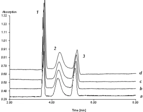
Figure 2. The electrophoregram of ß-lactoglobulin analysis at (a) 1st (b) 10th (c) 20th (d) 30th at pH 4.0 with the presence of PEG 32 mg/mL using a bare fused-silica capillary. Peak 1: neostigmine bromide; peak 2: ß-lactoglobulin; peak 3: acetanilide. Acetate buffer pH 4.0 (50 mM), V = 18 kV, I ~ 46 µA, additional pressure: 100 mbar
at this pH due to the long-term analysis that might change in protein surface. The number of negative charges of ß-lactoglobulin decreased. It causes a faster mobility. Nevertheless, the formation of protein layer on the capillary was not found in this experiment. This was shown by an excellent reproducibility of apparent EOF mobility with RSD 0.61% (Figure 1). Interactions between ß-lactoglobulin and capillary wall could therefore be prevented by the addition of PEG with a concentration of 32 mg/mL.
The benefit of the presence of PEG 32 mg/mL was also investigated at the different pH values, especially at the pH close to protein’s pI. The possibility of protein interaction on capillary wall becomes higher at a pH
Figure 3. Apparent EOF mobility of ß-lactoglobulin, cytochrome c and ß-casein analysis at pH 4.0 using a bare fused-silica capillary
lower than protein’s pI. ß-lactoglobulin was analyzed at 50 mmol/L acetate buffer pH 5.0. It was performed after one series of ß-lactoglobulin analysis at pH 6.0 by using of the same capillary. Before this capillary was used for next experiment, it was reconditioned by the same manner as new capillary. It was previously conditioned with 1 mol/L NaOH for 2 h, continued by a rinsing with buffer for 30 min (1200 mbar), then equilibrated for 2 h with the applied voltage and was afterwards directly used for protein analysis at pH 5.0. Surprisingly, a good stability of internal standard, ß-lactoglobulin and EOF marker was observed, even overlapping between ß-lactoglobulin and EOF marker peak was observed. The successful use of PEG in this experiment is also confirmed by an excellent reproducibility of apparent EOF mobility with RSD 1.2%.
In order to confirm the benefit of PEG to resist protein adsorptions, ß-lactoglobulin as an acidic protein, cytochrome c as a basic protein and ß-casein as a more easily denaturing protein were investigated in 50 mmol/L acetate buffer pH 4.0. Figure 2 shows the electrophoregram of ß-lactoglobulin analysis in the 1st, 10th, 20th, and 30th runs at pH 4.0. The analysis of ß-lactoglobulin at pH 4.0 shows an optimal condition of analysis showed by a good stability of migration time and a good resolution of each analyte peak were observed at pH 4.0. The excellent reproducibility of apparent EOF mobility was observed with RSD 0.52% (Figure 3).
Indo. J. Chem., 2009, 9 (3), 410 - 413
Adhitasari Suratman and Hermann Waetzig
413
experiment which was confirmed by the reproducibility of the apparent EOF mobility (RSD 0.41%, Figure 3).
The effectiveness of 32 mg/mL PEG to resist protein adsorption was also evaluated for ß-casein (pI 4.6) analysis at pH 4.0. ß-casein analysis at pH 4.0 even with the addition of PEG into buffer solution was not successful in resolving and detecting a protein peak. It was probably due to the fact that ß-casein undergoes the conformational structure change at pH 4.0. Nevertheless, the apparent EOF mobility remains stable with RSD 1.05% (Figure 3).
In general, the presence of 32 mg/mL PEG to 50 mmol/L phosphate buffer pH 6.0 and acetate buffer pH 5.0 and 4.0 has proven to be an effective way to suppress protein adsorption in long-term measurement.
CONCLUSION
Poly(ethylene glycol) was added into buffer solution in order to stabilize the proteins native state and coat the bare fused-silica capillary surface. The presence of 32 mg/mL PEG in protein and buffer solution in a range of pH 6.0 to 4.0 was successful to suppress protein adsorption during the separation in capillary zone electrophoresis. It can also be confirmed with the reproducibility of apparent EOF mobility with percentile RSD less than 2% in long-term measurement (n=30).
REFERENCES
1. Landers, J. P., 1997, Handbook of capillary electrophoresis, 2nd ed, CRC press, N. Y.
2. Wätzig, H., Degenhardt, M., and Kunkel, A., 1998, Electrophoresis, 19, 2695-2752.
3. Corradini, D. and Cannarssa, G., 1996, LC-GC, 14, 326-330.
4. Dolník, V., 2006, Electrophoresis, 27, 126-141. 5. Mohabbati, S., Hjertén, S., and Westerlund, D.,
2004, J. Chromatogr. A, 1053, 201-216.
6. [6] Karlsson, M., Ekeroth, J., Elwing, H., Carlsson, U., 2005, J. Biol. Chem., 27, 25558-25564.
7. Costantino, H.R., Andya, J.D., Nguyen, P., Dasovich, N., Sweeney, T.D., Shire, S.J., Hsu, C.C., and Maa, Y., 1998, J. Pharm. Sci., 11, 1406-1411.
8. Tzannis, S.T. and Prestrelski, S.J., 1999, J. Pharm. Sci., 3, 351-359.
9. Maury, M., Murphy, K., Kumar, S., Mauerer, A., and Lee, G., 2005, Eur. J. Pharm. Biopharm., 59, 251-261.
10. Kane, R.S., Deschatelets, P., and Whitesides, G.M., 2003, Langmuir, 19, 2388-2391.
11. Moelbert, S., Normand, B., and De Los Rios, P., 2004, Biophys. Chem., 112, 45-57.
12. Czeslik, C., 2006, Chem. Unserer zeit, 40, 238-245.
13. Schrim, B. and Wätzig, H., 1998, Chromatographia, 48, 331-346.