www.elsevier.com / locate / livprodsci
The importance of cell division in udder development and
lactation
*
C.H. Knight
Hannah Research Institute, Ayr KA6 5HL, UK
Abstract
The mammary secretory cell population increases in an exponential fashion during pregnancy in all species studied, as a consequence of very high rates of cell division. After parturition the mitotic index drops dramatically, but a limited amount of proliferation does continue, at least until the time of maximum milk yield. This is particularly true of rodents but also occurs in dairy species. During declining lactation apoptosis exceeds cell division, so the size of the cell population falls and it is this decrease which is responsible for the reduction in milk yield. Many factors influence cell division. In addition to well known hormones such as ovarian steroids, prolactin and growth hormone, and growth factors such as epidermal growth factor and insulin-like growth factor-1, there are also effects of milking frequency and nutrition. Some of these same factors are now also known to regulate apoptosis. The challenge for the future is to understand more about the relationships between apoptosis and cell division in the mammary gland; for instance, are the two mutually exclusive and independent or is apoptosis important in preparing the gland for renewed cell division? To this end, we have developed a lactation rescue model which will allow us to study interactions between apoptosis and cell division in lactating mouse and cow mammary glands. 2000 Elsevier Science B.V. All rights reserved.
Keywords: Mammary gland; Development; Cell proliferation; Apoptosis; Lactation
1. Introduction have depressed milk yield, and research has shown this to be a consequence of low blood concentrations Growth of the mammary gland is termed mam- of growth hormone (GH) and insulin-like growth mogenesis. There are two distinct phases, growth factor-1 (IGF-1) reducing peripubertal mammary cell prior to first conception and then cyclical waves of proliferation (Weber et al., 1999). The GH / IGF axis proliferation, secretion and involution during recur- is not the only endocrine regulator of mammary ring lactation cycles. The most important part of the proliferation, but it is an important element. During first phase is the allometric growth which occurs just the life of the cow the mammary cell population before and around puberty, since inappropriate de- probably expands by more than 100-fold (although velopment at this stage can have long term repercus- precise quantification is difficult) with the vast sions (Sejrsen, 1994). Rapidly-reared heifers often majority of this proliferation taking place during pregnancy under the stimulatory influence of ovarian steroids and peptide mammogens, including GH. *Tel.: 144-1292-674-046; fax: 144-1292-674-047.
E-mail address: [email protected] (C.H. Knight). Treatment of late pregnant goats with exogenous GH
increases cell proliferation and subsequent milk yield much milk as the LGM cows, from 1.3-times as (Knight et al., 1994), and there is much evidence to much udder tissue (Sorensen et al., 1998). Clearly, show that the number of secretory cells present in the the amount of secretory tissue is an important mature udder is a primary determinant of milk yield determinant of milk yield, and selection for output (Sorensen et al., 1998). Using bromodeoxyuridine has indeed resulted in larger udders.
injections to ‘‘tag’’ proliferating cells and then Udder volume is a crude measurement of total counting these cells in histological sections obtained tissue mass, and provides no information on the by biopsy, we have observed labelling indices of relative proportions of secretory and non-secretory around 10% in late pregnancy, subsequently falling tissue. Different tissue types can be distinguished by precipitously after parturition; mammary cells do not imaging techniques such as X-ray, computed tomog-proliferate extensively during lactation, although raphy (CT) scanning and magnetic resonance imag-they retain the potential to divide and simple treat- ing (MRI). We have used MRI in vivo in goats ments such as frequent milking are mitogenic (Wilde (Fowler et al., 1990) and others have used CT et al., 1987). GH administered in early lactation does scanning in excised udders of heifers (Sorensen et not stimulate proliferation (Knight et al., 1994), but al., 1987), but there is no possibility of using either later in lactation its mitogenic capacity returns method for dairy cows in vivo because cows are (Capuco and Byatt, 1998). Whether other mitogens simply too big. The alternative approach is to are only active at certain stages of the lactation cycle measure the composition of the tissue by biochemi-is not known, but it biochemi-is apparent that observations cal or histological analysis. We now do this in needle made at one point in time cannot be extrapolated to biopsy samples using a mechanical biopsy tool
cover all circumstances. (Magnum; C.R. Bard, Covington, GA, USA) which
allows for repeated measurements with relatively little disturbance to the cow. If it is the number of
2. Importance of cell number: comparative secretory cells which ultimately determines yield,
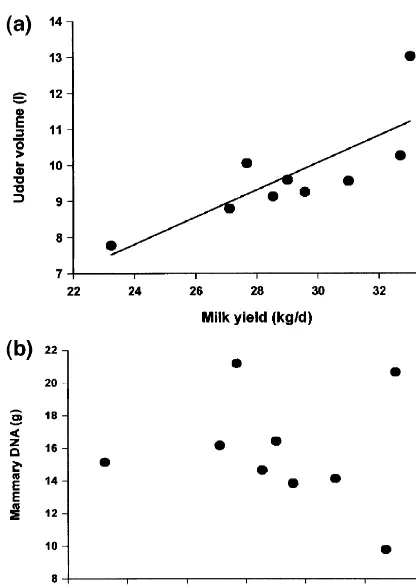
studies one would anticipate that milk yield would correlate more strongly with udder DNA content (a measure A positive relationship between mammary gland of cell number) than with crude udder volume. A size and milk yield has been recognised for many recent analysis shows that this is not necessarily the years (Linzell, 1966). This is easy to demonstrate in case; in a group of nine peak lactation cows we goats but less evident in cattle, due to the difficulty obtained a significant correlation between udder
2
of obtaining reliable data on udder size. However, volume and milk yield (r 50.63, P50.01, Fig. 1a) quick-setting polyurethane foam can be used to but absolutely no relationship between total DNA obtain casts of cow’s udders (Dewhurst et al., 1993), and milk yield (Fig. 1b). The data set was small and a technique which is relatively simple and which it is also important to remember that an additional produces an accurate measure of gross udder size. source of potential inaccuracy is introduced, since a Comparing a diverse group of cattle, we obtained a single biopsy may not always be fully representative. reasonable relationship between udder volume mea- Nevertheless, we have always maintained that milk sured in this way and milk yield (r50.704, P5 yield is actually determined by the function of cell 0.02) but further calculations revealed that we were number and cell activity, and there was indeed underestimating udder size in young heifers and evidence of greater enzyme activities (measured per overestimating it in old cows, due to the much more cell) in higher yielding cows.
DNA precursor, and proliferating cell nuclear an-tigen (PCNA), an endogenous proliferation-associ-ated protein (McCormick and Hall, 1992). Mathe-matical approaches are also possible: a model of mammary growth was published recently which inferred rates of cell proliferation and cell death from changes in DNA content (Dijkstra et al., 1997). In order to arrive at the estimates, the authors assumed that cell death did not occur during pregnancy, that the rate of cell proliferation fell off exponentially after parturition and that the rate of cell death during lactation was constant. Figures are quoted for the rate of cell proliferation at parturition which range from 3% per day (goats) to 39% per day (guinea pigs). An inverse relationship between gestation length and the mammary growth exponent [it is an exponential process: (Knight and Peaker, 1982)] is inevitable, since the growth rate achieved in mice would, if replicated throughout gestation in dairy cows, produce an udder capable of filling several football stadia (Sheffield and Anderson, 1985), so it is perhaps not surprising that proliferation rate was lowest in goats. Unfortunately, however, the model also predicted considerable variation between differ-ent reports within the one species, from 10.8 to Fig. 1. Relationship between milk yield and udder volume 33.4% in mice, for instance. Since one of these measured using polyurethane foam casting (a) or mammary DNA reports was from our own work (Knight and Peaker, content determined in biopsy samples (b) for nine peak lactation
1982), I have calculated predicted mammary cell 2
cows. Relationship with udder volume was significant (r 50.63,
population size at parturition based on the three P50.01), while the relationship with DNA content was not.
proliferation rates and our measurement of DNA content at day 12 of gestation. The lowest prolifer-lactation, without any change in cell activity (Wilde ation rate gave an accurate prediction (3.9 mg DNA
et al., 1986). vs. our measured value of 3.85 mg), while the other
two overestimated size by up to 3.7-fold. It would be nice to think that the lowest proliferation rate was
next 2 days in the mouse (Knight and Peaker, 1982). fold higher than apoptosis (1.4% vs. 0.08%). If one Dijkstra et al. (1997) only quote a proliferation rate assumed that these figures apply throughout lacta-for one time point (parturition, a bad choice, given tion, then by drying-off time the cow’s udder would the extreme variability inherent around this time), not have regressed at all, but would actually have although they do state that the rate varied during more than doubled in size! Two things are apparent. pregnancy. Since the highest parturient proliferation Firstly, apoptosis is almost certainly not constant rate was associated with the lowest achieved size throughout lactation and cell proliferation continues (based on our data set), one would assume that throughout lactation at a slow (but significant) rate, proliferation must previously have been much lower. so the model’s assumptions are inaccurate. Secondly, The reverse was actually the case; we observed when using snapshot measurements, it is very im-higher rates of cell proliferation at all other stages of portant to know the ‘‘shutter speed’’, i.e., the period pregnancy, and the model is almost certainly mis- of time over which the desired event is being appropriating the very high rates of proliferation detected. Consider the analogy of a photographer at a which we observed postpartum. Mechanistically, this grand prix car race. If he takes a series of randomly-is a very mrandomly-isleading error and a good example of timed shots standing at a slow bend, his chances of why ‘‘snapshot’’ data should not be inferred from getting good pictures of cars will be much greater mathematical modelling. But, it is also an excellent than if he were to do the same thing half way down example of how very crucial cell proliferation around the fastest section of track. My (inaccurate) sug-parturition is to mammary development. Because of gestion that the cow’s udder might grow rather than the exponential nature of mammogenesis, a doubling regress during declining lactation assumed that pro-of cell population occurring in the first few days liferation and apoptosis were being detected at the postpartum (if it were to happen) would give a net same point on the track, be it the hairpin or the increase in secretory cells of around 2.5-fold (mouse) straight, but we do not know that this is so. We used or almost five-fold (goat) greater than if the same PCNA to measure proliferation and, in this case, the event happened at mid-gestation. critical window is probably close to 1 day, since this This all shows that snapshot measurements of endogenous protein is up-regulated during DNA mammary cell proliferation must be interpreted with synthesis with a half-life of around 20 h (McCor-caution. Exactly the same arguments can be applied mick and Hall, 1992). Therefore, PCNA may give a to apoptosis, which is usually measured by detection good approximation of a daily proliferation rate. of the fragmented DNA typical of non-necrotic cell Apoptosis was detected using a commercial kit death. The mathematical model assumed a constant (Apoptag; Oncor, Gaithersburg, MD, USA) and, in rate of apoptosis during lactation which, in the case this case, the fragmented DNA on which the method of the goat, was 0.4% of cells per day (Dijkstra et relies is only present for a few hours (Hande et al., al., 1997). This seems a sensible figure. We have 1998), meaning that the apparent rate may only be measured mammary DNA content at peak lactation one-tenth (or thereabouts) of the daily rate. Even so, in cows and obtained a figure of around 15 g (C.H. the observed rates in our peak lactation cows would Knight, S. Robertson and A. Sorensen, unpublished still suggest a (small) day-on-day increase in mam-observations), which would fall to about 5 g at mary cell number rather than a decrease. We know drying-off at this rate of loss. This would equate well that this does not happen, so the measurements made with our belief that cell number and milk yield at peak lactation do not reflect later events.
not have a shortened life expectancy). As yet, very few data have been generated in this way.
4. Lactation rescue: a model system for
investigating relationships between proliferation and apoptosis
For many years cell proliferation was intensively studied in the mammary gland and elsewhere, where-as for the lwhere-ast few years it hwhere-as almost been forgotten in favour of apoptosis. However, it must be re-emphasised that changes in cell number result from the balance between the two processes (assuming necrotic cell death is minimal), and so it is very important to understand the relationship between the
Fig. 2. Litter weight gain during lactation rescue in mice. Pups two. As we have just seen, simply recording rates of were removed to a foster mother for 2 days on day 6 of lactation proliferation and apoptosis in normal mammary and then reunited with their mother. During weight loss (day 1 of resuckling) pups spent part of their time with the foster mother to tissue can lead to erroneous results, so a model
ensure they were fed; the data shown is for the period with their system is required in which the two events can be
own mother. Control mice are shown by the continuous line. turned on and off such that their time courses and
Values are means6standard error (S.E.), n56. spatial relationships can be studied. The majority of
apoptosis occurs during post-lactational involution,
so a logical starting point would be to arrest lactation We then repeated the 48-h separation, but this time so as to turn on apoptosis and, having done so, with administration of bromodeoxyuridine (to mea-attempt to reverse the process. If lactation were to sure cell proliferation) during either the separation or restart, would it require renewed cell proliferation? the resuckling period. Apoptosis was measured using We have done exactly this in mice (Sorensen and a commercial kit (Apoptag) as before. Compared
Knight, 1997,1998). with control mice with normal lactation, apoptosis
The effects of removing pups from a day 6 was greatly increased during litter removal and then lactating mouse, placing them with a foster mother quickly returned to normal levels during resuckling and then replacing them with their own mother 48 h (Table 1). This shows that the absence of a suckling later are shown in Fig. 2. For the first day after stimulus results in involution and cell loss which can replacement, weight gain was negative during the be reversed if the stimulus is reapplied. The highest period when the pups were with their own mother level of bromodeoxyuridine incorporation was ob-(they spent 12 h with the foster mother to ensure served during the resuckling period, indicating that, they were fed). From the second day onwards the
Table 1 pups spent all their time with their own mother and
Cell proliferation and apoptosis in mammary glands of mice either did gain weight and, by the fourth day, their weight lactating normally (control), separated from their young for 2 days gain had recovered to normal levels. At this age (11 (Separated) or separated for 2 days and then reunited for 3 days
a (resuckled) days) mouse pups are still entirely dependent on
maternal milk, so it was evident that lactation had Proliferation (%) Apoptosis (%) been successfully restored. Shorter and longer (up to
Control 4.6060.53 0.2360.05 72 h) periods of separation resulted in faster and Separated 9.5060.81** 4.6060.55*** slower restoration, respectively, and lactation was Resuckled 13.7662.50* 0.6960.24 also recoverable (but with greater difficulty) follow- a
Values are means6S.E. for groups of six mice.
ing separation later in lactation (Sorensen and *: P,0.05, **: P,0.01, ***: P,0.001, all with respect to
not only was apoptosis turned off, but cell prolifer- tricably linked and that induction of apoptosis re-ation was also turned on in order to recover the quires not only an initial signal causing cells to enter lactation. This very neat picture is, however, incom- the cell cycle but also the subsequent absence of a plete. Whilst bromodeoxyuridine incorporation was survival signal which would otherwise ‘‘rescue’’ the highest during resuckling, it was also elevated above cell. This reduces the likelihood of inappropriate control levels during the separation period, i.e., when proliferation (i.e., cancer) since two mutations would apoptosis was up-regulated and, we would have have to occur to achieve the desired combination of anticipated, proliferation was firmly turned off signals, but it also provides the necessary means by (Table 1). This raises the intriguing possibility that, which mammary involution can be rapidly reversed. faced with the absence of her pups, the lactating
mouse decides to turn off lactation but does so in
such a way that a fresh population of secretory cells 5. Cell survival and lactation length
will be readily recruitable should she have the need
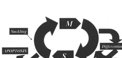
of them. Whilst it is possible that the same cue which During the separation period, milk collected from starts apoptosis also starts a totally different series of the mice contained high levels of one particular IGF events leading to cell proliferation, a more likely binding protein, IGFBP-5. This binding protein has explanation is given in Fig. 3, which puts apoptosis previously been observed in involuting mammary into the context of the cell cycle. It may be that tissue (Tonner et al., 1995) and was not present in terminally-differentiated cells can enter directly into milk from control or resuckled mice. IGF-1 is a apoptosis, but there is also evidence to show that known mammary mitogen and has been implicated they do not do this, but rather that they re-enter the as a mammary cell survival factor (see Flint and cell cycle, synthesise DNA (at which point they Knight, 1997), but its concentration is paradoxically would incorporate bromodeoxyuridine) and then elevated during post-lactational involution. In the undergo apoptosis (Colombel et al., 1992). Assum- presence of IGFBP-5, however, the cell survival ing there is a ‘‘switch’’ at this point, the resumption function is lost. The post-lactational appearance of of suckling would act to divert these cells away from IGFBP-5 is prevented by administration of the the apoptotic path and keep them in a proliferative suckling-related hormone, prolactin (Tonner et al., cell cycle. The ‘‘dual-signal’’ hypothesis of Evan and 1995), so it would appear that all the necessary Littlewood (1993) envisages exactly this. They claim elements of the dual-signal hypothesis are present. that apoptotic and proliferative pathways are inex- IGF-1 (possibly produced locally via GH stimulation of mammary endothelial cells) causes cells to enter the cell cycle and, if suckling occurs, prolactin is released, IGFBP-5 is down-regulated and the cell survival activity is turned on. When suckling is prevented, the cells lose their survival signal and apoptosis results, to be replaced by survival when suckling is restored (Fig. 3).
Although this model has been developed in ro-dents, there is no reason to believe that it does not also operate in dairy species, although it is important to recognise that the time course of events may be very different, since mammary involution is a much Fig. 3. Schematic representation of the cell cycle to show the slower process in cows than in mice (Wilde et al., possible routes to apoptosis. M: Mitosis. S: DNA synthesis. In the 1997). We are beginning to study lactation rescue in absence of continued suckling, differentiated mammary secretory
cows. The potential benefits of being able to unravel cells may enter directly into apoptosis or, more likely, re-enter the
the mechanisms controlling cell proliferation and cell cycle, synthesise DNA and then either undergo apoptosis or,
stated earlier, apoptosis usually exceeds proliferation, ADAS Bridgets Research Centre, funded by the Milk so there is a gradual loss of secretory cells and a Development Council and BOCM Pauls Ltd. consequent reduction in milk yield. For this reason,
dairy farmers have to rebreed their cows regularly
and the cows have to cope with the dual metabolic References
burdens of lactation concurrent with gestation and
the risks introduced by parturition. If we could move Capuco, A.V., Byatt, J., 1998. Cell turnover in the mammary gland. J. Dairy Sci. 76, 224, (abstr.).
to a scenario where proliferation matched involution
Colombel, M., Olsson, C.A., Ng, P.-Y., Buttyan, R., 1992. such that the population of secretory cells was stable,
Hormone-regulated apoptosis results from re-entry of differen-then milk yield should be sustainable at high levels tiated prostate cells onto a defective cell cycle. Cancer Res. 52, and the cow would not need to be rebred so often (or 4313–4319.
even at all). The benefits of extended lactation have Dewhurst, R.J., Mitton, A.M., Knight, C.H., 1993. Calibration of a polyurethane foam casting technique for estimating the weight been considered previously (Knight, 1997).
of bovine udders. Anim. Prod. 56, 444, (abstr.).
Dijkstra, J., France, J., Dhanoa, M.S., Maas, J.A., Hanigan, M.D., Rook, A.J., Beever, D.E., 1997. A model to describe growth patterns of the mammary gland during pregnancy and lactation.
6. Conclusions
J. Dairy Sci. 80, 2340–2354.
Evan, G., Littlewood, T., 1993. Curr. Opin. Genet. Dev. 3, 44–49. Cell division is crucial to the mammary gland’s Flint, D.J., Knight, C.H., 1997. Interactions of prolactin and preparation for lactation. It is a major player in the growth hormone (GH) in the regulation of mammary gland function and epithelial cell survival. J. Mammary Gland Biol. determination of milk yield, but is not the only
Neoplasia 2, 41–48. important factor; cellular differentiation also has a
Fowler, P.A., Knight, C.H., Cameron, G.G., Foster, M.A., 1990. big influence. Cell lifetime must be taken into Use of magnetic resonance imaging in the study of goat account, and it is lamentable that we still know very mammary glands in vivo. J. Reprod. Fertil. 89, 359–366. little about average life expectancy for individual Hande, S., Notidis, E., Manser, T., 1998. Bcl-2 obstructs negative
selection of autoreactive, hypermutated antibody V regions mammary secretory cells. When considering changes
during memory B cell development. Immunity 8, 189–198. in milk yield during the course of lactation and our
Knight, C.H., 1997. Biological control of lactation length. Livest. ability to manipulate these changes, proliferation and Prod. Sci. 50, 1–3.
apoptosis must be taken into account together and Knight, C.H., Brown, J.R., Sejrsen, K., 1994. A comparison of regarded as the whole process through which the cell growth hormone-induced mammogenesis in pregnant and
lactating goats. Endocr. Metab. 1 (Suppl. B), 52, (abstr.). population is modulated. I have argued for some
Knight, C.H., Dewhurst, R.D., 1994. Once daily milking of dairy time and continue to believe that it is during lactation
cows; relationship between yield loss and mammary cistern itself that the greatest strides could be made towards capacity. J. Dairy Res. 61, 441–449.
more efficient and welfare-friendly dairying through Knight, C.H., Peaker, M., 1982. Mammary cell proliferation in an improved understanding of the control of both mice during pregnancy and lactation in relation to milk yield.
Q. J. Exp. Physiol. 67, 165–177. apoptosis and cell proliferation.
Linzell, J.L., 1966. Measurement of udder volume in live goats as an index of mammary growth and function. J. Dairy Sci. 49, 307–311.
McCormick, D., Hall, P.A., 1992. The complexities of
prolifer-Acknowledgements
ating cell nuclear antigen. Histopathology 21, 591–594. Sejrsen, K., 1994. Relationships between nutrition, puberty and The Hannah Research Institute is supported by mammary development in cattle. Proc. Nutr. Soc. 53, 103–111. Grant-in-Aid from the Scottish Executive Rural Sheffield, L.G., Anderson, R.R., 1985. Interspecies variation in mammary gland growth rate; relation to gestation length. J. Affairs Department. Data for lactation rescue in mice
Dairy Sci. 68, 2571–2579. were collected by Annette Sorensen, funded by the
Sorensen, A., Knight, C.H., 1997. Restoration of lactation in mice Andersen, J., Akers, R.M., Sejrsen, K., 1999. Contribution of after litter removal for various lengths of time. J. Reprod. insulin-like growth factor (IGF-I) and IGF-binding protein-3 to Fertil. Abstr. Ser. 19, 46. mitogenic activity in bovine mammary extracts and serum. J. Sorensen, A., Knight, C.H., 1998. Mammary apoptosis and cell Endocrinol. 161, 365–373.
proliferation during lactation rescue in mice. J. Reprod. Fertil. Wilde, C.J., Addey, C.V.P., Li, P., Fernig, D.G., 1997. Programmed Abstr. Ser. 21, 43. cell death in bovine mammary tissue during lactation and Sorensen, M.T., Sejrsen, K., Foldager, J., 1987. Estimation of involution. Exp. Physiol. 82, 943–953.
pubertal mammary development in heifers by computed Wilde, C.J., Henderson, A.J., Knight, C.H., 1986. Metabolic tomography. J. Dairy Sci. 70, 265–270. adaptations in goat mammary tissue during pregnancy and Tonner, E., Quarrie, L., Travers, M., Barber, M., Logan, A., lactation. J. Reprod. Fertil. 76, 289–298.