DENTAL AND ORAL MANAGEMENT IN BETA MAJOR THALASSEMIA IN CHILDREN
ERISKA RIYANTI
ABSTRACT
Thalassemia beta major is a hereditary hemolytic anemia disease with various grades of severity, which can be found with no or less globin chain qualitative synthesis. The patient often experiences hepatosplenomegaly, growth retardation and bone disorder and the thalassemia facies/chipmunk face protrusive premaxillae due to erythroid hyperplasia with depressed bridge of the nose. The dentition shows protrusion, flaring and spacing of the maxillary anterior teeth, open bite that leads to malocclusion. The anemic condition makes the patient is difficult to do all oral hygiene instruction thus caries index will increase. Dental practitioners especially pediatric dentists are required to have awareness towards the nature of the disease and its implication on dental care. Coollaboration with hematologist has to be made in every dental treatment.
Key words: Thalassemia beta major, orofacial disorders, thalassemia facies
INTRODUCTION
Recently, thalassemia has been found in several countries. The main distribution of this disease includes boundary areas of the Mediteranian Sea, most of Central Africa, Middle East, India sub-continent and South East Asia including Indonesia with an incidence of 5-20%. 1-3 Currently, in Indonesia it is estimated that there are around 5,000 beta major thalassemia patients with around 380 patients are treated in Dr. Hasan Sadikin General Hospital Bandung. Six to ten of 100 Indonesian carry the gene for this disease. The Ministry of Health estimates that one of 1,600 newborn in Indonesia suffers from severe thalassemia and 200,000 of all newborn suffers from minor thalassemia. The Thalassemia Center in Cipto Mangunkusumo General Hospital Jakarta serves 1,200 patients every month, while the thalassemia clinic at Dr. Hasan Sadikin General Hospital serves about 380 patients. The data shows that thalassemia disease prevalence in Indonesia is quiet high.
among children because the patients of this type of thalassemia rarely reach adulthood. In Ferrara, the thalassemia patients who reach the age of six years are only 9% in the period of 1949 to 1957. In the half 1970’s, half the of the patients in Italy died before the age of 12. About 3% of the world population, i.e. 150 millions, have beta major thalassemia gene. The genetic factors appears to trigger thalassemia, which is in line with the findings reported by the researchers from Lembaga Biologi Molekular Eijkman (Eijkman biology Molecular Center) in Sumatera and East Nusa Tenggara that shows a small percentage in South Sumatera may reach 15 percent; in Sumba East Nusa Tenggara, it reaches 36%.
Meanwhile, stated that thalassemia is a typical genetic disease of the tropical population such as Sardinia, Italy, Cyprus, Mediteranian, and all Asian country to Papua New Guinea. This difference shows and evident the relationship between the genetic factors and thalassemia. 4
In general, this disease shows severe chronic anemia that is characterized by paleness, weakness, fatigue and malaise. The most severe symptoms are found in beta major thalassemia. This type of thalassemia is also known as Cooley anemia, which is the most severe congenital hemolytic anemia. The chronic hemlytic anemia symptoms in patients progressively appear since the age of 3-6 months leading to the need of routine blood transfusion. This condition makes the patients lazy to clean their teeth leading to poor oral hygiene and high caries index. While the typical thalassemia patients have a particullarly face called as facies rodent because of the protrusive anterior teeth and disturbed maxilla growth that may cause malocclusion. In this case, it is no doubt that thallasemia may give alteration of oral cavities condition.
is reduced; and beta zero, where there is no beta chain production found. 2-7 This beta zero thalssemia is known as beta major thalassemia and beta plus thalassemia is known as intermediate beta thalassemia. 4,7,9 Beta major thalassemia is a homozygote beta thalassemia disorder that is often called Cooley anemia. Its clinical manifestation usually appears after 4-6 months of life 7,9,13 and the patients experience severe anemia with less than 20% hematocryte that leads to dependency towards blood transfusion. 7,9,11 It shows that this kind of thalassemia is the most severe form of thalassemia.
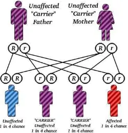
In fact, this disease is a recessive autosomal familial disease and particularly attacks beta globin gene. 12-14 When both parents are carrier of the disease, there is a possibility that one of their four children has thalaassemia, two are carriers and of of them is normal 14 and is often caused by the inheritance of two different mutation, each recognizes beta globin synthesis (mixed heterozygote). Therefore, the pathogenesis of beta thalassemia includes genetic mutation leading to beta polypeptide chain connection deviation in globin that affects beta chain synthesis. 7-13 Globin chains disorder may happen in the alpha or beta chains and may appear in the homozygote individuals and heterozygone individuals. 6-10 AS a result, globin beta chain synthesis is reduced.
Kleihauer test result that determines HbF using acid elution method is used to support diagnosis. 12-6
Talking about decreased hemoglobin due to beta chain deficiency, there is compensation from decreased HbA level and increased production of HbA2 and HbF will lead to various clinical signs. 5,9,14 The most common symptoms are spleen and liver enlargement, delayed bone growth and changes in bones. 7 The changes in bones are due to the bone marrow hyperactivity that causes excessive growth in frontal bone, zygomatic, and maxillary protrusion. This changed form leads to a typical facial appearance called thalassemia facies. The dental growth of the beta major thalassemia is poor and is accompanied by jaw bone refraction. 14 Beside, beta major thalassemia patients have a short life expectancy. The most severe patients rarely reach adulthood. In addition, the repeated big amount of blood transfusion accompanied by abnormal increased of iron will create a new comlication. 15-6
Meanwhile, there are two factors related to anemia pathogenesis in beta thalassemia are reduced beta globin synthesis and beta thalassemia hemolytic components. Beta globin synthesis reduction continues into abnormal erythroblast formation so that the total hemoglobin level for each cell is lower and the cell looks hypochromic. 13-15 The beta thalassemia hemolysis component is not cuased by beta globin deficiency but due to the excessive globin alpha chain with normal synthesis. The alpha free chain forms insoluble pool and creates sediment in the erythrocyte. This input damages the cell layer, reduce the flexibility and decrease the red cells. 16
The clinical manifestation of beta major thalassemia
anemia due to the absence of HbA synthesis. 17-18 Babies with beta mayor thalassemia will look pale with extended abdominal area due to the splenomegaly and failures in growth and development. 13-19 The child’s weight is usually lower than the average weight for his/her age. Fever, diarrhea and various gastrointestinal disorders are usually found during the first year of life. 14 The skin looks pale and yellowish and will become dark due to the iron sedimentation caused by repeated transfusion. 7
While the bone abnormalities happen mostly due to the erythroid marrow hypertrophy and expansion that leads to widen bone marrow, thin cortex and osteoporosis. 15, 19 The bone abnormalities are found initially in the metatarsal and metacarpal bones in the shape of rectangular convex site caused by increased erithropoiesis leading to bone marrow widening. 19,23
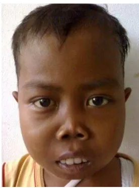
Older patients commonly show a typical face called facies Cooley with abnormal facial and cranial bone growth. The nose looks flat without nasal bridge, the distance between eyes is wide and the forehead bone is wide too. There is a thickening of cheek bones and deep nose bridge that leads to the typical features of facies Cooleh. The teeth look protrusive and the disturbed maxillary growth will create malocclusion. 19-24
Moreover, medullar tubercle gives a mosaic feature of the bone. Osteoporosis and cortical thinning may lead to pathological fracture in long bones and shortened arms due to irregular fusion of the humerus proximal area. 36, 39, 42 The ribs are widened especially in the area where the vertebras meet that leads to increased para vertebrae mass and pressure to the cord (cord compression). 19-20 The characteristic and the severity of the bone abnormalities increase with age. In older patients, the bony changes are more commonly found in the distal areas such as legs and arms. 31, 46, 48
routing transfusion. 2,7,9 Hemocyderosis evokes arrhythmia that also causes sudden death. The abnormality that is caused by severe anemia is hearth enlargement that can be accompanied by congestive heart failure. 11, 13, 17 Pericarditis may happen due to the severe iron sedimentation in the pericardium as well as the bacterial infection. 20
Beta mayor thalassemia patient growth and developmenbt are commonly disturbed even though the relative intelligence level development is not affected. This obstructed growth is caused by the severe anemia and low Hb level leading to tissue hypoxia. The secondary sexual growth is disturbed by the endocrine disorders that puberty is reached later and the growth accelartion is delayee or even does not occur. Menstruation in girls and secondary sexual signs in boys are delayed. 17
While in the older patients, Diabetes mellitus and hypoparatiroidism is commonly found due to the endocrine disorder. 12,16,17 The beta mayor thalassemia patients are also medically compromised patients because they are susceptible to infections. The kidney is usually enlarged as a result from extra medullar hematopoiesis with dark brown urine cased by heme catabolism product excretion. 17-20
Gambar 2. Fasies Cooley
DISCUSSION
Growth and development of oral cavity in beta mayor thalassemia patients show several differences compared to normal children especially in terms of facial bone structures, dental arrangement and gingival color.
Beta mayor thalassemia craniofacial growth and development characteristics
Gambar 3. (A) Gambaran Hair on End Pada Kranium dan (B) Honeycomb pada foto panoramik19,24
Radiographic characteristics of beta mayor thalassemia
The changes in beta mayor thalassemia patients bones that are caused by bone marrow hyperplasia can be observed in the radiographic images. Diploe in the cranial bones experience expansion that the inter plate space becomes widened while the external and internal part of the plate becomes thinner. The frontal bone creates a vertical tubercle that is parallel with the diploe plate external plate and, due to erythroid hyperplasia, the parietal bone has a porous part in the diploe plate external part that creates hair on end or sun ray appearance. 3, 20-8 The tubercle on the jaw bone can be seen in the panoramic image and gives Honeycomb appearance. 19-24
Oral cavity characteristics of beta mayor thalassemia patients
faster jaw growth. The periodontal ligament will seem widened as a result from Class II Division 1 malocclusion. The gingiva is pale especially when the patient’s Hb drops to below 8 gr/dl. The color of the gingiva sometimes tends to be dark, which is caused by the high ferritin level in the blood. The tongue size is bigger/macroglossi as a result from bigger arches.
Considerations during dental treatment
The treatment for beta mayor thalassemia is performed routinely especially when patients experience decreasing hemoglobin level. Therefore, the dental treatment should be performed by paying attention to these following considerations. 24-29
Consult all dental treatments planned with the hematologist. The dental treatment should be performed in a short time as possible and should be done after the patient receives blood transfusion. Do not do dental treatment when the hemoglobin level is less than 100 gl/l. Provide antibiotics prophylaxis especially for children who have undergone splenectomy. If an orthodontic treatment is needed, the dental movement should be watched closely because there may be faster movement compared to the normal situation. The retention phase is also more difficult in these patients.
CONCLUSION
REFFERENCES
1. Leavell, B.S.; O.A. Thorup. Fundamentals of Clinical Hematology. Philadelphia: W.B Saunders Company. 1960.
2. Robbins, S.L.; V. Kumar. Buku Ajar Patologi II. Diterjemahkan dari Basic Pathology Part II. Oleh Staf Pengajar Laboratorium Patologi Anatomik Fakultas Kedokteran Universitas Airlangga. Jakarta: EGC. 1995.
3. Nelson, W.E; R.E. Behrman; R.M. Kliegman et al. Nelson Ilmu Kesehatan Anak. Edisi 15. Diterjemahkan dari Nelson Textbook of Pediatrics. Oleh A. Samik Wahab. Jakarta: EGC. 1999.
4. Sjahruddin, L.D. Indeks kelainan dentofasial dan maturasi tulang vertebra servikal pada
penderita thalassemia beta hemoglobin E serta hubungannya dengan beberapa faktor risiko. Disertasi. Jakarta: Universitas Indonesia. 2004. 3,47-48,51
5. Gandy A. Thalassemia-Beta. Available at
http://www.icondata.com/health/pedbase/file/THALASSE.HTM. 1994.
6. Behrman, R.E. et al. Williams Hematology. 5th edition. New York: McGrow Hill, Inc. 1995.
7. Greer, J.P.; J. Foerster; J.N. Lukens. et al. Wintrobe’s Clinical Hematology. 11th ed. Philadelphia: Lippincott Williams & Wilkins. 2004.5,8,10,12,17-18,22,48
8. Beutler, E.; M.A. Lichtman; B.S. Coller. et al. William’s Hematology. 5th ed. USA: Mc Graw Hill. 1995.12,14-15,17
9. Miller, D.R.; R.L. Baehner; L.P. Miller. Blood Diseases of Infancy and Childhood. 7th ed. St. Louis: Mosby-Year Book, Inc. 1995.8-9,18-19
10. Lee, G.R.; J. Foerster; J.N. Lukens. et al. Wintrobe’s Clinical Hematology. 10th ed. Philadelphia: Lippincott Williams & Wilkins. 1999.1,5,9,14-15,17,19,48
11. Nathan, D.G.; F.A. Oski. Hematology of Infancy and Childhood. 4th ed. Philadelphia: W.B Saunders Company. 1993.22
12. Benz, E. J. and P. J. V. Giardina. Thalassemia syndromes. In: Miller, D. R., et al. Blood
of Infancy and Childhood. 7th ed. United States of America: Mosby. 1995.
13. Thein, S.L. Genetic Insights into The Clinical Diversity of Beta Thalassaemia. British
Journal of Haematology. 2004. 124, 264-274.
14. _________. Pathophysiology of Beta Thalassemia – A Guide to Molecular Therapies.
The American Society of Hematology. 2005.
16. Weatherall, D.J. The Thalassemia Syndromes. 3rd edition. Oxford: Blackwell Scientific Publications Limited. 1981.
17. ______________. Fortnightly Review : The Thalassaemias. BMJ 314:1675. 1997. 18. Kumar, S et al. Beta Globin Gene and Related Diseases : A Review. Int J Hum Genet,
2(3): 139-152. 2002.
19. Cohen, A. and E. Schwartz . Practice of Pediatrics. Volume 5. Philadelphia : Harper and Row. 1986.
20. Lynch, M.A., et al. Burkett’s Oral Medicine, Diagnosis and Treatment. 9th edition. Philadelphia: J.B. Lippincott Company. 1994.
21. Al-Wahadni, A., M.A. Qudeimat, and M. Al-Omari. Dental arch morphological and dimensional characteristics in Jordanian children and young adults with beta thalassemia major: International Journal of Pediatric Dentistry2005.
22. Alhaija, , E. S. J. A.; F. N. Hattab; and M. A. O. Al-Omari. Cephalometric measurement and facial deformities in subjects with beta-thalassaemia major. European J. of
Orthodontics, 2002. 24, 9-10.
23. Hazza’a, AM. and G. Al-Jamal. Radiographic feature of the jaws and teeth in thalassaemia major. Dentomaxillofacial Radiology, 2006.35, 283-288.
24. Greenberg, M. S. and M. Glick. Burket’s Oral Medicine. 10th ed. Spain: BC Decker Inc. 2003.
25. Wonke, B. Bone Disease in Beta Thalassaemia Major. British Journal of Haematolgy. 1998.103, 897-901.
26. Cao, A. Thalassemia:As A Model. Available at
http://www.charite.de/ch/medgen/eumedis/medgen/eumedis/medgen05/thalassemia-amodel.html. 2005.
27. Elstrom R. Clinical Thalassemia (Major and Minor). http://www.nhlbi.nih.gov/Ency/article/000587.htm. 2001.
28. Cannel, H. The development of oral and facial signs in beta thalassemia major. Br. Dent.
J. 1988.164, 50–51.