AND
Staphylococcus aureus
Aktivitas Antibakteri Ekstrak Metanol Kulit Batang Kamboja
(Plumeria acuminata) Terhadap Escherichia coli dan Staphylococcus aureus
Harwoko*, Nuni Anindita**, Eka Prasasti Nurrachmani*
*Pharmaceutical Biology Laboratory, Faculty of Health Sciences, Universitas Jenderal Soedirman, Purwokerto, Central Java
**Quality Assurance Division of Ferron Par Pharmaceuticals, Cikarang-Bekasi,West Java Email: [email protected]
ABSTRAK
Penyakit infeksi merupakan salah satu masalah kesehatan di Indonesia. Hal ini disebabkan oleh bakteri seperti
Escherichia coli dan Staphylococcus aureus. Salah satu tanaman berpotensi sebagai antibakteri adalah Plumeria acuminata (Kamboja). Tujuan dari penelitian ini adalah untuk mengidentifikasi kandungan fitokimia dan untuk mengetahui aktivitas antibakteri terhadap E. coli dan S. aureus dari ekstrak metanol kulit batang P. acuminata (MEPAS). Kulit batang P. acuminata dimaserasi dengan metanol selama 3x24 jam dan kemudian diidentifikasi kandungan fitokimia menggunakan kromatografi lapis tipis. Uji antibakteri dilakukan dengan metode difusi
Kirby-Bauer menggunakan empat kelompok perlakuan, kontrol negatif (media) dan sefotaksim sebagai kontrol positif.
Zona hambat dianalisis dengan analisis probit untuk mendapatkan nilai IC50. Hasil penelitian menunjukkan bahwa MEPAS terdapat alkaloid, terpenoid, flavonoid, dan saponin. Nilai IC50 MEPAS terhadap E. coli dan S. aureus adalah
2.325 ppm dan 1.800 ppm. Pada konsentrasi yang sama, kontrol positif menunjukkan lebih aktif daripada ekstrak.
Kata Kunci: Plumeria acuminata, antibakteri, Escherichia coli, Staphylococcus aureus.
ABSTRACT
Infectious disease is one of health problem in Indonesia. It is caused by bacteria such as Escherichia coli and
Staphylococcus aureus. One of potential plant as antibacterial is Plumeria acuminata (Kamboja). The aim of this
study is to identify the phytochemical contents and to examine antibacterial activity against E. coli and S. au-reus from methanolic extract of P. acuminata stembark (MEPAS). The stembark of P. acuminata was macerated
by methanol for 3x24 hours and then identified for phytochemical compound by Thin Layer Chromatography. Antibacterial assay was done by Kirby-Bauer method using four treatment groups, negative control (media), and cefotaxime as positive control. The inhibition zone was analyzed by probit analysis to obtain the IC50 value. The results showed that MEPAS contained alkaloid, terpenoid, flavonoid, and saponin. The IC50 value of MEPAS
against E.coli and S. aureus was 2,325 ppm and 1,800 ppm, respectively. In the same concentration, the positive
control showed more active than the extract.
Keywords:Plumeria acuminata, antibacterial, Escherichia coli, Staphylococcus aureus.
INTRODUCTION
Food and water contamination by
bacte-ria such as Escherichia coli and Staphylococcus
aureus can cause infectious diarrhea. Diarrhea is an endemic disease in Indonesia and poten-tially as extraordinary events which is often
cause of death. The basic health research 2007
conducted in Indonesia reported that diarrhea
is the first cause of death in neonatus (31.4%) and infants (25.2%), whereas in all age groups is the fourth cause of death (13.2%) (Balitbang
-kes, 2008). The need of antibiotics for infection
therapy is still relatively high, but on the other side appears pathogenic microorganisms that are resistant to antibiotics. Therefore, we need to explore new antibiotics, especially from natural product as an alternative therapy for infectious diseases.
Kamboja (Plumeria acuminata) have been used to treat rheumatism, ulcers, sores, and swelling from many parts of plant such as
flowers, leaves, exudates, and also stembark.
The chemical constituents of P. acuminata are
fuvoplumierin, geraniol, farnesol, sitronellol,
linallol, alkaloid, saponin, flavonoid, bitter substances, and resin (Hariana, 2008). Report
-edly, P. acuminata bark extract has antifungal
activity against Aspergillus and Candida
spe-cies that caused otomycosis (Villanueva et al.,
2008; Boncalon et al., 2009). Priwanda (2006)
showed that chloroform extract of P.
acumi-nata stembark has antiangiogenic properties.
Prihandono (1996) also reported that petro -leum ether,
chloroform, and methanol extracts of P.
acum-inata flower exhibited antibacterial activ -ity against E. coli and S. aureus. In the
pres-ent study, methanolic extract of P. acuminata
stembark (MEPAS) was used as a sample to be identified the phytochemical compounds and
was examined its antibacterial activity against
E. coli and S. aureus.
MATERIALS AND METHOD Materials
Kamboja (P. acuminata) stembark was
collected from Kaliputih, Purwokerto, Indone -sia. Nutrient agar and nutrient broth as media,
E. coli and S. aureus, silica gel F254, Dragendorff
and Vanillin-sulfuric acid reagent, cefotaxime,
methanol, chloroform, ethyl acetate, n-hexane, n-butanol, and glacial acetic acid.
Methods
Preparation of MEPAS
Kamboja stembark powders as much as 400 g was macerated with methanol (1:5) for 3x24 hours, filtered, and the filtrates were
evaporated, then concentrated on waterbath until getting solvent-free and viscous extract.
Phytochemical identification of MEPAS
All samples were spotted on silica gel
F254 plate and developed in mobile phase then
the samples were sprayed with specific re
-agents (Table 1). These spots were observed under UV254 and UV366 lights, then its hRf val
-ues were determined.
Table 1. TLC system on phytochemical identification of MEPAS
Antibacterial assay by Kirby-Bauer method
Each extract was diluted in distilled
water to obtain concentrations of 1,000; 2.000; and 3.000 ppm. Controls used were
media+bacteria (negative control) and
media+antibiotic+bacteria (positive control).
Nutrient broth and bacterial suspension (1.5 x 10⁸ CFU/mL) were added to reach a total of 100 μL. Incubation was taken place at 37°C for 24 hours. Experiments were done in triplicate
and then were measured the inhibition zones against E. coli and S. aureus.
Data analysis
The growth inhibition of MEPAS against
E. coli and S. aureus was calculated with the
following equation (Ishikawa et al., 2001).
Explanation:
I : growth inhibition (%)
d1 : diameter of paper disc (6 mm) d2 : diameter of clear zone (mm)
Furthermore, it was made the linear regression
equation y = a + bx (x as the concentration and
y is the probit number), then being determined
the IC50 value (Inhibitory Concentration) by probit analysis (Mursyidi, 1985).
RESULTS AND DISCUSSION
Methanolic extract of kamboja (P.
acuminata) stembark has dark brown colour
and 14.1% of rendemen.
Methanol is used as a solvent because it can dissolve most of the secondary metabolite
groups and the most frequently used in the
natural product isolation (Darwis, 2000).
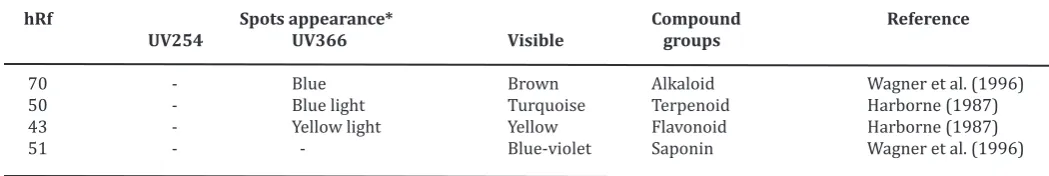
The TLC profile showed that phytochemical contents of MEPAS were flavonoid (hRf 43), terpenoid (hRf 50), saponin (hRf 51), and alkaloid (hRf 70) (Table 2). A tube test by mixing MEPAS with distilled water and then
shaken, the result showed a stable persistent
froth that confirm the presence of saponin in MEPAS.
Table 2. The phytochemical contents of MEPAS by TLC method
hRf Spots appearance* Compound Reference
UV254 UV366 Visible groups
70 - Blue Brown Alkaloid Wagner et al. (1996) 50 - Blue light Turquoise Terpenoid Harborne (1987) 43 - Yellow light Yellow Flavonoid Harborne (1987) 51 - - Blue-violet Saponin Wagner et al. (1996)
*after being sprayed with specific reagent.
Methanolic extract of P. acuminata
stem-bark showed antibacterial activity against E.
coli and S. aureus at concentrations of 1.000;
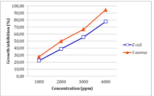
2.000; 3.000; and 4.000 ppm based on the in -hibition zone (Table 3). The higher concentra-tion of the extract, the higher percentage of growth inhibition which indicated that
anti-bacterial effect of MEPAS was dose-dependent (Figure 1). MEPAS at the concentration ranging between 250 and 1,000 ppm showed inhibito -ry activity against all tested bacteria, including
E. coli and S. aureus (Gupta et al., 2008).
Table 3. The result of inhibition zone in all samples against E. coli and S. aureus
Samples Diameter of inhibition zone ± SE (mm, n=3)
E. coli S. aureus
Negative control (media) ND ND
MEPAS 1000 ppm 7,33 ± 0,33* 7,67 ± 0,33*
MEPAS 2000 ppm 8,33 ± 0,33* 9,00 ± 0,00*
MEPAS 3000 ppm 9,33 ± 0,33* 10,00 ± 0,58*
MEPAS 4000 ppm 10,67 ± 0,33* 11,67 ± 0,33*
Positive control (Cefotaxim) 19,00 ± 0,00 18,00 ± 0,00
Note: MEPAS = Methanolic extract of P. acuminata stembark, ND = Not detected, ø paper disc = 6 m * : significantly difference compared to positive control (LSD test, p<0.05)
Based on the probit analysis, the IC50 value of MEPAS against E. coli was 2325 ppm
and S. aureus was 1800 ppm (Figure 1). Anti -bacterial activity of methanolic extract against
S. aureus was greater than that of E. coli. Gupta
et al. (2008) reported that methanolic extract
of P. acuminata Ait. leaves was more sensitive
to Gram positive than Gram negative bacteria. This could be caused by differences in extracts
penetration through the bacteria cell wall.
Both strains of these bacteria have different
cell wall composition.
Figure 1. Growth inhibition activity of methanolic extract of P. acuminata stembark against E. coli (blue) and S. aureus (red)
Staphylococcus aureus is a Gram positive bac -teria group that has a simple structure with peptidoglycan layer more than the lipid
lay-er, whereas the cell wall structure of E. coli is
relatively complex. Cell wall of Gram negative
bacteria composed of three layers, that is lipo-protein (outer), lipopolysaccharide (middle),
The IC50 value of MEPAS was used to
determine the concentration of the cefotaxime
antibiotic as a positive control. Cefotaxime was
the third-generation cephalosporin who has a broad spectrum, that is active on a wide range
of Gram positive and Gram negative bacterial
strains, especially aminoglycosides resistant strain. Mechanism of action of cefotaxime through inhibition of bacteria cell wall synthe-sis by binding to one or more penicillin binding
proteins (PBPs) which will inhibit the trans -peptidase synthesis on peptidoglycan layer of
bacteria cell wall (Bijie et al., 2005). MEPAS
able to inhibit the growth of E. coli and S.
au-reus bacteria, but its antibacterial activity was
lower than cefotaxime. It could be caused by crude extract and there are ballast substances that can inactivate the antimicrobial substance
that decreases its effectiveness (Pelczar and Chan, 2005).
Methanolic extract of P. acuminata
stembark has been shown to contain alkaloid,
terpenoid, flavonoid, saponin that is potential as an antibacterial with a different mechanism
of action. Alkaloid has antibacterial activity through the inhibition mechanism by interfer-ing with components of bacterial peptidogly-can in the cell so that the cell wall layers are not fully formed and caused the death of these
cells (Robinson, 1995). Terpenoid were able to
inhibit the transduction of a growth factor into cells so that cell proliferation is hampered due to the formation of the cell surface receptor
agonist (Fatoni et al., 2005). The mechanism of
inhibition of bacteria by saponin is through the incorporation of saponin which is polar group with a phospholipid layer that also is polar so can damage the permeability of bacteria cell
membrane (Lay and Hastowo, 1995).
CONCLUSION
Methanolic extract of P. acuminata
stem-bark (MEPAS) contains alkaloid, terpenoid, fla
-vonoid, and saponin. The IC50 value, a param
-eter of extract potency, of MEPAS against
E. coli was 2.325 ppm and S. aureus was 1.800
ppm.
ACKNOWLEDGEMENT
The authors express thanks to Pharma -cy Department Faculty of Health Sciences
Uni-versitas Jenderal Soedirman, Purwokerto.
REFERENCES
Badan Penelitian dan Pengembangan
Kesehatan. 2008. Riset Kesehatan Dasar (RISKESDAS) 2007. Balitbangkes.
De-partment of Health Republic of Indone -sia. Jakarta.
Bijie H., Kulpradist S., Manalaysay M. and Soebandrio A. 2005. In vitro activity, pharmacokinetics, clinical efficacy, safe -ty and pharmacoeconomics of ceftriax-one compared with third and fourth
generation cephalosporins. J. Chemoth
-er., 17(1): 3-24.
Boncalon RMV., Arugay MAV. and Ramos RZH. 2009. A preliminary study on the effica
-cy of Plumeria acuminata (Kalachuchi)
bark extract ointment versus clotrima-zole cream in the treatment of
otomy-cosis. Philippine Journal of
Otolaryngol-ogy-Head and Neck Surgery., 24(1): 5-8.
Darwis D. 2000. Teknik dasar laboratorium dalam penelitian senyawa bahan alam
hayati. Workshop pengembangan
sum-ber daya alam dalam bidang kimia or-ganik bahan alam hayati. FMIPA Uni
-versitas Andalas. Padang.
Fatoni A., Lestari P. and Diastuti H. 2005. Uji toksisitas ekstrak spong asal Cilacap
terhadap Artemia salina Leach. Majalah
Ilmiah UNSOED 3: 33-43.
Gupta M., Mazumder UK., Gomathi P. and
Selvan VT. 2008. Antimicrobial activity
of methanol extract of Plumeria
acumi-nata Ait. leaves and Tephrosia purpurea
(Linn.) Pers roots. Natural Product Ra -diance., 7(2): 102-105.
Harborne. 1987. Metode fitokimia, penuntun
cara modern menganalisis tumbuhan,
translation by Padmawinata, K. and So
-ediro, I. ITB Press. Bandung, 4-7, 69-73, 123-127, 151-156, 234-239.
Hariana A. 2008. Tumbuhan obat dan khasiatnya. Penebar Swadaya. Jakarta, 7-8.
Hugo WB. and Russell AD.1998. Pharmaceuti calmicrobiology. Sixth edition. Black-well Science. Oxford.
Ishikawa NK., Kasuya MCM. and Vanet
ti MCD. 2001. Antibacterial activity of Lentinus edodes grown in liquid medi -um. Brazilian Journal of Microbiology.
32: 206-210.
Lay BW. and Hastowo S. 1995. Mikrobiologi.
Rajawali Press. Jakarta, 67-70. Mursyidi A. 1985. Statistika Farmasi dan Biologi.
Ghalia Indonesia. Jakarta, 101-102. Pelczar MJ. and Chan CS. 2005. Dasar-dasar mikrobiologi. Universitas
Indone-sia Press. Jakarta, 447-458, 949, 954-955.
Prihandono IW. 1996. Isolasi dan uji aktivitas antibakteri kandungan daun P lume-ria acuminate, Ait beserta profil kro
-matografinya. Skripsi. Fakultas Farmasi
Universitas Gadjah Mada. Yogyakarta. Priwanda R. 2006. Efek antiangiogenik
ekstrak kloroform kulit batang
kam-boja (Plumeria acuminata, Ait) pada
Membran Korio Alantoin (CAM) embrio ayam yang terinduksi bFGF. Skripsi. Fakultas Matematika dan Ilmu Pengeta -huan Alam Universitas Islam Indonesia. Yogyakarta.
Robinson T. 1995. Kandungan organik
tumbuhan tinggi. Sixth edition,
trans-lation by Padmawinata, K. ITB Press. Bandung, 139-181, 191-216.
Villanueva JM., Arugay MAV. and Ramos RZH. 2008. In vitro antimycotic activity of
four medical plants versus clotrimazole in the treatment of otomycosis: a pre-liminary study.
Philippine Journal of Otolaryngology-Head and Neck Surgery., 23(1): 5-8.
Wagner H., Bladt S. and Zgainski EM. 1996.
Plant drug analysis: a thin layer chroma -tography atlas. Springer. New York, 24,