79
V. SIMPULAN DAN SARAN
A.Simpulan
Berdasarkan penelitian aktivitas antibakteri ekstrak daun parijoto (Medinilla speciosa) terhadap Escherichia coli dan Staphylococcus aureus
yang telah dilakukan, diperoleh kesimpulan bahwa:
1. Ekstrak daun parijoto (Medinilla speciosa) dengan pelarut etil asetat dan metanol memiliki aktivitas antibakteri terhadap Escherichia coli dan
Staphylococcus aureus. Tidak ada beda nyata pada luas zona hambat yang dihasilkan oleh ekstrak dengan metanol dan etil asetat daun parijoto, tetapi zona hambat terluas diperlihatkan oleh ekstrak dengan pelarut metanol. 2. Bakteri Gram positif yang diwakili oleh Staphylococcus aureus lebih
sensitif terhadap aktivitas antibakteri ekstrak daun parijoto dibandingkan dengan bakteri Gram negatif yang diwakili oleh Escherichia coli.
3. Nilai Konsentrasi Hambat Minimum (KHM) ekstrak metanol daun parijoto terhadap Escherichia coli adalah 50 mg/ml, sedangkan nilai KHM ekstrak metanol daun parijoto terhadap Staphylococcus aureus adalah 12,5 mg/ml. B.Saran
1. Pembuatan serbuk ekstrak dengan ukuran partikel yang lebih kecil (>35 mesh) dapat dilakukan agar ekstraksi maserasi dapat berlangsung lebih optimal.
2. Penelitian lanjutan berupa uji fitokimia kuantitatif terutama untuk senyawa tanin, perlu dilakukan untuk mengidentifikasi dan mengetahui komposisi senyawa-senyawa fitokimia yang terkandung dalam daun parijoto.
3. Metode pemurnian saponin dan atau tanin dari daun parijoto dapat dikaji lebih lanjut untuk mendapatkan aktivitas antibakteri yang lebih kuat.
4. Pengujian aktivitas antijamur ekstrak daun parijoto dapat dilakukan untuk mengkaji manfaat lain dari daun parijoto.
5. Aplikasi daun parijoto sebagai antibakteri alami misalnya pengembangan serbuk daun parijoto sebagai bahan obat kumur anti-sariawan dapat dikaji lebih lanjut.
81
DAFTAR PUSTAKA
Abel, E. E., Poonga, P. R. J., dan Panicker, S. G. 2014. Effects of different solvent extracts of Cassia tora leaves against Gram positive bacteria.
International Journal of Pharmacy and Life Science 5(4): 3436-3439. Adegoke, O. A., Analytical, biochemical and synthetic applications of
para-dimethylaminobenzaldehyde. International Journal of Pharmaceutical Science Review and Research 11(2): 17-29.
Ajayi, A. A., Aiyedun, B. T., dan Olasehinde, G. I. 2013. The effect of hand treatments on Staphylococcus aureus: a normal flora of the human palms.
Advances in Bioscience and Bioengineering 1(2): 44-53.
Akiyama, H., Fujii, K., Yamasaki, O., Oono, T., dan Iwatsuki, K. 2001. Antibacterial action of several tannins against Staphylococcus aureus.
Journal of Antimicrobial Chemotherapy 48:487-491.
Akoue, G. N., Obame, L. C., Ondo, J. P. Brama, I., Nang, E. S. O., Tapoyo, S. Y., Souza, A. 2013. Phytochemical composition and antiradical activity of
Sakersia Africana Hook. f. medicinal plant from Gabon. International Journal of Biomolecules and Biomedicine 3(3): 1-8.
Alabri, T. H. A., Musalami, A. H. A. S. A., Hossain, M. A., Weli, A. M., dan Al-Riyami, Q. 2014. Comparative study of phytochemical screening, antioxidant, and antimicrobial capacities of fresh and dry leaves crude plant extract of Datura metel L. Journal of King Saud University 26:237-243.
Alhanout, K., Malesinki, S., Vidal, N., Peyrot, V., Rolain, J. M., dan Brunel, J. M. 2010. New insights into the antibacterial mechanism of action of squalamine. Journal of Antimicrobial Chemotherapy
doi:10.1093/jac/dkq213.
American Society for Microbiology. 2005. Manual of Antimicrobial Susceptibility Testing. ASM, New York. Halaman 53-59.
Anandhi, D., Srinivasan, P. T., Kumar, G. P., dan Jagatheesh, S. 2014. Influence of flavonoids and glycosides from Caesalpinia coriaria wild as bactericidal compound. International Journal of Current Microbiology and Applied Sciences 3(4): 1043-1051.
Aniszewski,T. 2007. Alkaloids-Secret of Life: Alkaloid Chemistry, Biological, Significance, Applications and Ecological Role. Elsevier, Oxford. Halaman 6-12,130, dan 187.
Arora, D. S. dan Bhardwaj, S. K. 1997. Antibacterial activity of some medicinal plants. Geobios 24: 127-131. diacu dalam Parvez, S., Begum, F., Neela, F. A., dan Alam, M. F. 2015. Screening of MDR-bacteria from fecal specimens of AAD patient and inhibit them using fruits extrats of
Moringa oleifera Lam. International Journal of Bioscience 6(3): 402-409.
Ashour, M., Wink, M., dan Gershenzon, J. 2010. Biochemistry of Terpenoids: Monoterpenes, Sesquiterpenes, and Diterpenes. Dalam: Wink, M. Annual Plant Reviews Volume 40: Biochemistry of Plant Secondary Metabolism
second edition. Blackwell Publishing, West Sussex. Halaman 258-263. Ayoola, G. A., Coker, H. A. B., Adesegun, S. A., Bello, A. A. A., Obaweya, K.,
Ezennia, E. C., dan Atangbayila, T. O. 2008. Phytochemical screening and antioxidanr activities of some selected medicinal plants used for malaria therapy in Southwestern Nigeria. Tropical Journal of Pharmaceutical Research 7(3): 1019-1024.
Baccou, J. C., Lambert, F., dan Sauvaire, Y. 1977. Spectrophotometric method for determination of total steroidal sapogenin. Analyst 102: 458-465.
Barrett, L. 2015. Olive Leaf Extract: The Mediterranean Healing Herb. Healthy Living Publications, Summertown. Halaman 1-3.
Burke, K. A. dan Lascelles, J. 1975. Nitrate reductase system in Staphylococcus aureus wild type and mutants. Journal of Bacteriology 123(1): 308-316. Cennimo, D. J., Koo, H., Mohamed, J. A., Huang, D. B., Chiang, T.
Enteroaggregative Escherichia coli: A review of trends, diagnosis, and treatment. Infections in Medicine March 2007: 100-110.
Centers for Disease Control and Prevention (CDC). 2013. Antibiotic Resistance Threats in the United States 2013. CDC, Georgia. Halaman 77.
Clinical Laboratory Standards Institute. 2015. Performance Standards for Antimicrobial Susceptibility Testing; Twenty Fifth Informational Supplement, CLSI document M100-S25. Wayne, CLSI. Halaman 44-46 dan 64-66.
Cseke, L. J., Kirakosyan, A., Kaufman, P. B., Warber, S. L., Duke, J. A., dan Brielmann, H. L. 2006. Natural Products from Plants. CRC Press, Florida. Halaman 17-18.
Cunha, W. R., Matos, G. X., Souza, M. G., Tozatti, M. G., Silva, M. L. A., Martins, C. H. G., Silva, R., dan Filho, A. D. S. 2010. Evaluation of the antibacterial activity of the methylene chloride extract of Miconia ligustroides, isolated triterpene acids, and ursolic acid derivatives.
Pharmaceutical Biology 48(2): 166-169.
Cushnie, T. P. T., Cushnie, B., dan Lamb, A. J. 2014. Alkaloids: an overview of their antibacterial, antibiotic-enhancing and antivirulence activities.
International Journal of Antimicrobial Agents 44:377-386.
Departemen Kesehatan Republik Indonesia. 1985. Cara Pembuatan Simplisia. Direktorat Jendral Pengawasan Obat dan Makanan, Jakarta. Halaman 2, 7-12, dan 26.
Departemen Kesehatan Republik Indonesia. 2000. Parameter Standar Umum Ekstrak Tumbuhan Obat. Direktorat Jendral Pengawasan Obat dan Makanan, Jakarta. Halaman 7-12.
Devi, A. S., Rajkumar, J., Modilal, R. D., Ilayaraja, R. 2012. Antimicrobial activities of Avicennia marina, Caesalpinia pulcherima, and Melastoma malabathricum against clinical pathogens isolated from Uti.
International Journal of Pharma and Bio Sciences 3(3): 698-705.
Dickson, C. 2014. Experiments in Pharmaceutical Chemistry Second Edition. CRC Press, Florida. Halaman 84.
Drugeon, H. B., Juvin, M. E., Caillon, J., dan Courtieu, A. L. 1987. Assessment of formulas for calculating critical concentration by the agar diffusion method. Antimicrobial Agents and Chemotherapy 31(6): 870-875. Ecosystem Research and Development Bureau. 2012. Philippine Country Report
on Forest Genetic Resources. Ecosystem Research and Development Bureau, Laguna. Halaman 10.
Evans, W. C. 2009. Trease and evans pharmacognosy. Saunders Elsevier, Edinburgh. Halaman 223, 336-337, dan 543.
Garrity, G. M., Brenner, D. J., Krieg, N. R., dan Staley, J. T. 2009 a. Bergey's Manual of Systematic Bacteriology Second Edition Volume Two: The
Proteobacteria. Springer, New York. Halaman 607-623.
Garrity, G. M., Brenner, D. J., Krieg, N. R., dan Staley, J. T. 2009 b. Bergey's Manual of Systematic Bacteriology Second Edition Volume Three: The Firmicutes. Springer, New York. Halaman 392-401.
Gartner, T. K. dan Riley, M. 1965. Isolation of mutants affecting tryptophanase production in Escherichia coli. Journal of Bacteriology 89(2): 313-318. Gaylord Chemical Company. 2007. Dimethyl Sulfoxide (DMSO) Solubility Data.
GCC Bulletin 102 B, Los Angels. Halaman 1.
Gilbert, A., Herve, T. T., William, Y. N., Leonard, S. F., Roger, K. J., Albert, K. 2014. Antidiarrhoeal and antibacterial activity of aqueous and methanolic leaves extracts of Dissotis thollonii Cogn. (Melastomataceae). Asian Pacific Journal of Tropical Biomedicine 4(2): S672-S678.
Glaser, A. N. 2001. High Yield Biostatistics. Lippincott Williams and Wilkins, Philadelphia. Halaman 42-44, dan 58-61
Green, J. 2011. The Herbal Medicine Maker's Handbook. Crossing Press, New York. Halaman 310.
Grinsted, B. dan Bennet, P. M. 1988. Methods in Microbiology Volume 21. Academic Press, London. Halaman 108.
Grotewold, E. 2006. The Science of Flavonoids. Springer, New York. Halaman 1-2, 47-51, dan 73-75.
Hajnos, M. W., Sherma, J., dan Kowalska, T. 2008. Thin Layer Chromatography in Phytochemistry. CRC Press, Florida. Halaman 529.
Handa, S. S. 2008. An Overview of Extraction Techniques for Medicinal and Aromatic Plants. Dalam: International Centre for Science and High Technology. 2008. Extraction Technologies for Medicinal and Aromatic Plants. International Centre for Science and High Technology, Trieste. Halaman 31, 61, dan 69-72.
Harborne, J. B. 1998. Phytochemical Methods Third Edition. Thomson Publishing, London. Halaman 4-6, 60-63, 108, 132, 135, 188, 208-209, dan 291.
Hassan, S. M., Haq, A. U., Berhow, M. A., Catwright, A. L., dan Bailey, C. A. 2010. Haemolytic and antimicrobial activities of saponin-rich extract from guar meal. Food Chemistry 119:600-605.
Hikosaka, K., Nagamatsu, D., Ishii, H. S., dan Hirose, T. 2002. Photosynthesis-nitrogen relationships in species at different altitudes on Mount Kinabalu, Malaysia. Ecological Research 17: 305-313.
Hooker, W. J. 1847. Curtis's Botanical Magazine Volume LXXIII. London. 4321. Isaza, J. H., Ito, H., dan Yoshida, T. 2004. Oligomeric hydrolyzable tannins from
Monochaetum multiflorum. Phytochemistry 65: 359-367.
Jacob, S. W. dan de la Torre, J. C. 2015. Dimethyl Sulfoxide (DMSO) in Trauma and Disease. CRC Press, Boca Raton. Halaman 1-4.
Jork, H., Funk, W., Fischer, W., dan Wimmer, H. 1990. Thin-Layer Chromatography: Reagents and Detection Methods Volume 1. Verlagsgesellschaft, Weinheim. Halaman 195-197.
Kalt, F. R. dan Cock, I. E. 2014. Gas chromatography-mass spectroscopy analysis of bioactive Petalostigma extract: Toxicity, antibacterial, and antiviral activities. Pharmacognosy Magazine 10(37): S37-S49.
Kaufman, P. B., Kirakosyan, A., McKenzie, M., Dayanandan, P., Hoyt, J. E., dan Li, C. 2006. The Uses of Plant Natural Products by Humans and Risks Associated with Their Use. Dalam: Cseke, L. J., Kirakosyan, A., Kaufman, P. B., Warber, S. L., Duke, J. A., dan Brielmann, H. L. 2006.
Natural Products from Plants Second Edition. CRC Press, Florida. Halaman 42.
Kementerian Negara Riset dan Teknologi. 2015. Medinilla speciosa. http://www.warintek.ristek.go.id. Diakses pada tanggal 23 Februari 2015. Kementerian Pertanian Republik Indonesia. 2012. Standar Operasional Prosedur
Pascapanen Tanaman Obat Daun. Kementerian Pertanian Direktorat Jendral Hortikultura Direktorat Budidaya dan Pascapanen Sayuran dan Tanaman Obat, Jakarta.
Khardori, N. 2006. Antibiotics: Past, present, and future. Medical Clinic Journal of North America 90(1): 1049–1076.
Kreis, W. dan Mueller-Uri, F. 2010. Biochemistry of Sterols, Cardiac Glycosides, Brassinosteroids, Phytoecdysteroids, and Steroid Saponins. Dalam: Wink, M. Annual Plant Reviews Volume 40: Biochemistry of Plant Secondary Metabolism second edition. Blackwell Publishing, West Sussex. Halaman 343-344.
Leandro, L. M., Vargas, F. S., Barbosa, P. C. S., Neves, J. K. O., Silva, J. A., dan Veiga-Junior, V. F. 2012. Chemistry and biological activities of terpenoids from copaiba (Copaifera spp.) oleoresins. Molecules 17: 3866-3889.
Lide, D. R. 2005. Handbook of Chemistry and Physics. CRC Press, Boca Raton. Halaman 43
Maatalah, M. B., Bouzidi, N. K., Bellahouel, S., Merah, B., Fortas, Z., Soulimani, R., Saidi, S., dan Derdour, A. 2012. Antimicrobial activity of the alkaloids and saponin extracts of Anabasis articulata. Journal of Biotechnology and Pharmaceutical Research 3(3): 54-57.
Madigan, M. T., Martinko, J. M., Bender, K. S., Buckley, D. H., dan Stahl, D. A. 2015. Brock Biology of Microorganism Fourteenth Edition. Pearson Education, Boston. Halaman 171-178.
Madland, E. 2013. Extraction, isolation, and structure elucidation of saponins from Herniaria incana. Department of Chemistry of Norwegian Universitu of Science and Technology, Trondheim. Halaman 5 dan 11. Maria, C., Erszebet, B., dan Denisa, H. 2012. Medinilla: An exotic and attractive
indor plant with great value. Journal of Horticulture, Forestry, and Biotechnology 16(2): 9-12.
Marliana, S. D., Suryanti, V., dan Suyono. 2005. Skrining fitokimia dan analisis kromatografi lapis tipis komponen kimia buah labu siam (Sechium edule
Jacq. Swartz.) dalam ekstrak etanol. Biofarmasi 3(1):26-31.
Mazimba, O, Wale, K., Kwape, T. E., Mihigo, S. O., dan Kokengo, B. M. 2015.
Cinnamomum verum: Ethylacetate and methanol extracts antioxidant and antimicrobial activity. Journal of Medicinal Plants Studies 3(3): 28-32. Melander, R. J., Minvielle, M. J., dan Melander, C. 2014. Controlling bacterial
behavior with indole-containing natural products and derivatives.
Tetrahedron 70:6363-6372.
Monsquera, O. M., Correra, Y. N., dan Nino, J. 2008. Antioxidant activity of plant extracts from Colombian flora. Brazilian Journal of Pharmacognosy
19(2A): 382-387.
Morello, J. A., Granato, P. A., dan Mizer, H. E. 2003. Laboratory Manual and Workbook in Microbiology. McGraw-Hill, New York. Halaman 313-314. Morse, M. L., Hill, K. L., Egan, J. B., dan Hengstenberg, W. 1968. Metabolism of
lactose by Staphylococcus aureus and its genetic basis. Journal of Bacteriology 95(6): 2270-2274.
Mulyati, E. S. 2009. Uji aktivitas antibakteri ekstrak etil asetat daun ciremai (Phyllanthus acidus L. Skell) terhadap Staphylococcus aureus dan
Escherichia coli dan bioautografinya. Naskah Skripsi S-1. Fakultas Farmasi Universitas Muhamadiyah Surakarta, Surakarta.
National Development Planning Agency (BAPPENAS). 1993. Biodiversity Action Plan for Indonesia, Jakarta.
Niranjan, K., Sathiyaseelan, V., dan Jeyaseelan, E. C. 2013. Screening for anti-microbial and phytochemical properties of different solvents extracts of leafs of Pongamia pinnata. International Journal of Scientific and Research Publications 3(1): 1-3.
Niswah, L. 2014. Uji aktivitas antibakteri dari ekstrak buah parijoto (Medinilla speciosa Blume) menggunakan metode difusi cakram. Naskah Skripsi S-1. Fakultas Kedokteran dan Ilmu Kesehatan Universitas Islam Negeri Syarif Hidayatullah Jakarta, Jakarta.
Omojate, G. C., Enwa, F. O., Jewo, A. O., dan Eze, C. O. 2014. Mechanism of antimicrobial actions of phytochemicals against enteric pathogens: A review. Journal of Pharmaceutical, Chemical, and Biological Science
2(2): 77-85.
Onyegbule, A. F., Anowi, C. F., Gugu, T. H., dan Uto-Nedosa, A. U. 2011. Evaluation of antimicrobial properties of ethyl acetate extract of the leaves of Napoleoneae imperalis family Lecythiaceae. International Journal of Drugs Research and Technology 1(1): 45-51.
Osbourn, A. E. 2003. Molecules of interest: Saponins in cereals. Phytochemistry
62: 1-4.
Pengelly, A. 2004. The Constituents of Medicinal Plants second edition. Allen and Unwin, Crows Nest. Halaman 29-37, 45-53, dan 74-81.
Promega. 2009. Technical bulletin: Griess reagent system. Promega corporation, Madison. Halaman 2 dan 6.
Purves, B. dan Sadava, D. 2003. The Life Science of Biology Seventh Edition. Sinauer Associates Inc., New York. Halaman 528-529.
Rahim, G., Qureshi, R., Arshad, M., dan Gulfraz, M. 2013. Phytochemical analysis and antioxidant properties of Teucrium stocksianum flower from Malakand Division, Pakistan. International Journal of Agriculture and Biology 15(2): 337-381.
Ringoringo, V. S., Suwarno, E. dan Chandra, Y. A. 2008. Bioavailabilitas
Komparatif Tiga Preparat Tablet Ampisilin 500
mg.http://www.kalbe.co.id. Diakses pada tanggal 29 April 2015.
Robinson, T. 1995. Kandungan Organik Tumbuhan Tinggi. Penerbit ITB, Bandung. Halaman 157.
Sadek, P. 2002. The HPLC Solvent Guide. Wiley Interscience, New York. Halaman 22-24.
Semmelweis University. 2014. Crude drugs containing tannins. http://Semmelweis.hu/farmakognozia/files/2014/03/tannins.pdf. Diakses pada tanggal 27 Januari 2016.
Sen, S., Makkar, H. P. S., Muetzel, S., dan Becker, K. 1998. Effect of Quillaja saponaria saponins and Yucca schidigera plant extract on growth of Escherichia coli. Letters in Applied Microbiology 27:35-38.
Shanmugam, S., Kumar, T. S., dan Selvam, K. P. 2010. Laboratory Handbook on Biochemistry. PHI Learning, New Delhi. Halaman 130-132.
Sher, A. 2009. Antimicrobial activity of natural products from medicinal plants.
Gomal Journal of Medical Sciece 7(1): 72-78.
Showe, M. K. dan deMoss, J. A. 1968. Localization and regulation of synthesis of nitrate reductase in Escherichia coli. Journal of Bacteriology 95(4): 1305-1313.
Sinaka, A. 2010. Formulasi tablet hisap ekstrak etanol daun ceremai (Phyllantus acidus) dengan amilum manihot sebagai pengikat serta uji aktivitas antibakteri terhadap Staphylococcus aureus. Naskah Skripsi-S1. Fakultas Farmasi Universitas Muhammadiyah Surakarta, Surakarta.
Smallwood, I. M. 1996. Handbook of Organic Solvent Properties. John Wiley and Sons, New York. Halaman 15, 61, 63, 247, dan 249.
Thermo Fisher Scientific. 2015 a. Nutrient Agar. http://www.oxoid.com/uk/blue /prod detail/prod detail.asp?pr=CM0309&org=107&c=uk&lang=en. Diakses pada tanggal 12 September 2015.
Thermo Fisher Scientific. 2015 b. Nutrient Broth. http://www.oxoid.com/UK/blue /prod_detail/prod_detail.asp?pr=CM0001&cat=&sec=1&c=uk&lang=en. Diakses pada tanggal 12 September 2015.
Tregoning, J. J. dan Poe, C. F. 1937. Production of variants of the colon and aerogenes groups in different media. Journal of Bacteriology: 465-473. Veerachari, U dan Bopiah, A. K. 2012. Phytochemical investigation of the
ethanol, methanol, and ethyl acetate leaf extracts of six Cassia species.
International Journal of Pharma and Bio Sciences 3(2): 260-270.
Volk, W. A. dan Wheeler, M. F. 1988. Dasar-Dasar Mikrobiologi Edisi ke-5 Jilid 1. Erlangga, Jakarta. Halaman 25-255.
Wachidah, L. N. 2013. Uji aktivitas antioksidan serta penentuan kandungan fenolat dan flavonoid total dari buah parijoto (Medinilla speciosa
Blume). Naskah Skripsi S-1. Fakultas Kedokteran dan Ilmu Kesehatan Universitas Islam Negeri Syarif Hidayatullah Jakarta, Jakarta.
Walsh, G. 2003. Biopharmaceutical: Biochemistry and Biotechnology second edition. John Wiley and Sons, West Sussex. Halaman 459.
Wei, S. E. 2011. Isolation and determination of anti-nutritional compounds from root and shell of peanut (Arachis hypogea). Skripsi-S1. Faculty of Science Universiti Tunku Abdul Rahman, Malaysia.
Wibowo, H. A., Wasino, dan Setyowati, D. L. 2012. Kearifan lokal dalam menjaga lingkungan hidup (Studi kasus masyarakat di Desa Colo Kecamatan Dawe Kabupaten Kudus). Journal of Educational Social Studies 1(1): 25-30.
Widjanarko, M. 2013. Jelajah Muria. Muria Research Center, Kudus. Halaman 2. Widjanarko, M. dan Wismar'ein, D. 2011. Identifikasi sosial potensi ekowisata
berbasis peran masyarakat lokal. Jurnal Psikologi UNDIP 9(1): 333-38. Wiegand, I., Hilpert, K., dan Hancock, R. E. W. 2008. Agar and broth dilution
Xia, J. Y., Zuo, G. Y., Wang, G. C., Xu, G. L., dan Zhao, Y. B. 2009. Screen of chinese herbal medicines originated in Yunnan Province against drug resistant, Escherichia coli producing ESBLs in vitro. Medical Journal of National Defending Forces in Southwest China 19(7): 664-666.
Yulianti, L. I. M. 2014. Biostatistika. Graha Ilmu, Yogyakarta. Halaman 60 dan 70-76.
Zearah, S. A., Al-Fartosy, A. J. M., dan Al-Kanany, G. F. 2013. Antibacterial activity of the glycosidic extract from Citrus laurantifoia L. fruits. Der Pharma Chemica 5(6):73-78.
Zuhud, E. A. M., Sinroyo, Sandra, E., Hikmat, A., dan Adhiyanto, E. 2014. Buku Acuan Umum Tumbuhan Obat Indonesia Jilid VI. Dian Rakyat, Jakarta. Zumadhl, S. S. dan Zumadhl, S. A. 2010. Chemistry. Cengage Learning, Belmont.
Halaman 25 dan 532.
91 LAMPIRAN
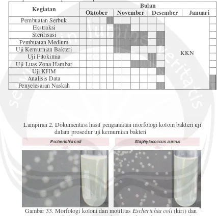
Lampiran 1. Jadwal pelaksanaan penelitian
Kegiatan Bulan
Oktober November Desember Januari Pembuatan Serbuk
KKN
Ekstraksi
Sterilisasi Pembuatan Medium Uji Kemurnian Bakteri Uji Fitokimia Uji Luas Zona Hambat
Uji KHM
Analisis Data Penyelesaian Naskah
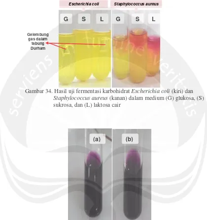
Lampiran 2. Dokumentasi hasil pengamatan morfologi koloni bakteri uji dalam prosedur uji kemurnian bakteri
Escherichia coli Staphylococcus aureus
Gambar 33. Morfologi koloni dan motilitas Escherichia coli (kiri) dan
Lampiran 3. Dokumentasi hasil uji sifat biokimia isolat bakteri uji dalam prosedur uji kemurnian
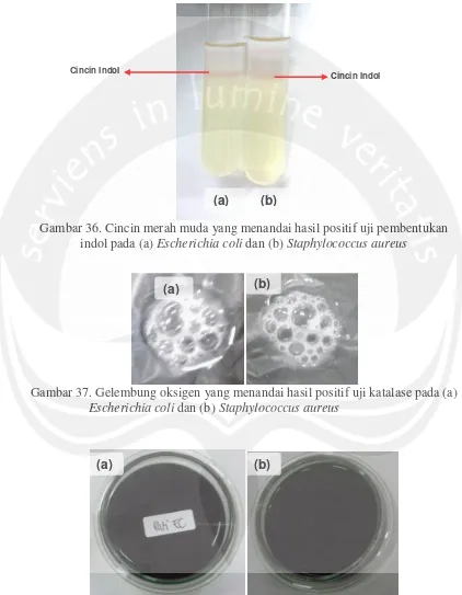
Gambar 34. Hasil uji fermentasi karbohidrat Escherichia coli (kiri) dan
Staphylococcus aureus (kanan) dalam medium (G) glukosa, (S) sukrosa, dan (L) laktosa cair
Gambar 35. Warna magenta yang menandai uji positif reduksi nitrat pada (a) Escherichia coli dan (b) Staphylococcus aureus
Escherichia coli Staphylococcus aureus
G S L G S L
Gelembung gas dalam
tabung Durham
Lanjutan Lampiran 3. Dokumentasi hasil uji sifat biokimia isolat bakteri uji dalam prosedur uji kemurnian
Gambar 36. Cincin merah muda yang menandai hasil positif uji pembentukan indol pada (a) Escherichia coli dan (b) Staphylococcus aureus
Gambar 37. Gelembung oksigen yang menandai hasil positif uji katalase pada (a)
Escherichia coli dan (b) Staphylococcus aureus
Gambar 38. Warna biru pekat tanpa adanya halo transparan yang menandai hasil negatif uji hidrolisis amilum pada (a) Escherichia coli dan (b)
Staphylococcus aureus
(a) (b) Cincin Indol
Cincin Indol
(a) (b)
Lampiran 4. Raw data luas zona hambat ekstrak daun parijoto terhadap
Escherichia coli dan Staphylococcus aureus
Perlakuan Ulangan
Bakteri
Escherichia coli Staphylococcus aureus
Diameter (cm)
Luas (cm2)
Diameter (cm)
Luas (cm2) Ekstrak Daun Parijoto dengan Pelarut Metanol
1 1,50 1,766 1,50 1,766
2 1,40 1,539 1,80 2,543
3 1,40 1,539 1,70 2,269
4 1,50 1,766 1,90 2,834
5 1,40 1,539 1,80 2,543
Rata-rata 1,44 1,630 1,74 2,390
Ekstrak Daun Parijoto dengan Pelarut Etil Asetat
1 1,40 1,539 1,40 1,539
2 1,20 1,130 1,50 1,766
3 1,25 1,227 1,40 1,539
4 1,20 1,130 1,40 1,539
5 1,50 1,766 1,40 1,539
Rata-rata 1,31 1,360 1,42 1,580
Kontrol Negatif (DMSO)
1 0,80 0,502 0,80 0,502
2 0,80 0,502 0,80 0,502
3 0,80 0,502 0,80 0,502
4 0,80 0,502 0,80 0,502
5 0,80 0,502 0,80 0,502
Rata-rata 0,80 0,502 0,80 0,502
Kontrol Positif (Ampicillin)
1 1,30 1,327 2,20 3,799
2 1,40 1,539 2,40 4,522
3 1,50 1,766 2,50 4,906
4 1,30 1,327 2,50 4,906
5 1,30 1,327 2,50 4,906
Rata-rata 1,36 1,460 2,42 4,610
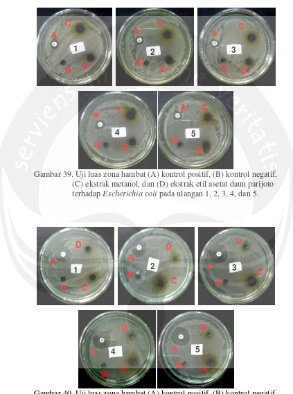
Lampiran 5. Uji aktivitas antibakteri terhadap Escherichia coli dan
Staphylococcus aureus
Gambar 39. Uji luas zona hambat (A) kontrol positif, (B) kontrol negatif, (C) ekstrak metanol, dan (D) ekstrak etil asetat daun parijoto terhadap Escherichia coli pada ulangan 1, 2, 3, 4, dan 5.
Gambar 40. Uji luas zona hambat (A) kontrol positif, (B) kontrol negatif, (C) ekstrak metanol, dan (D) ekstrak etil asetat daun parijoto terhadap Staphylococcus aureus pada ulangan 1, 2, 3, 4, dan 5.
A C
B D
A B C D A B C D A B C D A B C D
1 2 3
4 5
A
B C
D A B C D A B C D A
B C
D A B C D 5 4
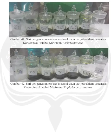
Lampiran 6. Seri pengenceran dalam penentuan Konsentrasi Hambat Minimum
Gambar 41. Seri pengenceran ekstrak metanol daun parijoto dalam penentuan Konsentrasi Hambat Minimum Escherichia coli
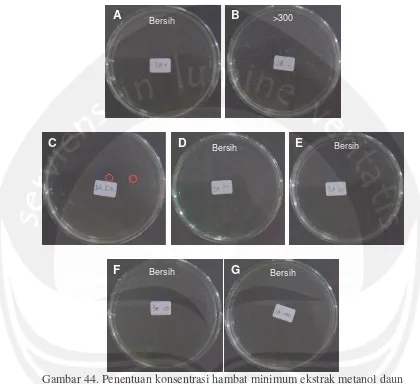
Lampiran 7. Penentuan Konsentrasi Hambat Minimum ekstrak metanol daun parijoto terhadap Escherichia coli
Gambar 43. Penentuan konsentrasi hambat minimum ekstrak metanol daun parijoto terhadap Escherichia coli dengan metode dilusi agar Keterangan: (A) Kontrol positif, (B) kontrol negatif, (C) konsentrasi ekstrak 6,25
mg/ml, (D) konsentrasi ekstrak 12,5%, (E) konsentrasi ekstrak 25%, (F) konsentrasi ekstrak 50%, dan (G) konsentrasi ekstrak 100%
Bersih
Spreader
Bersih Bersih
A B
C D E
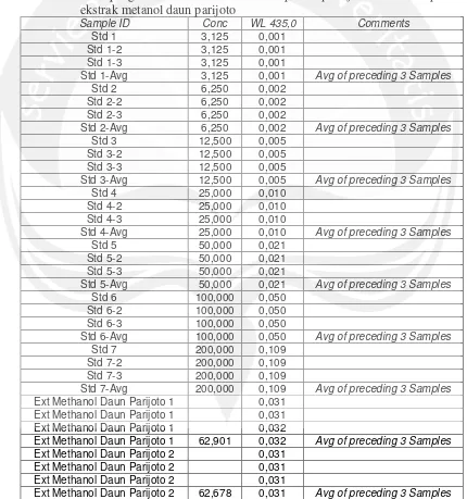
Lampiran 8. Penentuan Konsentrasi Hambat Minimum ekstrak metanol daun parijoto terhadap Staphylococcus aureus
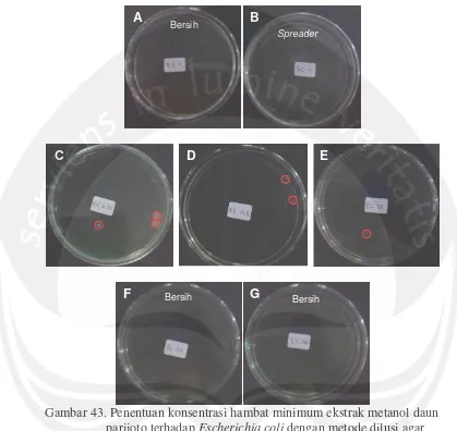
Gambar 44. Penentuan konsentrasi hambat minimum ekstrak metanol daun parijoto terhadap Staphylococcus aureus dengan metode dilusi agar Keterangan: (A) Kontrol positif, (B) kontrol negatif, (C) konsentrasi ekstrak 6,25 mg/ml, (D) konsentrasi ekstrak 12,5%, (E) konsentrasi ekstrak 25%, (F) konsentrasi ekstrak 50%, dan (G) konsentrasi ekstrak 100%
Bersih >300
Bersih Bersih
Bersih Bersih
A B
C D E
Lampiran 9. Hasil ANAVA luas zona hambat ekstrak metanol dan etil asetat daun parijoto terhadap Escherichia coli dan Staphylococcus aureus
Variabel Bebas:LZH
Sumber Jumlah Kuadrat Tipe II
Derajat
Bebas Rerata Kuadrat F Sig.
Model Terkoreksi 44,178a 4 11,044 21,663 ,000
Intercept 123,071 1 123,071 241,402 ,000
Bakteri 10,701 1 10,701 20,990 ,000
Perlakuan 33,477 3 11,159 21,888 ,000
Galat 17,844 35 ,510
Total 185,092 40
Total Terkoreksi 62,021 39
1. Bakteri
Variabel Bebas: LZH
Bakteri Rerata Std. Error Taraf Kepercayaan 95% Batas Bawah Batas Atas
Escherichia coli 1,237 ,160 ,913 1,561
Staphylococcus aureus 2,271 ,160 1,947 2,595
2. Perlakuan
Variabel Bebas:LZH
Perlakuan Rerata Std. Error Taraf Kepercayaan 95% Batas Bawah Batas Atas
Ekstrak Metanol 2,010 ,226 1,552 2,469
Ekstrak Etil Asetat 1,471 ,226 1,013 1,930
Kontrol Negatif ,502 ,226 ,044 ,960
Lampiran 10. Hasil DMRT luas zona hambat ekstrak metanol dan etil asetat daun parijoto terhadap Escherichia coli dan Staphylococcus aureus
Perlakuan
LZH
Duncana,b
Perlakuan N Subset
1 2 3
Kontrol Negatif 10 ,50200
Ekstrak Etil Asetat 10 1,47140
Ekstrak Metanol 10 2,01040
Kontrol Positif 10 3,03250
Sig. 1,000 ,100 1,000
Lampiran 11. Perhitungan kadar saponin dalam ekstrak metanol daun parijoto pada uji aktivitas zona hambat
Kadar saponin dalam ekstrak kental : 1,1% (b/b)
Asumsi ekstrak yang dibuat : 1 g ekstrak dalam 10 ml pelarut
Volume DMSO : 1 ml
Berat ekstrak metanol : 0,1 gram = 100 mg Berat saponin dalam 1 ml DMSO : 1,1%× 100 mg = 1,1 mg
Lampiran 14. Kurva standar dan data hasil pengukuran absorbansi
Gambar 45. Kurva standar pengukuran kadar saponin kuantitatif Tabel 11. Hasil pengukuran absorbansi standar saponin quillaja bark dan sampel
ekstrak metanol daun parijoto
Sample ID Conc WL 435,0 Comments
Std 1 3,125 0,001
Std 1-2 3,125 0,001
Std 1-3 3,125 0,001
Std 1-Avg 3,125 0,001 Avg of preceding 3 Samples
Std 2 6,250 0,002
Std 2-2 6,250 0,002
Std 2-3 6,250 0,002
Std 2-Avg 6,250 0,002 Avg of preceding 3 Samples
Std 3 12,500 0,005
Std 3-2 12,500 0,005
Std 3-3 12,500 0,005
Std 3-Avg 12,500 0,005 Avg of preceding 3 Samples
Std 4 25,000 0,010
Std 4-2 25,000 0,010
Std 4-3 25,000 0,010
Std 4-Avg 25,000 0,010 Avg of preceding 3 Samples
Std 5 50,000 0,021
Std 5-2 50,000 0,021
Std 5-3 50,000 0,021
Std 5-Avg 50,000 0,021 Avg of preceding 3 Samples
Std 6 100,000 0,050
Std 6-2 100,000 0,050
Std 6-3 100,000 0,050
Std 6-Avg 100,000 0,050 Avg of preceding 3 Samples
Std 7 200,000 0,109
Std 7-2 200,000 0,109
Std 7-3 200,000 0,109
Std 7-Avg 200,000 0,109 Avg of preceding 3 Samples
Ext Methanol Daun Parijoto 1 0,031
Ext Methanol Daun Parijoto 1 0,031
Ext Methanol Daun Parijoto 1 0,032
Ext Methanol Daun Parijoto 1 62,901 0,032 Avg of preceding 3 Samples
Ext Methanol Daun Parijoto 2 0,031
Ext Methanol Daun Parijoto 2 0,031
Ext Methanol Daun Parijoto 2 0,031