CLINICAL ANDDIAGNOSTICLABORATORYIMMUNOLOGY,
1071-412X/01/$04.0010 DOI: 10.1128/CDLI.8.1.166–169.2001
Jan. 2001, p. 166–169 Vol. 8, No. 1
Copyright © 2001, American Society for Microbiology. All Rights Reserved.
Lateral-Flow Assay for Rapid Serodiagnosis of Human Leptospirosis
HENK L. SMITS,1* C. K. EAPEN,2SHEELA SUGATHAN,2MARIAMMA KURIAKOSE,2M. HUSSEIN GASEM,3CLAUDE YERSIN,4DAVID SASAKI,5BAMBANG PUJIANTO,3MARC VESTERING,1 THERESIA H. ABDOEL,1
ANDGEORGE C. GUSSENHOVEN1
Department of Biomedical Research, Royal Tropical Institute, Amsterdam, The Netherlands1; Malankara Orthodox Syrian
Church Medical Mission Hospital, Kolenchery, India2; Department of Medicine, Dr. Kariadi Hospital/Diponegoro
University, Semarang, Indonesia3; Department of Medicine, Victoria Hospital, The Seychelles4;
and Epidemiology Branch, Department of Health, Honolulu, Hawaii5
Received 12 June 2000/Returned for modification 20 September 2000/Accepted 19 October 2000
An assay device for the rapid detection ofLeptospira-specific immunoglobulin M (IgM) antibodies in human sera is presented. The sensitivity (85.8%) and specificity (93.6%) of the assay compared well (91.9% agreement) with those of an IgM enzyme-linked immunosorbent assay routinely used in the serodiagnosis of leptospirosis. The sensitivity of the assay varied with the stage of the disease. The assay uses stabilized components and is simply performed by the addition of serum and sample fluid to the sample well of the assay device. The assay is read after 10 min, and a positive result is obtained when staining of the test line is observed.
As the clinical symptoms and signs of leptospirosis often are nonspecific, the disease is easily mistaken for other major in-fectious diseases. Manifestations of leptospirosis may vary, and different types of disease may be observed, from relatively mild influenza-like symptoms to severe disease with renal failure, liver impairment, and haemorrhage (Weil’s syndrome). Men-ingismus and (aseptic) meningitis can be observed as well. Because of the wide variety of symptoms, leptospirosis is easily confused with many other fibril illnesses including haemor-rhagic fevers, e.g., dengue fever (7). Laboratory testing to confirm the clinical diagnosis thus is essential for optimal treat-ment and patient managetreat-ment. The laboratory diagnosis of leptospirosis mainly depends on serology (8). The microscopic agglutination test (MAT) (5, 31) is considered the reference test for leptospirosis, but the enzyme-linked immunosorbent assay (ELISA) (2, 15–17, 19, 32, 34, 36) and a number of other tests including the immunofluorescent-antibody test (IFAT) (3), the slide agglutination test (9), the macrocapsule aggluti-nation test (4), and the hemagglutiaggluti-nation test (13, 28, 29) can be used as well. Drawbacks of the standard diagnostic assays like MAT, ELISA, and IFAT are that they are not very easy to perform, require special and expensive equipment, depend on the availability of electricity and refrigeration, or can be ap-plied only by trained personnel. Hence, these assays are avail-able only in a few specialized laboratories. MAT, which is considered the reference test for leptospirosis, is rarely per-formed by routine diagnostic laboratories.
Leptospirosis has been reported from countries all over the world (1). Sporadic cases of leptospirosis may occur in coun-tries with moderate climates. The disease, however, can be endemic in countries with wet and warm climates. People living under poor socioeconomical and hygienic conditions are at particular risk of getting the disease. Outbreaks have been reported (6, 11, 12, 14, 20–23, 27, 30, 33). Most people at risk
cannot depend on health care facilities supported by laborato-ries capable of performing the more complicated standard laboratory assays. We previously developed a dipstick assay for the detection ofLeptospira-specific immunoglobulin M (IgM) antibodies in human sera (10, 20, 24–26, 35). This assay can be used outside the specialized laboratory and may even be used in the field. As the assay takes 3 h to develop, we have aimed at developing an assay which gives a quicker result. Here we describe a lateral-flow assay for the detection ofLeptospira -specific IgM antibodies in human sera. The assay uses a broad-ly reactive leptospiral antigen to bind to Leptospira-specific antibodies present in the serum and a colloidal gold-labeled anti-human IgM antibody as the detection reagent. The assay is performed by the addition of serum and sample fluid and can be read after 10 min. An immunoblot assay for the detection of
Leptospira-specific IgM antibodies with a gold-labeled
conju-gate has been reported (18).
MATERIALS AND METHODS
Lateral-flow assay.The lateral-flow assay consists of a detection strip made of nitrocellulose that is flanked at one end by a reagent pad that contains the dried colloidal gold-labeled anti-human IgM antibody and at the other end by an absorption pad. A sample application pad in turn flanks the reagent pad. Heat-resistant antigen was prepared from a nonpathogenic leptospiral strain (strain Patoc I) by boiling of a washed and concentrated bacterial culture. The boiled suspension was centrifuged to remove cell debris, and the supernatant containing the antigen was filtered. The antigen was deposited as a 1-mm narrow line onto the nitrocellulose strip. Human IgM was deposited in a second line parallel to the antigen line to function as a reagent control. The composite was backed by a support and was cut into 5-mm-wide test strips to fit a plastic housing with a round sample application well positioned above the sample pad and a square detection window positioned above the detection strip. The amounts of antigen and detection reagent were optimized in a step-by-step procedure with a panel of positive and negative control sera. The assay is performed by the addition of 5ml of undiluted serum followed by the addition of 130ml of sample fluid. The sample fluid consists of phosphate-buffered saline containing 0.66 mg of bovine serum albumin per ml and 3% Tween 20. The assay is scored positive when a distinct staining of the antigen line is observed. When no staining is observed the test is negative. To increase stability, the devices are individually packed in a moisture-resistant sachet made from plastic-coated aluminum foil. Sealed assay devices can be stored for at least 1 year between 4 and 28°C and for 6 months at 45°C without showing a loss of activity.
* Corresponding author. Mailing address: Department of Biomedi-cal Research, Royal TropiBiomedi-cal Institute, Meibergdreef 39, 1105 AZ Amsterdam, The Netherlands. Phone: 31 20 5665470. Fax: 31 20 6971841. E-mail: [email protected].
166
by on September 10, 2009
cvi.asm.org
Evaluation studies.The sensitivity and specificity of the lateral flow assay were determined by testing single and paired samples from(i) 135 patients with lab-oratory-confirmed cases of leptospirosis (case patients; 268 samples), (ii) 138 controls (212 samples), and (iii) 147 patients with various diseases other than leptospirosis (167 samples), including patients with human immunodeficiency virus infection, hepatitis A, hepatitis B, syphilis, malaria, toxoplasmosis, menin-gitis, meningococcal meninmenin-gitis, Lyme borreliosis, hantavirus infection, and au-toimmune disease and rheumatoid factor-positive patients. The samples from the case patients and controls had been referred for confirmation because of suspicion of leptospirosis to laboratories in hospitals in Hawaii (15 case patients and 36 controls), Indonesia (41 case patients and 15 controls), The Netherlands (37 case patients and 62 controls), and the Seychelles (42 case patients and 25 controls). The suspected patients had been stratified as case patients and controls on the basis of the results of the MAT, which was performed and whose results were interpreted by routine diagnostic procedures. Paired samples from some (57.0%) of the case patients showed seroconversion or a fourfold or greater rise in MAT titer, and paired samples from some (41.4%) of the case patients showed MAT titers of$1:160 without showing a fourfold or greater rise. Two (1.6%) case patients for which single samples were available had MAT titers of$1:160. MATs for the samples from Indonesia and The Netherlands were performed at the laboratory of the Royal Tropical Institute in Amsterdam. All samples were also tested by the IgM ELISA performed with antigen prepared from strain Wijnberg (serovar copenhageni). The IgM ELISA and lateral-flow test were performed at the laboratory of the Royal Tropical Institute. Culture was per-formed for the samples from The Netherlands only and confirmed the results of the MAT for five patients.
To study the clinical utility of the lateral-flow assay, the assay was applied in a district hospital in India to 101 samples collected during the first week of hos-pitalization from 90 consecutive patients admitted with clinical suspicion of leptospirosis in November and December 1999. As a confirmatory test the IgM ELISA with antigen prepared from strain Patoc I was applied.
RESULTS AND DISCUSSION
A positive result in the lateral-flow assay for one or both samples was obtained for 116 of the 135 case patients, for 9 of the 138 controls, and for 19 of the 145 patients with a disease other than leptospirosis. The overall sensitivity of the lateral-flow assay thus was calculated to be 85.8% (95% confidence interval [CI], 79 to 91%), and the overall specificity was 93.6% (95% CI, 88 to 97%). The selectivity of the assay as calculated for the group of patients with a disease other than leptospirosis was 88.4% (95% CI, 82 to 93%). Cross-reactivity at a weak staining intensity in particular was observed for samples from patients with meningitis and for rheumatoid factor-positive samples. For comparison, the overall sensitivity of the IgM ELISA was 89.3% (95% CI, 82 to 94%), and the overall spec-ificity of this assay was 94.2% (95% CI, 89 to 97%). The sensitivity of the lateral-flow assay for patients showing sero-conversion or a fourfold or greater rise in MAT titer was higher than the sensitivity for the group of case patients not meeting these criteria. The sensitivities for the two groups were 90.9 and 79.3%, respectively. Similar differences in sensitivity of 92.2 and 82.7%, respectively, for the two groups of patients were noted for the IgM ELISA. Demonstration of seroconver-sion or a fourfold or greater rise in titer by MAT is consistent with acute disease. Agglutination in the MAT at an elevated level for paired sera without a fourfold or greater rise in titer is consistent with leptospirosis but does not provide evidence of acute disease. Specific IgM antibodies usually appear 5 to 6 days after the onset of the disease and remain present at ele-vated levels for a few months. Agglutinating antibodies react-ing in the MAT may remain present for a much longer period. The lateral-flow assay like the IgM ELISA demonstrates the presence of specific IgM antibodies and aims at the identifica-tion of patients with acute or recent leptospirosis. The absence
of specific IgM antibodies as demonstrated by the IgM ELISA in some of the patients who did not show seroconversion in the MAT or a fourfold or greater rise in MAT titer may indicate that the samples had been collected relatively late in the dis-ease or that these patients suffered from a disdis-ease other than leptospirosis but still had specific agglutinating antibodies from a previous infection.
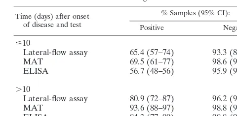
The sensitivity of the lateral-flow assay was 65.9% for sam-ples collected during the first 10 days of the disease and 80.9% for samples collected 10 to 30 days after the onset of the disease (Table 1). The specificities for these two groups were 93.3 and 96.2%, respectively. This result shows that the lateral-flow assay has a relatively high sensitivity for samples collected early in the course of disease. The sensitivity of the lateral-flow device was higher than that of the IgM ELISA and was only slightly lower than that of MAT for samples collected early in the course of disease (Table 1). The detection rate of the lateral-flow assay for samples collected more than 10 days after the onset of the disease, however, was lower than those of the IgM ELISA and MAT. Compared with MAT and the IgM ELISA, the lateral-flow assay had a somewhat larger number of false-positive results. The results of the lateral-flow assay and the IgM ELISA showed 91.9% agreement (for agreement beyond chance, kappa50.83 and 95% CI50.74 to 0.92%). The maximum value of kappa is 1.0. A kappa value of.0.8 demonstrates almost perfect agreement.
Application of the lateral-flow assay in a district hospital in India to a group of consecutively admitted patients with clin-ical suspicion of leptospirosis again revealed a high rate of concordance with the results of the IgM ELISA (90.1% agree-ment; kappa, 0.80 [95% CI, 0.62 to 0.98%]). In this study the lateral-flow assay gave a correct positive result for the samples collected from 90.7% (39 of 43) of the patients with a positive result (titer, $1:80) by the IgM ELISA. A correct negative
result was obtained for 87.2% (41 of 47) of the patients with a negative result (titer,,1:80) by the IgM ELISA. A positive result by the lateral-flow test was obtained for a total of 50 samples, of which 35 gave strong staining and 15 gave weak staining. For comparison, by the IgM ELISA moderate to high titers (.1:80) were obtained for 32 samples, a borderline titer (1:80) was obtained for 17 samples, and a titer just below the cutoff value was obtained for 4 samples. Of the 15 samples that gave weak staining by the lateral-flow assay, 2 tested negative by the IgM ELISA, 2 had titers below the cutoff value, 6 had borderline titers, and 3 had titers above the cutoff value. These TABLE 1. Test results of the LEPTO lateral-flow device according
to duration of disease and comparison with results of MAT and IgM ELISA
Lateral-flow assay 65.4 (57–74) 93.3 (88–98)
MAT 69.5 (61–77) 98.6 (94–100)
ELISA 56.7 (48–56) 95.9 (90–99)
.10
Lateral-flow assay 80.9 (72–87) 96.2 (90–99)
MAT 93.6 (88–97) 98.8 (92–100)
ELISA 84.3 (77–90) 98.8 (92–100)
VOL. 8, 2001 SERODIAGNOSIS OF HUMAN LEPTOSPIROSIS 167
by on September 10, 2009
cvi.asm.org
results demonstrate that the lateral-flow assay allows rapid confirmation of results for patients with symptoms suspicious for leptospirosis even when samples from a collection of sam-ples with a high proportion with borderline or low titers are tested. As the result of the lateral-flow assay can be obtained shortly after drawing of the blood sample, application of the assay will likely improve the treatment of the patients by al-lowing a better diagnosis to be made and treatment to be started promptly.
Many different serovars of Leptospira exist, and serovar-specific antibodies can be detected in agglutinating antibody assays such as MAT that use live leptospiras as antigen. The so-called genus-specific antibody assays such as ELISA that are based on denatured antigens and that are aimed at the detec-tion of IgM antibodies react with antibodies to many serovars. The results of this study show that the lateral-flow test reacts with antibodies to at least serovars australis, autumnalis, bata-viae, canicola, celledoni, cynopteri, grippotyphosa, icterohae-morrhagiae, javanica, pomona, sejroe, shermani, and tarassovi. As the lateral-flow test is read by visual inspection for stain-ing of the antigen line, readstain-ing of the test is subjective for samples giving a weak staining. A weak positive result may be due to cross-reactivity or may correlate with low or borderline titers in the IgM ELISA. Weak positive results by the lateral-flow assay, like low or borderline in an ELISA, should be confirmed by testing of a second sample collected at a later stage to look for an increase in antibody level. The lateral-flow assay has some major advantages compared with the standard reference tests. The lateral-flow assay is quick and can be performed by modestly trained personnel simply by following the instructions provided in a short instruction leaflet. The assay does not require expensive equipment, and as the com-ponents are stabilized, they do not depend on refrigeration for storage. No electricity is required to perform the assay. Taken together, these characteristics make the assay ideal for use in situations in which adequate laboratory facilities for perfor-mance of the more complicated standard confirmatory assays are lacking. The lateral-flow assay potentially can be used outside the laboratory and can be used in district hospitals and primary health posts or even in the field.
The result of the lateral-flow assay should be interpreted with respect to the clinical findings. As seroconversion usually takes place 5 to 7 days after the onset of the disease, the sensitivity and negative predictive value are relatively low for samples collected early in the course of the disease. From the results of this study a sensitivity of 65.9% was calculated for samples collected during the first 10 days after the onset of illness. The negative predictive value at this stage of the disease was calculated to be 68.3%. The sensitivity (80.9%) and neg-ative predictive value (73.5%) increase for samples collected at a later stage. Therefore, it is advisable that a second serum sample drawn one or a few days after collection of the first sample be tested when a negative result is obtained with the first sample but when clinical suspicion of leptospirosis re-mains. The epidemiological situation should also be consid-ered when interpreting the assay result. As the specificity of the assay was calculated to be high, the positive predictive value is likely to be high as well in situations in which the prevalence of leptospirosis among patients with suspected leptospirosis is high. From the results of this study the positive predictive value
was calculated to be 93.7% for samples collected during the first 10 days of the disease and 98.1% for samples collected at a later stage. In situations in which leptospirosis is rare, how-ever, the positive predictive value is likely to be lower, and in that case a positive result ideally should be confirmed by fur-ther laboratory testing, preferably by MAT.
REFERENCES
1.Anonymous.1999. Leptospirosis worldwide, 1999. Wkly. Epidemiol. Rec.74: 237–242.
2.Adler, B., A. M. Murphy, S. A. Locarnini, and S. Faine.1980. Detection of specific anti leptospiral immunoglobulins M and G in human serum by solid-phase enzyme-linked immunosorbent assay. J. Clin. Microbiol.11:452– 457.
3.Appassakij, H., K. Silpapojakul, R. Wansit, and J. Woodtayakorn.1995. Evaluation of the immunofluorescent antibody test for the diagnosis of human leptospirosis. Am. J. Trop. Med. Hyg.52:340–343.
4.Arimitsu, Y., E. Kmety, Y. Anayina, G. Baranton, I. R. Ferguson, L. Smythe, and W. J. Terpstra.1994. Evaluation of the one-point microcapsule agglu-tination test for the diagnosis of leptospirosis. Bull. W. H. O.72:393–399. 5.Dikken, H., and E. Kmety.1978. Serological typing methods of leptospires.
Methods Microbiol.11:259–294.
6.Easton, A.1999. Leptospirosis in Philippine floods. Br. Med. J.319:212. 7.Faine, S.1982. Guidelines for the control of leptospirosis. World Health
Organization, Geneva, Switzerland.
8.Farr, R. W.1995. Leptospirosis. Clin. Infect. Dis.21:1–8.
9.Galton, M. M., D. K. Powers, A. M. Hall, and R. G. Cornell.1958. A rapid microcapsule-slide screening test for the serodiagnosis of leptospirosis. Am. J. Vet. Res.19:505–512.
10. Gussenhoven, G. C., M. A. W. G. van der Hoorn, M. G. A. Goris, W. J. Terpstra, R. A. Hartskeerl, B. W. Mol, C. W. van Ingen, and H. L. Smits. 1997. Lepto dipstick, a dipstick assay for detection ofLeptospira-specific immunoglobulin M antibodies in human sera. J. Clin. Microbiol.35:92–97. 11. Jayaraman, K. S.1998. India urged to act against leptospirosis. Nature392:4. 12. Ko, A. I., M. Galvao Reis, C. M. Ribeiro Dourado, W. D. Johnson, and L. W. Riley.1999. Urban epidemic of severe leptospirosis in Brazil. Lancet354: 820–825.
13. Levett, P. N., and C. U. Whittington.1998. Evaluation of the indirect hem-agglutination assay for diagnosis of acute leptopsirosis. J. Clin. Microbiol. 36:11–14.
14. Lupido, R., M. Cinco, D. Balinzin, E. Delprete, and P. E. Varaldo.1991. Serological follow-up of patients in a localized outbreak of leptospirosis. J. Clin. Microbiol.29:107–109.
15. Mailloux, M., Y. Dufresne, J. Mazzonelli, and G. T. Dorta de Mazzonelli. 1984. Interet de la methode ELISA dans le diagnostic des leptospirosis. Med. Mal. Infect.14:107–109.
16. Milner, A. R., K. B. Jackson, K. Woodruff, and I. L. Smart.1985. Enzyme-linked immunosorbent assay for determining specific immunoglobulin M in infections caused byLeptospira interrogansserovarhardjo. J. Clin. Microbiol. 22:539–542.
17. Park, K.-H., W.-H. Chang, J.-S. Lee, K.-W. Choi, K.-H. Park, and H.-B. Oh. 1986. Diagnosis of leptospirosis by enzyme-linked immunosorbent assay. J. Korea Soc. Microbiol.21:181–189.
18. Petchlai, B., S. Hiranras, and U. Potha.1991. Gold immunoblot analysis of IgM-specific antibody in the diagnosis of human leptospirosis. Am. J. Trop. Hyg.45:672–675.
19. Ribeiro, M. A., C. S. N. Assis, and E. C. Romero.1994. Serodiagnosis of human leptospirosis employing immunodominant antigen. Serodiagn. Im-munother. Infect. Dis.6:218–221.
20. Sanders, E. J., J. G. Rigua-Perez, H. L. Smits, C. C. Deseda, V. A. Vorndam, T. Aye, R. A. Spiegel, R. S. Weyant, and S. L. Bragg.1999. Increase in leptospirosis in dengue-negative patients, after a hurricane in Puerto Rico, 1996. Am. J. Trop. Med. Hyg.61:399–404.
21. Sehgal, S. C.1996. Human leptospirosis—an emerging public health prob-lem in India. Trans. R. Soc. Trop. Med. Hyg.10:477–478.
22. Sehgal, S. C., M. V. Murhekar, and A P. Sugunan.1995. Outbreak of leptospirosis with pulmonary involvement in north Andaman. Ind. J. Med. Res.102:9–12.
23. Sehgal, S. C., P. Vijiyachari, M. V. Murhekar, A. P. Sugunan, S. Sharma, and S. S. Singh.1999. Leptospiral infection among primitive tribes of An-daman and Nicobar islands. Epidemiol. Infect.122:423–428.
24. Sehgal, S. C., S. Vijayachari, S. Sharma, and A. P. Sugunan.1999. LEPTO Dipstick: a rapid and simple method for serodiagnosis of acute leptospirosis. Trans. R. Soc. Trop. Med. Hyg.93:161–164.
25. Smits, H. L., Y. V. Ananyina, A. Chereshsky, L. Dancel, R. F. M. Lai-A-Fat, H. D. Chee, P. N. Levett, T. Masuzawa, Y. Yanagihara, M. A. Muthusethu-pathi, E. J. Sanders, D. M. Sasaki, H. Domen, C. Yersin, T. Aye, S. L. Bragg, G. C. Gussenhoven, M. A. G. W. Goris, W. J. Terpstra, and R. A. Hartskeerl. 1999. An international evaluation of the clinical utility of a dipstick assay for the detection of leptospira-specific immunoglobulin M antibodies in human
168 SMITS ET AL. CLIN. DIAGN. LAB. IMMUNOL.
by on September 10, 2009
cvi.asm.org
serum specimens. J. Clin. Microbiol.37:2904–2909.
26. Smits, H. L, R. A. Hartskeerl, and W. J. Terpstra.2000. An international multi-centre evaluation of a dipstick assay, a quick and easy test for the serodiagnosis of acute human leptospirosis. Trop. Med. Int. Health5:124– 128.
27. Suarez Hernandez, M., R. Martinez Sanches, P. E. Posada Fernandez, I. Vidal Garcia, F. Bravo Fleites, and A. Sanchez Sibello.1999. Brotes de leptospirosis humana en la provincia de Ciego de Avila, Cuba. Rev. Soc. Bras. Med. Trop.32:13–18.
28. Sulzer, C. R., J. W. Glosser, F. Rogers, W. L. Jones, and M. Frix.1975. Evaluation of an indirect hemagglutination test for the diagnosis of human leptospirosis. J. Clin. Microbiol.2:218–221.
29. Sulzer, C. R., and W. L. Jones.1973. Evaluation of a hemagglutination test for human leptospirosis. Appl. Microbiol.26:655–657.
30. Tangkanakul, W., and D. Kingnate.1998. Leptospirosis epidemic in north-eastern provinces of Thailand, 1997. Health Sci.7:386–395.
31. Terpstra, W. J., G. S. Ligthart, and G. J. Schoone.1980. Serodiagnosis of human leptospirosis by enzyme-linked-immunosorbent-assay (ELISA).
Zentbal. Bakteriol. Mikrobiol. Hyg. Abt. I Orig. A247:400–405. 32. Terpstra, W. J., G. S. Ligthart, and G. J. Schoone.1985. ELISA for the
detection of specific IgM and IgG in human leptospirosis. J. Gen. Microbiol. 131:377–385.
33. Trevejo, R. T., J. G. Rigua-Perez, D. A. Ashford, E. M. McClure, C. Jarquin-Gonzalez, J. J., Amador, J. O. de los Reyes, A. Gonzales, R. S. Nasci, R. S. Weyant, C. A. Bolin, S. L. Bragg, B. A. Perkins, and R. A. Spiegel.1998. Epidemic leptospirosis associated with pulmonary hemorrhage—Nicaragua, 1995. J. Infect. Dis.178:1457–1463.
34. Winslow, W. E., D. J. Merry, M. L. Pire, and P. L. Devine.1997. Evaluation of a commercial enzyme-linked immunosorbent assay for the detection of immunoglobulin M antibodies in diagnosis of human leptospirosis. J. Clin. Microbiol.35:1938–1942.
35. Yersin, C., B. Bovet, H. L. Smits, and P. Perolat.1999. Field evaluation of a one-step dipstick assay for the diagnosis of human leptospirosis in the Sey-chelles. Trop. Med. Int. Health4:38–45.
36. Zichowski, W. J., S. A. Waitkins, and M. F. Palmer.1987. The use of ELISA in the diagnosis of human leptospirosis. Isr. J. Vet. Med.43:330–339.
VOL. 8, 2001 SERODIAGNOSIS OF HUMAN LEPTOSPIROSIS 169
by on September 10, 2009
cvi.asm.org