www.elsevier.com / locate / bres
Short communication
Subcutaneous administration of nicotine changes dorsal raphe
serotonergic neurons discharge rate during REM sleep
a b,c a b,d
´
´
´
Ruben Guzman-Marın , Md. Noor Alam
, Stefan Mihailescu , Ron Szymusiak
,
b,c e ,
*
´
´
Dennis McGinty
, Rene Drucker-Colın
a
´ ´ ´ ´
Departamento de Fisiologıa, Facultad de Medicina, Universidad Nacional Autonoma de Mexico, AP 70-250, 04510 Mexico, D.F., Mexico
b
Veterans Affairs Greater Los Angeles HealthCare System, 16111 Plummer Street, North Hills, CA 91343, USA
c
Department of Psychology, University of California, Los Angeles, CA, USA
d
Department of Medicine, School of Medicine, University of California, Los Angeles, CA, USA
e
´ ´ ´
Departamento de Neurociencias, Instituto de Fisiologıa Celular, Universidad Nacional Autonoma de Mexico, Apdo. Postal 70-600, ´
04510 Mexico, D.F., Mexico
Accepted 10 October 2000
Abstract
In the present study nicotine (0.1 mg / kg, s.c.) increased discharge rate of putative dorsal raphe (DRN) serotonergic neurons of behaving rats during REM sleep (362.61%), without any significant change during waking and non-REM sleep. Since serotonergic DRN neurons gate PGO onset, these results suggest that nicotine-induced suppression of PGO spikes during REM sleep previously reported is achieved through stimulation of dorsal raphe serotonergic cells. 2001 Elsevier Science B.V. All rights reserved.
Theme: Neural basis of behavior
Topic: Biological rhythms and sleep
Keywords: Nicotine; Serotonergic neuron; Dorsal raphe nucleus; REM-off neuron; Single-unit recording
In previous studies we have shown that nicotine sup- In this study we hypothesize that nicotine increases presses the ponto-geniculo-occipital spikes (PGOs) of discharge rate of putative serotonergic neurons (PSNs) rapid eye movement (REM) sleep [21] and improves mood during REM sleep. This may account for PGO spikes in patients with major depression [17]. It is possible that suppression [21] and mood improvement previously re-these effects are serotonin-mediated since pharmacological ported [17]. This hypothesis is supported by the presence [8] and electrophysiological [9] studies indicated that of nicotinic receptors in the DRN of rats [18] and by serotonergic neurons from the dorsal raphe nucleus (DRN) reports showing anatomical projections [5] from PSNs to play a permissive role in PGO spikes generation and most the PGO wave generator [4].
of treatments currently employed in depression increase The present study, therefore, examined the effects of brain serotonin (5-HT) traffic [2]. In vitro experiments systemic nicotine administration on single-unit activity of have shown that nicotine increases discharge rate of PSNs in undrugged and unrestrained rats during the sleep– serotonergic DRN neurons and 5-HT release in a dose- wake cycle and especially during REM sleep.
dependent manner [12,15]. Systemic administration of Experiments were conducted in six Sprague–Dawley nicotine in rats facilitates 5-HT release as measured by rats (300–350 g). Under deep anesthesia (ketamine [80 microdialysis in frontal cortex [19]. However less is mg / kg]1xylazine [10 mg / kg], i.p.) and aseptic conditions, known regarding nicotine actions on serotonergic DRN rats were surgically prepared for chronic recording of DRN neurons in behaving rats. unit activity along with electroencephalogram (EEG) and electromyogram (EMG) across the sleep–wake cycle, as previously described [7].
*Corresponding author. Tel.:152-5-550-6662; fax:152-5-550-0904. ´
E-mail address: [email protected] (R. Drucker-Colın). In order to record DRN unit activity, stainless steel
microelectrodes (20 mm) inserted into a microdrive were mean discharge rate was calculated in each third as well as sterereotaxically aimed at the DRN (AP 27.8, L 0.0, H the initial 20–25 s of REM sleep.
4.5). The microdrive was advanced in steps of 25 mm, The final position of the microwires was histologically until stable single-units (signal:noise, 3:1) were found. For identified in Nissl-stained sections.
each cell, recordings were performed across 2 to 3 sleep– A total of 36 cells were recorded from the DRN. Sixteen waking complete cycles for identification of the REM-off of these cells were classified as PSNs according to the
pattern. criteria previously established [7]. The mean duration of
After baseline recording, nicotine (bitartrate Sigma 0.1 the action potential was 2.7460.11 ms. Firing rates mg / kg, dissolved in sodium chloride 0.9%) was adminis- declined progressively across the sleep–wake cycle tered s.c. in a volume less than 1 ml [10]. After nicotine (1.2860.19 spikes / s during waking, 0.4560.06 during administration recordings continued for 2 h. Action po- NREM sleep and 0.1060.01 in REM sleep). The mean tential waveform of individual neurons was continuously percentage of reduction in discharge rate from waking to monitored during the experiments to ensure that the same REM sleep was290.3961.61%. In addition in six of these
cell was recorded. presumed serotonergic neurons systemic administration of
Mean discharge rate per second (spikes / s) was calcu- (6)8-OH-DPAT displayed a marked suppression of unit lated from 30 s epochs both during baseline and after activity (294.5563.31%, P#0.01, paired t-test).
nicotine injection. Cells were classified as wake-related, The remaining 20 neurons did not meet the criteria for sleep-related and as state-indifferent according to its PSNs and were classified as sleep-related (n53), state-NREM / Wake ratio as previously established [1]. indifferent (n55) and wake-related neurons (n512).
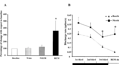
Since previous analysis of the DRN PSNs stated that The effect of systemic administration of nicotine was these cells cease discharge before the onset of PGO spikes examined in all 16 PSNs. Nicotine administration in-during the NREM–REM transition, we carried out an creased discharge rate during REM sleep by analysis of this transition [7]. In order to achieve this goal 362.616145.29% (P,0.05, repeated measures ANOVA we selected periods of NREM sleep (60–75 s in duration) followed by Bonferroni test). No statistical differences which preceded a REM sleep period (defined by the were observed in the mean discharge rate during waking presence of desynchronization and atonia longer than 1 and NREM sleep (Fig. 1A). Fig. 2A and B illustrates min), being divided into thirds (20–25 s each one). The polysomnographic recordings both during baseline and
Fig. 2. Polygraph recording and unit activity of a putative serotonergic neuron. The profile discharge of a presumed serotonergic neuron across the sleep–wake cycle. Discharge rate is displayed as unit. (A) Polygraph plot during baseline. Note that during REM sleep this cell almost ceases firing. (B) Polygraph plot, showing activity from the same cell recorded in A. During REM sleep there is an increase in discharge rate following nicotine administration (0.1 mg / kg, s.c.). Abbreviations: EEG, electroencephalogram; EMG, electromyogram.
after nicotine administration of PSNs recorded across the unpaired t-test) and the 20–25 initial seconds of the REM
sleep–wake cycle. sleep stage (P,0.05 unpaired t-test).
significant. For those cells classified as wake-related the paradoxical, since DRN serotonergic neurons inhibit [20] percentage of reduction during REM sleep after nicotine the pedunculopontine (PPT) and laterodorsal tegmental administration was 63.95617.33, 77.22645.03 in sleep- (LDT) neurons, generators of REM sleep [6]. In our related neurons and 49.45610.91 in state-indifferent neu- experimental conditions however it may be assumed that
rons. nicotine increased REM sleep by directly stimulating PPT
Additionally we measured the mean duration of each and LDT neurons. The previously reported PGO spike REM sleep episode. Nicotine induced an increase in suppression by nicotine may be explained by serotonergic duration of REM sleep episodes from 118.0963.62 to inhibition of PGO wave generator located in rats in the 138.0163.51 s (P,0.05, unpaired t-test) without signifi- subcoeruleus nuclei [4].
cantly affecting the frequency of events (2.2460.16 during In sum, our study shows that nicotine increases dis-baseline vs. 2.3860.23 after nicotine administration). charge rate selectively during REM sleep in PSNs. This The principal finding of this report is that nicotine (0.1 effect could be responsible at least in part for mood mg / kg) increased the discharge rate of PSNs during REM improvement in-patients with major depression having sleep. In addition there was a significant increase in firing nicotine patches [17] and suppression of PGO spikes [21] rate during the NREM–REM sleep transition. Taken in cats after nicotine administration.
together these data support the view that PGO spikes suppression [21] and mood improvement by nicotine [17]
is mediated by its stimulatory effect on DRN PSNs during Acknowledgements REM sleep.
This interpretation is consistent with the observation of This work was supported by V.A. Medical Research Jacobs et al. [9], that electrical stimulation of the DRN Service. RGM is recipient of a scholarship from the during REM sleep suppresses the presence of the PGO Consejo Nacional de Ciencia y Tecnologıa (CONACYT)´
spikes. and DGEP-UNAM. Support from the UC-Mexus Program
Aside from the increase in discharge rate during REM to RD-C is also acknowledged. We wish to thank Janice sleep, as mentioned above, it was observed that nicotine King, Darrell Thomson and Lindsay Chiu for their excel-increased discharge rate in the DRN PSNs in the 20–25 s lent technical support, and Mrs Ma. Teresa Torres-Peralta prior to REM sleep. It has been shown that in cats PGO for typing the manuscript.
spikes appear 30–60 s before REM sleep onset [3], coincidentally with the lowest discharge level of 5-HT DRN cells [13,14]. Therefore it is possible that the
References increase in discharge rate of DRN PSNs induced by
nicotine during the NREM–REM sleep transition accounts
[1] M.N. Alam, D. McGinty, R. Szymusiak, Neuronal discharge of for the inhibition of PGO spikes. This is consistent with
preoptical anterior hypothalamic thermosensitive neurons: relation to the report that sertraline (an 5-HT reuptake inhibitor) NREM sleep, Am. J. Physiol. 269 (1995) R1240–R1249. decreases the presence of PGO spikes in the NREM–REM [2] P. Blier, C. de Montigny, Possible serotonergic mechanisms
under-laying the antidepressant and anti-obsessive-compulsive disorder sleep transition [16].
responses, Biol. Psychiatry 44 (1998) 313–323. Our study also is in agreement with previous reports in
[3] D.C. Brooks, E. Bizzi, Brain stem electrical activity during deep vitro, showing that nicotine increases PSNs discharge rate
sleep, Arch. Ital. Biol. 101 (1972) 648–665.
as well as serotonin release [15]. [4] S. Datta, D.F. Siwek, E.H. Patterson, P.B. Cipolloni, Localization of As concerns sleep-related, state indifferent and wake- pontine PGO wave generation sites and their anatomical projections related neurons, the consistent finding in this study was a in the rat, Synapse 30 (1998) 409–423.
[5] S. Datta, E.H. Patterson, D. Siwek, Brainstem afferents of the reduction in the discharge rate along the sleep–wake cycle
cholinoceptive pontine wave generation sites in the rat, Sleep Res. which, however, was not statistically significant. It is
Online 2 (1999) 79–82.
evident that these neurons represent different neuronal [6] J.C. Gillin, R. Salin-Pascual, J. Velazquez-Moctezuma, P. Shiromani, populations, which agrees with reports showing that DRN R. Zoltoski, Cholinergic receptor subtypes and REM sleep in neuronal composition is not homogeneous and serotonergic animals and normal controls, Prog. Brain Res. 98 (1993) 379–387.
´ ´
[7] R. Guzman-Marın, Md. Noor Alam, R. Szymusiak, R. Drucker-neurons represent only one third of the Drucker-neurons in this
Colin, H. Gong, D. McGinty, Discharge modulation of rat dorsal nucleus [11].
raphe neurons during sleep and wakefulness: effects of preoptic / As concerns the influence of nicotine on sleep architec- basal forebrain warming, Brain Res. 875 (2000) 23–34.
ture, the mean duration of each REM sleep period was [8] B.L. Jacobs, S.J. Henriksen, W.C. Dement, Neurochemical basis of increased without affecting the frequency of episodes. No the PGO wave, Brain Res. 48 (1972) 406–411.
[9] B.L. Jacobs, R. Asher, W.C. Dement, Electrophysiological and significant changes were observed during waking and
behavioral effects of electrical stimulation on the raphe nuclei in NREM sleep. Similar findings were reported previously
cats, Physiol. Behav. 11 (1973) 489–495.
postnatal rat serotoninergic neurons in dissociated cell culture, [17] R.J. Salin-Pascual, R. Drucker-Colin, A novel effect of nicotine on Neuroscience 63 (1994) 775–787. mood and sleep in major depression, NeuroReport 9 (1998) 57–60. [12] X. Li, D.G. Rainnie, R.W. McCarley, R.W. Greene, Presynaptic [18] M. Segal, Y. Dudai, A. Amsterdam, Distribution of bungarotoxin-nicotinic receptors facilitate monoaminergic transmission, J. Neuro- binding cholinergic nicotinic receptor in rat brain, Brain Res. 148
sci. 18 (1998) 1904–1912. (1978) 105–119.
[13] R. Lydic, R.W. McCarley, A. Hobson, The time-course of dorsal [19] K.L. Summers, E. Giacobini, Effects of local and repeated systemic raphe discharge. PGO waves, and muscle tone averaged across administration of (2)nicotine on extracellular levels of acetylcho-multiple sleep cycles, Brain Res. 274 (1983) 365–370. line, norepinephrine, dopamine and serotonin in rat cortex, Neuro-[14] D.J. McGinty, R.M. Harper, Dorsal raphe neurons: depression of chem. Res. 20 (1995) 753–759.
firing during sleep in rats, Brain Res. 101 (1976) 569–575. [20] M. Thakkar, R.E. Strecker, R.W. McCarley, Behavioural state [15] S. Mihailescu, M. Palomero-Rivero, P. Meade-Huerta, A. Maza- control through differential serotonergic inhibition in the mesopon-Flores, R. Drucker-Colin, Effects of nicotine and mecamylamine on tine cholinergic nuclei: a simultaneous unit recording and mi-rat dorsal raphe neurons, Eur. J. Pharmacol. 360 (1998) 31–36. crodialysis study, J. Neurosci. 18 (1998) 5490–5497.