Retrospective study of presumably allergic dogs examined over a
one-year period at the Veterinary Faculty, University of Ljubljana,
Slovenia
Tina Kotnik*
small animal Clinic, Veterinary Faculty, University of Ljubljana, slovenia
KoTniK, T.: Retrospective study of presumably allergic dogs examined over a one-year period at the Veterinary Faculty, University of Ljubljana, Slovenia. Vet. arhiv 77, 453-462, 2007.
ABSTRACT
Two hundred and twelve animals examined over a one-year period at the Dermatology Department of Small Animal Clinic, Veterinary Faculty, University of Ljubljana, were included in a retrospective study. For further evaluation dogs with flea allergy dermatitis (FAD), Canine atopic disease (CAD) and Food hypersensitivity as the first differential diagnosis (n = 126) were considered. Among the clinical features in these dogs, erythema was most frequently found (83.4%). Owners (23.9%) were able to provide us with the familial history of their dog and 13.6% described allergic skin disease symptoms in one or more of their dogs’ relatives. After initial diagnostic procedures, i.e. skin scrabs, cytology and bacteriology, accurate initial therapy was constituted when a relevant elimination diet was prescribed for 3 months. Evaluation of the diet trial was done by recording clinical improvement and by estimation of the pruritus intensity. In 37 (29.4%) dogs’ skin problem symptoms were effectively suppressed by elimination diet. The condition of 34 dogs improved with initial therapy or they were lost for further evaluation. Dogs that did not completely improve by elimination diet and initial therapy (n = 50) were submitted for intradermal (ID) allergic testing (46 dogs) or IgE measurement (4 dogs). Tested dogs were sensitised against multiple antigens in the majority of cases (47 of 50). A group of storage and house dust mites represented the most common antigens. The most frequent mite was acarus siro against which 71.7% of our ID tested dogs were sensitised.
Key words: allergies, skin, intradermal allergy tests, dogs introduction
The three most frequent allergic skin diseases in dogs are flea allergy dermatitis (FAD), Canine atopic disease (CAD) and Food hypersensitivity. Prevalence of FAD can be as high as 11.4% (PENALIGGON, 1997) whereas the prevalence of CAD is about 10% (MUELLER and BETTENAY, 1996). Food hypersensitivity can account for 5% of all skin *Contact address:
Tina Kotnik, PhD, DVM, Teaching Assistant, Veterinary Faculty of Ljubljana, C. v Mestni log 47, 1000 Ljubljana, Slovenia, Phone: + 386 1 4779 284; Fax: + 386 1 4779 349; E-mail: tina.kotnik@vf.uni-lj.si
diseases or 15% of allergic dermatoses (CARLOTTI, 1990). About 30% of dogs with food hypersensitivity can have FAD or CAD additionally (SCOTT et al., 1995) and as many as 75% of atopic dogs have concurrent FAD (SCOTT et al., 2001). A tentative diagnosis of one of the three allergic skin diseases can be based on history, clinical signs, and laboratory tests to rule out other possible diseases (such as scabies, bacterial folliculitis, Malassezia dermatitis and some uncommon hypersensitivities). Diagnosis is based on history, physical examination, diet trial, and intradermal (ID) testing or in vitro (serologic) allergy tests (SCOTT et al., 2001).
At the Small Animal Clinic of the Veterinary Faculty of Ljubljana we have been performing intradermal (ID) allergy tests since 1993 (OROŽIM, 1998). We began ID testing experimentally using 6 antigens. Today, we are using a total of 36 antigens. ID allergy tests are considered as a golden standard in diagnostics of CAD and are superior to in vitro testing (SCOTT et al., 2001), although some authors disagree (HILLER, 2002). in vitro tests are nevertheless easier to perform and can give useful information when performed and interpreted according to broadly defined accepted criteria, but can give false positive results due to their lower specificity (HILLER, 2002).
The aim of our retrospective study was to evaluate the results of diet trial and allergic testing in dogs with presumed allergic dermatosis, examined during a one-year period at the Dermatology Department of the Small Animal Clinic, Veterinary Faculty of Ljubljana.
Materials and methods
During a one year period (25. 05. 2004 to 25. 05. 2005) 212 animals (197 dogs and 15 cats) were examined at the Dermatology Department of the Small Animal Clinic, Veterinary Faculty of Ljubljana. Since the number of examined cats was small, only dogs were taken for further evaluation. At the first presentation an exhaustive history was compiled and clinical features were recorded. Initial diagnostic procedures, i.e. skin scrabs, cytology and bacteriology, were performed at the first visit. Based on these procedures, non-allergic disease was diagnosed in 71 dogs. A tentative diagnosis of flea allergy dermatitis (FAD), Canine atopic disease (CAD), and/or Food hypersensitivity was made in the remaining 126 dogs and they were submitted for the next procedure. According to the feeding habits of each individual dog, elimination diet was prescribed for 3 months. Initial therapy was constituted. Insecticides were used during the study for flea control (FrontlineR, Merial Animal Health Ltd or StrongholdR, Pfizer Animal Health). Antibiotics
(SynuloxR, Pfizer italiana) or antimycotics (ZonitonR, Krka) were used to eliminate
concurrent infections and antihistamines (TelfastR, Hoechst) or glucocoticoids (MedrolR,
Upjohn) to control pruritus. These agents were usually prescribed for the first 3 weeks, together with elimination diet, and their effect was evaluated at a control examination
at the end of the medical therapy. The effect of the diet was evaluated after a 3-month period at a control examination, and sometimes by a check over the phone. Evaluation was carried out by recording clinical improvement and estimation of pruritus intensity. Dogs whose condition did not completely improve by initial therapy and elimination diet were submitted to allergic testing. We used an ID set of 34 antigens and 2 controls (Artu Biologicals, Netherlands) in 46 dogs and sent the blood of 4 dogs for IgE measurement to the Alergovet Laboratory (Madrid, Spain).
Results
One of the three most important allergic diseases in dogs (CAD, FAD or Food hypersensitivity) was presumed in 126 dogs out of a total of 197 (64.0%). These dogs were considered for further evaluation. Elimination diet resolved skin problems in 37 (29.4%) dogs. These dogs were not subsequently tested against environmental allergens. Fifty (39.6%) dogs were tested, 46 by using ID tests and 4 by using IgE blood measurement. The condition of the remaining 34 dogs improved with initial therapy, or they were lost for further evaluation.
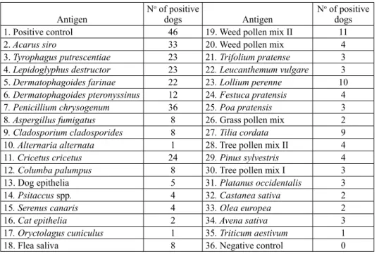
Table 1. Results of intradermal tests in 46 dogs tested over a one-year period at the Small Animal Clinic, Veterinary Faculty of Ljubljana
Antigen N
o of positive
dogs Antigen N
o of positive
dogs
1. Positive control 46 19. Weed pollen mix II 11
2. acarus siro 33 20. Weed pollen mix 4
3. tyrophagus putrescentiae 23 21. trifolium pratense 3
4. Lepidoglyphus destructor 23 22. Leucanthemum vulgare 3
5. Dermatophagoides farinae 22 23. Lollium perenne 10
6. Dermatophagoides pteronyssinus 12 24. Festuca pratensis 4
7. Penicillium chrysogenum 36 25. Poa pratensis 3
8. aspergillus fumigatus 8 26. Grass pollen mix 2
9. Cladosporium cladosporides 8 27. tilia cordata 9
10. alternaria alternata 1 28. Tree pollen mix II 4
11. Cricetus cricetus 24 29. Pinus sylvestris 4
12. Columba palumpus 8 30. Tree pollen mix I 3
13. Dog epithelia 5 31. Platanus occidentalis 3
14. Psitaccus spp. 4 32. Castanea sativa 2
15. serenus canaris 4 33. Olea europea 2
16. Cat epithelia 2 34. avena sativa 3
17. Oryctolagus cuniculus 1 35. triticum aestivum 1
Comments on Table 1: In all 46 tested dogs positive control was confirmed as positive and negative control was confirmed as negative. The dogs tested were sensitised against multiple antigens in the majority of cases (44 of 46). A group of storage and house dust mites (numbers 2 - 6) represented the most common antigens. Among fungi group (numbers 7 - 10) frequent positive reaction of injection site was established to Penicillium chrysogenum while it was infrequent to other representatives of the group. The same situation was concluded with epithelia group (numbers 11 - 17); positive skin reaction to hamster epithelia was frequent but infrequent to all others in the epithelia group. Among the remainder of the antigens (numbers 18 - 35) weed pollen mixture II, lollium perenne (grass), tilia cordata (linden), and flea saliva had been fairly frequently found to be positive. Definite diagnose of FAD was made in 8 of 46 (17.4%) tested dogs, and all of them were concurrently sensitised against multiple inhalant allergens.
We evaluated some important history data in dogs that had undergone ID allergic tests (n= 46). The majority of these dogs (29) were living in apartments with their owners. On average they spent about 20 hours a day in the apartment and went out only for a walk. Four out of 46 dogs had been living exclusively outdoors (8.7%), 13 dogs (28.3%) had been living somehow in combination - half of the year outside and half of the year inside (2 dogs), half of the day outside and half of the day inside (5 dogs), one-third of the day inside (3 dogs), one-third of the day outside (2). For one dog, exact data is unknown because it was found by the actual owner when it was about 9 years old.
All of the ID tested dogs expressed pruritus at the first visit. Pruritus intensity ranged from 2 - 10 points (median 6.9). Twenty-six (56.5%) dogs were never previously treated with glucocorticoids. Twelve dogs (26.1%) expressed mild pruritus (5 points or less). Among 20 dogs treated with glucocorticoids 17 were glucocorticoid responsive and 3 were not.
Of 46 dogs, 80.4% showed their first clinical symptoms between 1 and 3 years old, while 4 of 46 (8.7%) showed their first clinical symptoms before 12 months of age, and 5 of 46 (10.9%) after 3 years of age. At the time of their first presentation dogs showed next clinical manifestations: erythema (105; 83.4%), alopecia (38; 30.2%), seborrhoea (37; 29.4%), crusts (37; 29.4%), hyperpigmentation (36; 28.6%), papules (33; 26.2%), excoriations (17; 13.5%), lichenification (16; 12.7%), hypotrichia (13; 10.3%), epidermal collaretes (8; 6.3%), bullae (5; 3.9%), pustules (4; 3.2%), hyperkeratosis (3; 2.4%), ulcus (2; 1.6%) and erosio, rhagas, and bull’s eye (1; 0.8%) each.
Three-quarters of owners (35 of 46) did not know any of their dogs’ relatives and were unable to give us their familial history. Among the remaining 11 owners, 6 (13.6%) described skin disease in one or more of their dogs’ relatives.
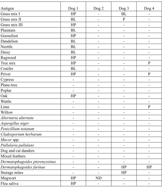
Table 2. Results of IgE measurement against different antigens, carried out at Alergovet Laboratory (Madrid, Spain) from the sera sent from the Small Animal Clinic, Veterinary Faculty
of Ljubljana over a one-year period
Antigen Dog 1 Dog 2 Dog 3 Dog 4
Grass mix I HP - BL
-Grass mix II BL - P
-Grass mix III HP - -
-Plaintain BL - - -Goosefoot HP - - -Dandelion BL - - -Neettle BL - - -Daisy BL - - -Ragweed HP - - -Tree mix HP - - P Conifer BL - - -Privet HP - - P Cypress - - - -Plane-tree - - - -Poplar - - - -Oak HP - - -Wattle - - - -Lime - - - P Willow - - - -alternaria alternate - - - -aspergillus niger - - - -Penicillium notatum - - - -Cladosporium herbarum - - - -Mucor spp. - - - -Pullularia pullulans - - -
-Dog and cat danders - - -
-Mixed feathers - - - -Dermatophagoides pteronyssinus - - - -Dermatophagoides farinae - - HP HP Storage mites - - HP -Mugwort HP ND - -Flea saliva HP - -
Comments on Table 2: Similar to the results of ID tests, dogs tested serologically were sensitised against multiple antigens in the majority of cases (3 of 4 dogs). One of them showed extremely multiple positive results (dog 1) and one test was negative (dog 2).
Discussion
During their first presentation at our dermatology department a high percentage of dogs (64%) with presumed allergic skin disease was evaluated, which shows that mainly dogs with chronic skin disease were presented. They were sent to our department from private practitioners, or else owners had brought their dogs in by themselves seeking a second opinion. Although we cannot gather the prevalence of allergic skin diseases in dogs in Slovenia from our data, our clinical impression is that the number of allergic dogs is increasing.
The prevalence of atopic diseases in humans has risen since World War II (LEUNG, 1999). According to the standpoint of the American College of Veterinary Dermatology Task force on Canine Atopic Dermatitis (HILLIER, 2001), most of the factors linked to the increasing incidence of atopic disease in humans are also consistent with the changing environment of dogs. Therefore, factors that may contribute to an increase in the incidence of canine atopic dermatitis in pet dogs are: an increased exposure to noxious pollutants; increasingly urbanized environment; dogs are spending more time indoors thus increasing exposure to common indoor allergens such as house dust mites; there is more wide-spread vaccination of puppies which may increase IgE antibody production; the practice of internal and external parasite control (parasitic infestations may be protective against the development of allergy (LYNCH et al., 1993) by dog owners is more common (HILLIER and GRIFFIN, 2001). Spending most of the time indoors was also characteristic of our dogs diagnosed as atopic. The number of the dogs that had spent most of their time in an apartment (about 20 hours daily) was 29 (63.0%). If we add the number of dogs that lived in combination (28.3%) the total percentage would be 91.3%. Therefore, the main factor of sensitisation in our dogs could be exposure to mites in apartments, and foods.
An elimination diet food trial should always be performed in allergic patients since serum testing is not helpful in diagnosing cutaneous adverse food reactions (MUELLER and TSOHALIS, 1998) neither it is helpful in determining protein and carbohydrate sources to prescribe a suitable elimination diet (HILLER, 2002). Elimination diet suppressed skin symptoms in 37 (29.4%) of our dogs, so they had stayed in remission for at least 3 months after the beginning of the elimination diet. Regarding the literature (SCOTT et al., 1995) it is known that cytokines, being produced as a consequence of food allergy can promote histamine release in the body for as long as 10-13 weeks after the last contact with an allergen. Probably for that reason, only 25% of dogs with food allergy would show an improvement in the first 3 weeks of elimination diet, and the remaining dogs as late as 10 weeks after introducing the diet (WILLS and HARVEY, 1994; ROSSER, 1993). Considering
these data we did not expect a rapid response to the diet, and much of the improvement during the first 3 weeks was considered a consequence of the drugs being concurrently used. We used these drugs to eliminate secondary bacterial or fungal infections when present, or to eliminate strong pruritus. Only with secondary infections eliminated can the response to elimination diet be evaluated. Whenever response to the diet was complete the owners were encouraged to perform a diet challenge (CESTNIK et al., 2001), and only when the symptoms returned food hypersensitivity was proven. Unfortunately, not all owners were willing to perform the challenge.
The use of antipruritic agents during the first 3 weeks seemed reasonable to us because the owners were all the more prepared to continue with the diet. If we were to leave a dog with a strong pruritus untreated, not many owners would be encouraged to perform an elimination diet for as long as three months. Of course, whenever the symptoms reappeared - and this happened soon after discontinuation of the antipruritic agent - this was a sign that diet had not solved the problem. In these cases we prolonged antipruritic therapy when needed. After 3 months of the elimination diet all dogs whose condition did not compltely improve were submitted to additional allergy testing (ID or IgE measurement). These dogs were considered atopic (no improvement) or having a combination of food hypersensitivity with FAD or CAD (moderate improvement, reaction to food challenge).
The procedure was recommended in the literature (MUELLER, 2000). Undergoing its recommendations, about one-third of the presumably allergic dogs (29.4%) did not need to be skin allergic tested, since the only true indication for allergy testing would be formulation of allergen-specific immunotherapy (HILLER, 2002; MACDONALD, 2006). The benefit was better compliance for the owner due to dog management and a favourable financial effect. Sensitivity of ID tests was high. Out of the 46 ID tests performed only 1 gave a negative result.
Intradermal (ID) tests are still considered as the golden standard in diagnostics of atopic dermatitis in dogs and cats. Therefore, we are using ID tests whenever possible. In any event, there are exceptional situations when IgE tests have an advantage. This could be the case with very old animals, or with cardiac patients that we do not want to put under sedation. This is also necessary in some dogs used for show performance that should not be shaved for aesthetic reasons. IgE tests are also a good choice whenever glucocorticoid therapy cannot be withdrawn for at least 3 weeks before ID testing. These were the main reasons for IgE testing in our patients. What antigens are really clinically important should always be considered in accordance with the history and clinical features of each dog, since we know that IgE measurement is a test prone to giving false positive results (SCOTT et al., 2001). This would also be an explanation for the extremely multiple positive result in one of our serology tested dogs. In any case, dogs were sensitised against multiple antigens in the majority of our cases (95.7%). This finding is in correlation with results of most authors in the United States and Europe (SCOTT et al., 2001).
Thirty-seven dogs in our study whose condition improved with elimination diet were not all food allergic. We know that challenge diet should be performed and allergic reaction should be shown to prove food allergy (CASE et al., 1995). In allergic dogs we can notice pruritus as soon as 12-72 hours (LEIB and AUGUST, 1989) or as late as 10-14 days (SCOTT et al, 1995) after exposure to the original diet. Accordingly, we suggested to dog owners to start adding a single component in small amounts from the original diet into the dog’s food each day for 7 to 10 days and observe the appearance of pruritus. Unfortunately, some of the dog owners were satisfied with the effect of the elimination diet and did not feel the need to challenge their dog. In these cases we were unable to prove food allergy.
Among our 37 dogs there were also some whom owners were still observing some pruritus at the end of elimination diet, but it was so mild that dogs tolerated it very well. We consider these dogs as having had a combination of food allergy and CAD or FAD, but were unable to prove it.
Referral dermatology clinics in Europe usually perform ID testing against 60 antigens. At our clinic we were obliged to make a compromise between a wide range of antigens and the financial constraints of the owners. For a certain assortment of antigens we decided to regard the most important and most prevalent antigens in our environment, and were guided by the acknowledged immunology professional (*Halliwell R. Personal communication).
With the testing of 50 dogs we obtained 9 positive reactions to flea saliva (see Tables 1 and 2). This result means that 18% of our atopic dogs concurrently had flea bite hypersensitivity. This result is not in accordance with data mentioned in the literature (SCOTT et al., 2001) that suggests a concurrency as high as 75%. Discrepancies might have appeared due to the fact that we did not test all 126 dogs with a presumed allergy. With testing all we would prove was FAD’s and we probably found more concurrent sensitisations to inhalant allergens, but estimating FAD epidemiology was not the goal of our study.
Although 73.9% of our ID tested dogs expressed moderate to strong pruritus only 43.5% were treated with glucocorticoids prior to their first visit to our department. This may show more rational use of glucocorticoids in the dogs with skin disease in Slovenia of late. Our clinical impression in the past was that these substances were abused many times over. Pruritus intensity generally depends on the phase of the disease (the later the phase, stronger the pruritus). It depends on the season of the year, when seasonal antigens participate. It also depends on the concurrent secondary bacterial or fungal infections, and on the use of antipruritic therapy. There has also been some influence of the breed described (SCOTT et al., 2001): English bulldogs, when allergic usually show little or no pruritus.
80.4% of our ID tested dogs showed their first symptom between 1 and 3 years of age. This finding is in correlation with data stated in the literature (SCOTT et al., 2001),
i.e. that about 70% of dogs first manifest clinical signs between 1 and 3 years of age. Erythema was a clinical feature most commonly seen in our allergic dogs (83.4%). In about 40% of cases alopecia, seborrhoea, crusts, papules and hyperpigmentation were present. Excoriations, lichenification and hypotrichia were found in about 15% of cases, while all the rest were presented in less than 10% of cases.
According to our data familial history in small animal dermatology seems less important compared to human dermatology. The majority of our clients (76.1%) were unable to provide us with any data about the relatives of their dogs. Making inquiries with breeders about chronic skin disease in their dogs might seem to be irrelevant. But if the owner is acquainted with similar symptoms in a related dog the information can be very important in certain cases, and so we keep collecting it.
Conclusions
From the results of our study we can conclude that the Dermatology Department of the Small Animal Clinic, Veterinary Faculty of Ljubljana, Slovenia, deals predominantly with allergic dogs. Atopic dogs are sensitised against multiple antigens in the majority of cases. A group of house dust and storage mites represents the most important antigens. The main clinical feature presented in our dogs was erythema. The main antigen against which 71.7% of our ID tested dogs were sensitised was acarus siro.
References
CARLOTTI, D. N. (1990): Food allergy in dogs and cats: A review and report of 43 cases. Vet. Dermatol. 1, 55-62.
CASE, L. P., D. P. CAREY, D. A. HIRAKAWA (1995): Canine and feline nutrition, Mosby, St. Louis. pp. 382-388.
CESTNIK, V., T. KOTNIK, Z. PAVLICA, D. PAVLIN, J. PEČAR, N. TOZON (2001): Alimentarne alergije pri psih in mačkah. In: Izbrana poglavja iz bolezni prebavil pri psu in mački. (Pavlica, Z., Ed. ). Veterinarska fakulteta. Ljubljana. pp. 42-47. (in Slovene).
HILLER, A. (2002): Allergy testing and treatment for canine atopic dermatitis. Vet. Med. 97, 210-224.
HILLIER, A., GRIFFIN, C. E. (2001): The ACVD task force on canine atopic dermatitis (I): incidence and prevalence. Vet. Immunol. Immunopathol. 81, 147-151.
LEIB, M. S., J. R. AUGUST (1989): Food hypersensitivity. In: Textbook of veterinary internal medicine (Ettinger S. J., Ed.), 3rd ed., Saunders, Philadelphia. pp. 194-197.
LEUNG, D. Y. M. (1999): Pathogenesis of atopic dermatitis. J. Allergy Clin. Immunol. 104, 99-108.
LYNCH, N. R., HAGEL, I., PEREZ, M., Di PRISCO, M. C., LOPEZ, R., ALVAREZ, N. (1993): Effect of anthelmintic treatment on the allergic reactivity of children in a tropical slum. J. Allergy Clin. Immunol. 92, 404-411.
MACDONALD, J. M. (2006): Allergen specific immunotherapy for atopy. In: Proceedings of the North american veterinary conference, January 7-11, 2006, Florida, 20; 396-398.
MUELLER, R. S. (2000): Dermatology made easy, manual for the small animal practice, Teton NewMedia, Jackson,WY.
MUELLER, R. S., S. V. BETTENAY (1996): Long-term immunotherapy in 146 dogs with atopic dermatitis - a retrospective study. Aust. Vet. Practit. 26, 128-132.
MUELLER, R. S., TSOHALIS, J. (1998): Evaluation of serum allergen-specific IgE for the diagnosis of food adverse reactions in dogs. Vet. Dermatol. 9, 167-171.
OROŽIM, E. (1998): Ugotavljanje atopij z intrakutanimi alergijskimi testi in zdravljenje atopij s pomočjo desenzibilizacije. In: Zbornik referatov 11. Simpozija o aktualnih boleznih malih živali, Poljče. pp. 12-16. (in Slovene)
PENALIGGON, J. (1997): Winter prevalence of flea infestation and flea allergy dermatitis in cats and dogs in Great Britain and Ireland. Proc. Annu. Memb. Eur. Soc. Vet. Dermatol. Eur. Coll. Vet. Dermatol. 14, 167.
ROSSER, E. J. (1993): Diagnosis of food allergy in dogs. J. Am. Vet. Med. Ass. 203, 259-262. SCOTT, D. W., W. H. MILLER, C. E. GRIFFIN (1995): Small Animal Dermatology, 5th ed.,
Saunders, Philadelphia.
SCOTT, D. W., W. H. MILLER, C. E. GRIFFIN (2001): Small Animal Dermatology, 6th ed.,
Saunders, Philadelphia.
WILLS, J., R. HARVEY (1994): Diagnosis and management of food allergy and intolerance in dogs and cats. Austral. Vet. J. 71, 322-326.
KoTniK, T.: Retrospektivni prikaz vjerojatno alergičnih pasa pretraženih u tijeku godine dana na Veterinarskom fakultetu Sveučilišta u Ljubljani. Vet. arhiv 77, 453-462, 2007.
SAŽETAK
U istraživanje je bilo uključeno 212 pasa pretraženih u tijeku godine dana na Dermatološkom odjelu Klinike za male životinje Veterinarskoga fakulteta Sveučilišta u Ljubljani. Razmatrani su bili psi s alergijskim dermatitisom na ubod buha, atopijskom bolesti i preosjetljivošću na hranu kao početnom diferencijalnom dijagnozom (n = 126). Eritem je ustanovljen kao najčešći (83,4%) klinički znak bolesti u tih pasa. Svega 23,9% vlasnika moglo je dati podatke o povijesti svoga psa, a 13,6% je opisalo znakove alergijske bolesti kože u jednog ili više srodnika njihovih pasa. Nakon početnoga dijagnostičkoga postupka, tj. uzimanja sastrugane kože, citološke i bakteriološke pretrage, određena je početna terapija s uskraćivanjem odgovarajuće hrane tijekom tri mjeseca. Učinak pokusne dijete razmotren je nakon postizanja kliničkog poboljšanja i to procjenom jačine svrbeža. Uvođenjem dijetne prehrane u 37 (29,4%) pasa poremećaji na koži bili su bitno blaži. Stanje se u 34 psa poboljšalo nakon početne terapije ili dalje nisu bili promatrani. Psi u kojih se stanje nije u potpunosti poboljšalo uskraćivanjem određene hrane i početne terapije (n = 50) bili su podvrgnuti intradermalnom alergijskom testiranju (46 pasa) ili mjerenju IgE (4 psa). Testirani psi bili su u većini slučajeva (47 od 50) preosjetljivi na više antigena. Ustanovljeno je da je skupina grinja iz kućne prašine najčešći antigen. Najčešća grinja bila je
acarus siro na koju je bilo senzibilizirano 71,7% testiranih pasa.
Ključne riječi: alergije, koža, pas, intradermalni alergijski testovi
Received: 14 November 2005 Accepted: 28 September 2007