Cytotoxic Activity, Proliferation Inhibition and Apoptosis
Induction of
Rhaphidophora Pinnata (L.F.) Schott Chloroform
Fraction to MCF-7 Cell Line
Masfria
1*, Urip Hara hap
1,
Maratua Pandapotan Nasution
1, Syafruddin Ilyas
21
Faculty of Pharmacy, University of Sumatera Utara, Indonesia.
2
Faculty of Mathematics and Natural Sciences, University of Sumatera Utara,
Indonesia.
*
Corres.author: [email protected]
Abstract:
Breast cancer is a type of cancer that has an ability to survive by continuously proliferate. Herbal plants are used for treatment various diseases. Ekor Naga plant (Rhaphidophora pinnata (Lf) Schott), Araceae family, has been used for antibacteria, anticancer, rheumatism and cough. Separation was done by column chromatography, and grouped based on thin layer chromatography profiles. There were nine isolates (A–I isolates) from the separation. The fractions were tested for their cytotoxicity, proliferation inhabition by using MTT method (3-(4,5-dimetiltiazol-2-yl)-diphenyl tetrazolium bromide-2.5, and apoptosis observation by usingacridine orange-ethidium bromide reagents to MCF -7 cancer cells.
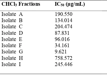
The cytotoxic activity results of the nine isolates (A - I) of cytotoxicity activity had IC50 score in the range of
190.550; 134.014; 204.474; 87.831; 96.016; 34.161; 9.621; 758.572; 245.446 ug/ml, four isolates (D-G) had IC50 score ≤ 100 mg / ml which function as anticancer. The result of proliferative test for isolate D it showed the
inhibition of cell proliferation cell and spur apoptosis of MCF-7 cells with a red-orange fluorescence, blebing nucleus also had been . Characterization result using infrared spectrophotometer showed the functional groups of O-H; C = O; C-O, and C = C which have function as anticancer.
Keywords:Rhaphidophora pinnata, "ekor naga", proliferation, apoptosis, MCF-7 cells.
Introduction
Chemotherapeutic agent for cancer treatment is still an option in cancer medication. Several studies had been done on the extraction testing of natural products potency as potential chemoprevention agents which has function as escorting agent of chemotherapy. So it could increase the sensitivity of cancer cells and reduced the side effects caused by chemotherapy agents1.
An effort of seeking chemotherapeutic agents from natural products is continuously developed at the target, the growth regulatory genes or cells with a minimum side effects, it is really necessary in cancer treatment. The extracts of several herbs have been used in a long time in cancer medication2. Ekor naga (Rhaphidophora pinnata (Lf) Schott), Araceae family, has been used as , antibacteria, anticancer, rheumatism and cough.
Pre elemintory research on ekor naga leaf showed the chemical compounds from class of alkaloids, terpenoid/steroid, saponins and polyphenol compounds. Fisrt screening test of toxicity ethanol extract (crude), extracts n-hexane and ethyl acetate of ekor naga leaf by using BST (Brine Shrimp Lethality Test) method to
Artemia salina Leach gave score LC50 of 19.686 µg/mL, 505.82 µg/mL, 28.84 µg/mL
4,5
. Cytotoxic activity of ethanol extract (crude), chloroform fraction and ethylacetate fraction to MCF-7 cancer cells has IC50 of 112.24
µg/mL; 59.082 µg/mL; 812.663 µg/mL, respectively6. Based on the tests which have done, it is necessary to do the next investigation from chloroform fractions of ekor naga leaf in the proliferacy cell inhibition and apoptosis induction of MCF-7 cancer cells byin vitro as well as functional groups in the fractions by infra red spectrophotometer.
Method
Chloroform fraction isolation by Column Chromatography
Chloroform fractions were separated by column chromatography using silica gel 60 as stationary phase and mobile phase obtained from the thin layer chromatography (TLC), chloroform: acetone (3:7). Silica gel was inserted into the column and added written mobile phase to solidify, then mobile phase was added until 2 cm from the surface of the column and left for 24 hours. A total of 5.0089 g of chloroform fraction was immersed slowly to the column. Faucet was opened slowly and mobile phase (a mixture of CHCl3-aceton; 100:0 to 0:100 /
gradient elution) was added portion by portion. Elution was collected every 20 ml. The separation results were grouped in the same spot pattern by thin-layer chromatography (TLC) of the eluate until nine isolates (A – I isolates) were obtained.
In vitro Cytotoxic activity test on MCF-7 cancer cells
Cytotoxic activity test done for chloroform fraction (A-I isolates). MCF-7 cells were implanted into 96 wellmicroplate and incubated at 5% CO2 incubator for 24 hours, to obtain confluent growth, in order to obtain
the density of 104cells/well. Then the media was replaced with the new ones and added with 100 µL of test solution with different concentrations (500, 250, 125, 62.5, 31.25 µg/mL). incubated again for 24 hours. At the end of incubation, removed the media was and then the cells were washed with a solution of Phosphate Buffer Saline (PBS). Each well was added with 100 µL of cultur media and 10 µL of MTT reagent (3-(4,5-dimetiltiazol-2-yl) -2.5-diphenyl tetrazolium bromide) 0.5%. Cells were incubated again for 4-6 hours in a 5% CO2 incubator at 37° C to form formazan. The reaction was stopped by 100 µL of MTT reagent stopper
(sodium dodecyl sulfate). The cells were incubated for one night at room temperature, and then analysed by ELISA reader at 595 nm7.
Proliferation kinetic assay (doubling time of cells)
Proliferation kinetic assay was performed on D isolates with three concentrations. MCF-7 cells were implanted into 96 well microplate and incubated at 5% CO2 incubator for 24 hours to obtain confluent growth,
in order to obtain the density of 5x10³ cells/well. Then the media was replaced with the new ones and added with 100 µL of test solution with different concentrations and incubated for 24, 48, and 72 hours. At the end of incubation, the media removed and then the cells were washed with solution of phosphate buffered saline (PBS). Each well was added with 100 µL of cultur media and 10 µL of reagent MTT (3 - (4,5-dimetiltiazol-2-yl) -2.5 -diphenyl tetrazolium bromide) 0.5%. Cells were incubated again for 4-6 hours in a 5% CO2 incubator
at 37°C to form formazan. The reaction was stopped by 100 µL of MTT reagent stopper (sodium dodecyl sulfate). The cells were incubated for one night at room temperature, and then analysed by ELISA reader at
595 nm7.
Apoptosis assay
Apoptosis assay was performed on D isolates with two concentrations (IC50 and ½ IC50). MCF-7 cells
were implanted in a 24 wells microplate using coverslips and incubated in 5% CO2 incubator for 24 hours to
Characterization using Infrared Spectrophotometer
Characterization using infrared spectrophotometer was performed to find out the functional groups in a compound. Characterization was done by mixing 1 mg of sample with 150 mg of potassium bromide using a mixture vibrate, (then the mixture was pressed into pellets at a pressure of 11.5 ton/cm2 to form KBr plate). KBr plate was inserted into the sample compartment of infrared spectrophotometer and the absorbance was measured at wave numbers 4000 - 400 cm-1.
Analysis Data
Absorbance data obtained from the kinetics of proliferation and cytotoxic assay were calculated by using the following formula:
% Living Cells = Absorbance Treatment – Absorbance Media Control
X 100 %
Absorbance Control cel – Absorbance Media Control
IC50concentrations were then calculated by probit analysis using SPSS 17, from the linear relationship
between log concentration and percentage of live cells. IC50 is the concentration that causes death of 50% cell
population.
Result and Discussion
Separation by column chromatography resulted nine isolates (A-I isolates), obtained from the grouping of the came spot pattern by thin layer chromatography. These fractions were tested for anticancer activity on MCF-7 cancer cells.
Cytotoxic Activity of resulted fractions.
Cytotoxic activity test was performed to determine the potential toxicity of A - I isolates. The cytotoxic activity result of the nine isolates (A-I) on MCF-7 cancer cells. Can be seen in Table 1.
Table 1. Cytotoxic activity of CHCl3 fractionation on MCF-7 cells
CHCl3 Fractions IC50 (µg/mL)
Referring to Table 1, it shows cytotoxic activity in D - G isolates gave IC50 values <100 µg/mL, which
Figure 1: Concentration of isolates vs mean of living cells percentage
The cell morphology after addition of A - I isolates on MCF-7 cells showed many dead cells. It can be seen in
Figure 2.
Figure 2: Morphology of MCF-7 cells after incubated with A – I isolates for 24 h (magnification 10 x 10). Living cells were indicated by black arrows ( ) while dead cells was shown by red arrows ( )
Isolate A
Isolate B Isolate C
Isolate D Isolate E Isolate F
Isolate G Isolate H Isolate I
Cytotoxic activity is used as a reference for tracking mechanism of D isolate in inhibiting the growth of cancer cells. Therefore, it is necessary to test the kinetic of cell proliferation and to do apoptosis observation to determine the profile of inhibition mechanism of cell growth by D isolate.
Antiproliperative effect of D fraction
Doubling time test was conducted to view the activity of MCF-7 cell proliferation treated by D isolate. Compounds that can extend the doubling time demonstrated the ability of these compounds to inhibit proliferation of cancer cells through a mechanism of cell cycle arrest. Doubling time test was performed by counting the number of live cells treated with D isolate in each unit of time. Doubling time is the time required by cells to double itself into two. The results showed that the level of 45 µg/mL D isolate was able to inhibit cell proliferation significantly. Mean while, the level of 90 and 180 µg/mL, the isolate could suppress the cell growth up to 100% (Figure 3).
Figure 3: Profile of MCF-7 cell growth due to the treatment of D isolate observed at 24, 48 and 72 hour.
Apoptosis Induction Activity of D Isolate
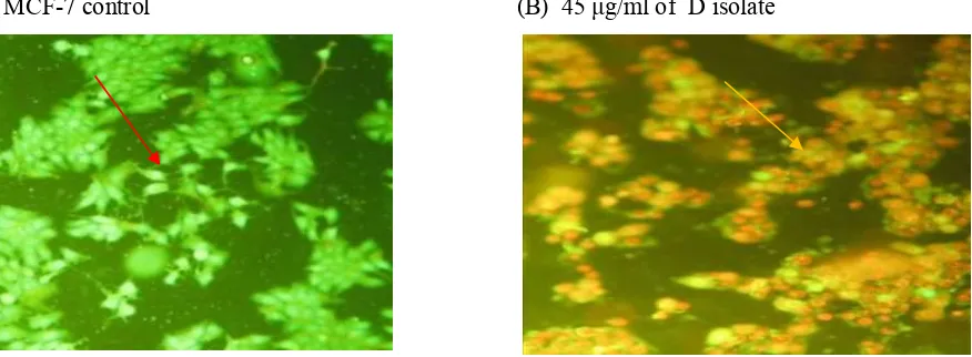
Observation of apoptosis was conducted using double staining gaveethidium bromide- akridine orange
(EB - OR) method. The results showed that the control cells green fluorescence which absorbed the acrydine orange reagent. For the treated cells, it showed yellow-orange red fluorescence. It indicated the loss of membrane permeability in some cells by giving D isolate, which caused ethidium bromide can enter the cell and caused orange red fluorescence as an indicator of cell death, also a fragmentation of nucleus was seen.
(A) MCF-7 control (B) 45 g/ml of D isolate
Figure 4:D isolate induced apoptosis in MCF-7 cells after 72 hours incubation viewed with a fluorescence microscope (100x magnification) control MCF-7 cells, the cells undergo apoptosis
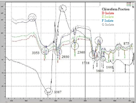
The results of characterization by Infrared Spectrophotometer
ray and will be recorded in the form of spectra. Infrared spectra of the CHCl3 fraction, D, E, F, and G isolates
are shown in the Figure 5.
Figure 5:Infrared spectrum of chloroform fraction,(D, E, F, and G isolates)
Discussion
Ekor naga leaf D isolate activity can inhibit the growth of cancer cells,tudy MCF-7. It provides excellent results with IC50 values less than 100 ug / ml which potential to be developed as anticancer. Based on
the observation of the kinetics of cell proliferation, D isolate showed an increase in number of dead cells with the increasing of incubation time (Figure 3). This suggests that D isolate can inhibit genes work or work of proteins that regulate cell cycle.
From figure 3, it showed that there was no decrease in the percentage of live cells within observation time (24, 48, and 72 hours). This suggests that D isolate have interacted with MCF-7 cells to inhibit the growth of these cells. According to Ueda8, if the extracts had IC50 values ≤ 100 mg/mL, it can be said to have the
potential of antiproliferation. Based on the above reference, it can be said that the D isolate with IC50 value of
58.567 µg/mL significantly inhibited cell proliferation in MCF-7 that needs to be followed up with a bioactive material isolation.
Death of MCF-7 cells cancer due to the treatment of ekor naga leaf D isolate could be investigated through apoptosis assay. Concentration used for the apoptosis assay was 90 and 45 µg/mL, both of the concentration were on the IC50and half IC50 value of MTT cytotoxicity assay method, therefore death cells and
live cells could be differentiated at observation time.
In DNA double staining method using acridine orange-ethidium bromide, both of the reagent were used simultaneously wihch can produce contrast colors, therefore making it easier to observe under a fluorescent microscope. Observation of cell morphology should be done immediately because if the double staining solution interact too long with cells, the life cells will die. Ethidium bromide-acridine orange can penetrate cell membranes and interact with the cell's DNA. The observation under a fluorescent microscope
Chloroform Fraction D Isolate
E Isolate
showed that control cells were green, and cells with treatment appeared orange red (figure 4). The nuclei were contracted, blebing was occurred and there were cells or incomplete cells which showed cells death were occurred by a mechanism of apotopsis. This indicated that the test substance was able to stimulate apoptosis of the MCF-7 cells. These results were in accordance with the results of the doubling time test which showed the level of 45 µg/ml of the test substance provided growth inhibition. It proved that the D isolate was capable to stimulate apoptosis and inhibit the growth of MCF-7cancer cells.
Based on the data from the infrared spectrum, chloroform fraction, (D, E, F, and G isolates) showed a sharp absorption at wavenumber region 3387-3230 cm-1 showing the O-H C-H aliphabetic group (CH3 and
CH2), strengthen with the absorption at 875 which is the -CH2 absorption. Wave number of 1709 cm -1
with strong intensity showed a carbonyl group (C=O), followed by wave numbers 1201 cm-1, indicating a C-O group, while thedouble bond duplicate at wavenumber 1676 cm-1 indicate the presence of group C= C aromatic followed by wave numbers 1445 cm-1 indicate the presence of aromatic C-C group9,10.
In MCF-7 control, apoptosis was not occured because the apotopsis regulator of MCF-7 cells, the p53 protein was bounded and degraded by E6 protein. Isolate with concentration of 45 µg/ml indicated the mechanism of cell cycle arrest and cell death was suspected. The DNA staining data could support the possibility of cell death through apoptosis mechanism
Conclusion
The result obtained by column chromatography was nine isolates that had cytotoxic activity. The ekor naga leaf (Rhaphidophora pinnata (Lf) Schott) D isolate had antiproliferative activity and promote apoptosis of the MCF-7 cells. The results of infrared spectrum were the functional groups of O-H; C-H, C = O, and C = C aromatic of A-I isolates that had anticancer properties.
References
1. Kitagawa, S., (2006), Inhibitor Effect of Polyphenols on P-Glycoprotein-Mediated Transport, Biol. Pharm. Bull, 29, 1-6.
2. Newman, D.J., Cragg, G.M. & Snader, K.M. (2000). The influence of natural products on drug discovery.Nat. Prod. Rep.17(3): 215-234.
3. Fisher, D.E., (1994), Apoptosis in Cancer Therapy: Crossing The Threshold, Cell, 78, 539-542.
4. Masfria, Harahap U, Nasution MP, Ilyas S. (2011), Cytotoxicity test of n-hexane extract, Ethyl Acetate and Ethanol ekor naga leaf (Rhaphidophora pinnata (L.f.) Schott) by Brine Shrimp Lethality Test methode (BSLT), Pharmacy Update3, Medan.
5. Aini MN. (2006). Anticancer activity test praskrining Aktivitas extrac n-hexane, dichloromethane and methanol leaf Rhaphidophora pinnata (L.f.) with Brine Shrimp Lethality Test Methods. http://www.unair.ac.id Undergraduate Theses Airlangga University.
6. Masfria, Harahap U, Nasution MP, Ilyas S. (2012), Cytotoxicity test of Etanol extrac and some Fractions ekor naga leaf (Rhaphidopora pinnata(L.f) Schott) with MTT method MCF-7 cell line, Seminar Nasional Biologi, Medan
7. Mosmann,T. (1983) Rapid colorimetric assay for cellular growth and survival: Application to proliferation and cytotoxicity assay.Journal of immunological Method,65, 65-69.
8. Ueda, J.Y., Tezuka, Y., Banskota A.H., Le Tran Q., Tran Q.K.,Harimaya Y., Saiki I., Kadota S.(2002), Antiproliferative activity of Vietnamese medicinal plants, Biol Pharm Bull; 25(6): 753-760.
9. Sastrohamidjojo, H. (1991).Basic of Spektroskopi. Yogyakarta: Liberty. Ed. II;80-100.
10. Unannnng Supratman, (2010), Elusidasi Struktur Senyawa Organic. Bandung: Widya Padjadjaran.; 94-102.