Vol.7, No.2, pp 238-242, 2014-2015
Cytotoxicity of “Ekor naga” Leaf (Rhaphidophora pinnata (Lf)
Schott) Chloroform Extract against T47D Cancer cells
Masfria, Aswita Hafni
Faculty of Pharmacy, University of Sumatera Utara
Abstract:Cancer is the second disease cause of mortality in USA, after cardiovascular diseases.
Nowadays, the current research tend to develop anticancer agents from plants. One of the plants that can be used for anticancer therapy is the “ekor naga” leaves (Rhaphidophora pinnata (Lf) Schott), which is empirically used by some communities as breast cancer treatment.
The extraction of the dried leaf was done by soxletation with n-hexane, chloroform, ethyl acetate, and ethanol solvents. The cytotoxic activity was performed using T47D and Vero cell by MTT assay (tetrazolium salt { (3- (4,5-dimetiltiazol-2-yl) -2.5-diphenyl tetrazolium bromid}) method, and to determine the value of the Selectivity Index (SI).
The results of cytotoxicity test of n-hexane, chloroform, ethyl acetate, and ethanol extracts showed IC50 values of 422.921, 69.067, 475.448, 10185.896 µg/mL, respectively. The
chloroform extract had IC50 values ≤ 100 µg/mL which had the anticancer activity on T47D
cells. The result of extract IC50 selectivity index value of cancer cells againsted normal cells,
showed that the chloroform extract had a high selectivity with value of the SI was 4.25, againsted T47D cancer cells.
Keyword: “ekor naga” leaf. T47D cell, Vero cell, Rhaphidophora pinnata.
Introduction
Indonesia is a rich country with diverse traditional medicinal plants that have anticancer activity. Among these plants is the leaves of ekor naga plant (Rhaphidophora pinnata (Lf) Schott), which has been used empirically by the Community for the treatment of several diseases, especially breast cancer. In the development of traditional medicine, it is needed a lot of researches to get accurate results of the efficacy of the plants.
The “Ekor naga” leaf is a plant that has been widely studied for its cytotoxic effects and can inhibit the growth of MCF-7 cancer cells. It contains chemical class of compounds such as saponin, tannin, alkaloids, steroid/triterpenoid, flavonoids, and glycoside, which play a role in the inhibition of cancer cells. Some studies of “ekor naga” leaf included: antibacterial activity of some extracts with different polarity against some bacteria (Kadriani1 and Masfria2 ,2009; 2013), the test of toxicity from ethanol extract (crude) and extracts (n-hexane, ethyl acetate and ethanol) by using Brine Shrimp Lethality Test (BST) method showed a cytotoxic result to
Artemia salina Leach (Masfria3, et al., 2011), and antioxidant activity test against DPPH (1,1-diphenyl-2-picrylhidrazyl) from n-hexane, ethyl acetate and ethanol extracts (Masfria4, et al., 2012). It suggested that the cytotoxic test method using BST and the antioxidant properties positively correlated with anticancer activity (Silalahi5, 2006).
Methodology
Preparation of “Ekor Naga Leave Extracts
A total of 10 parts of dried powder leaves extracted by soxcletation. Dried powder leaves were inserted to the soxcletation tool using n-hexane, chloroform, ethyl acetate, and ethanol solvents. The extraction was stopped when the solvent in tubes had been clear. The obtained extract was evaporated to get a thick extract and dried using a freez dryer. (Ditjen POM6, 1979).
Cytotoxic Test by MTT assay
The T47D cells were seeded in to 96 wells microplate in order to obtain the density of 1x 10⁴ cells/well and incubated for 24 hours to get a good growth. After that, the medium was replaced with a new one and then 100 µL of test solution in series of concentrations of 250, 125, 62.5, 31.25, 15.625 ug/mL was added, and incubated for 24 hours again. At the end of incubation, the medium was discarded and the cells were washed with PBS solution, then each of the wells was added with 0.5% MTT (10 µL reagent 3- (4,5-dimetiltiazol-2-yl) -2.5-diphenyl tetrazolium bromide) in PBS solution. Incubation was continued for 4 hours at 37°C to form formazan. The living cells would converted MTT to the dark blue formazan. MTT reaction was stopped by 100 µL of the stopper reagent (Sodium dodecyl sulphate = SDS), and then incubated overnight at room temperature. MTT assay was performed according to Mossman7 (1983) with the modification of stopper reagent.
Selectivity index
To determine the value of the Selectivity Index (SI), it was necessary to know the value of vero and T47D cells IC50 using MTT method.
Selectivity index was calculated using the following equation:
SI indicated the cytotoxic selectivity (security) of the extract against cancer cells versus normal cells, which was calculated by comparing IC50 of extract on normal cells and IC50 extract against cancer cells.
Analysis Data
The absorbance data obtained from the cytotoxic test was calculated by the following formula:
% Living Cells =
IC50 concentrations were calculated by probit analysis using SPSS 17 program, from the linear
correlation between log concentration and percentage of living cells. IC50 was the concentration that caused
death of 50% of cell population.
Results and Discussion
Cytotoxic Activity
Cytotoxic activity test was conducted to determine the potential toxicity of extracts (n-hexane, chloroform, ethyl acetate, and ethanol) with IC50 parameter. The concentration of the test materials with the
series of 15.625, 31.25, 62.5, 125, 250 µg/mL had a correlation between the concentration of the test solution with the resulting toxic effects.
Table 1.Results of Cytotoxic Activity Against Multiple Extracts on T47D and Vero Cells
IC50 (µg/mL)
No Extracts T47D cell Vero cell
1 n Hexane Extract 422,921 3417,897 2 Chloroform Extract 69,067 293,410 3 Ethyl Acetate Extract 475,448 1180,121 4 Ethanol Extract 10185,896
-Referring to Table 1, chloroform extract on T47D cells had IC50 values ≤ 100 µg/mL. It showed that
chloroform extract had anticancer activity because it was thought to contain a chemical class of compounds which had the anticancer activity against T47D cells with IC50 values ≤ 100 µ/mL.
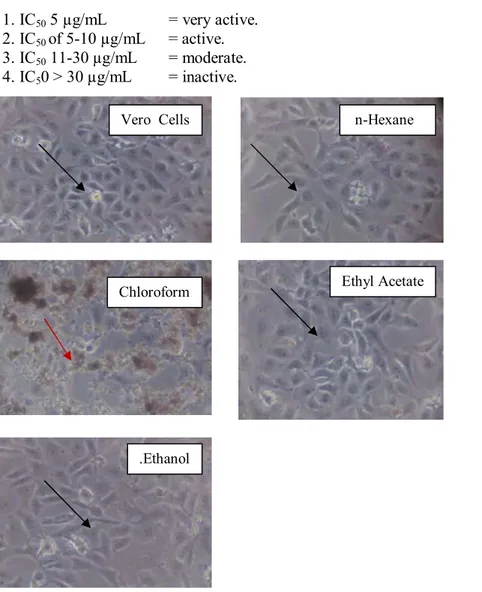
Figure 1. Morphology of T47D cells after incubated some extracts for 24 h (magnification 10 x 10). Living cells indicated by black arrows( ), while the dead cells was shown by red arrows ( )
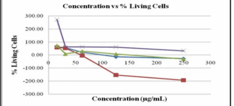
Figure 2. Graph of chloroform extracts concentration versus living cells
Treatment with some “ekor naga” leaf extracts on T47D cancer cells affected the morphology of the cell. Living cells appeared in the form of leaves and remained floating on the wells (Figure 1 and 3), whereas the death cell looked round and blackened. The chloroform, ethyl acetate and ethanol “ekor naga” leaf extracts
T47D Cells E. n-Hexane
Chloroform Ethyl Acetate
showed dose-dependent phenomenon where by percentage of living cells continued to decrease with the increasing of dose (Figure 2). It showed that the “ekor naga” leaf extract could induced cell death in T47D breast cancer cells.
The data showed that “ekor naga” chloroform extract had potential against T47D cells with IC50 value
of 69.067 µg/mL. It gave a great hope to the treatment of ER-positive breast cancer by the representation of T47D cells.
Probit analysis results showed that all the extracts against vero cells had IC50 values ≥ 100 µg/mL.
According to Ueda9,et al., (2002), the IC50 value as an anticancer was potential if the IC50 ≤ 100 µg/mL and by
Cho10, (1998), IC50 value > 30 µg/mL = not active, so that all extracts were non toxic to normal cells, with the
following criteria:
1. IC50 5 µg/mL = very active.
2. IC50of 5-10 µg/mL = active.
3. IC50 11-30 µg/mL = moderate.
4. IC50 > 30 µg/mL = inactive.
Gambar 2. Morphology of Vero cells after incubated with all extracts for 24 h (magnification 10 x 10). Living cells indicated by black arrows( ) , while the dead cells was shown by red arrows ( )
IC50 values can indicate the potential as a cytotoxic compound. Based on the IC50 values, the test
materials were safe against Vero cells with IC50 values ≥ 30 µg / mL.
Selectivity Index
The ekor naga chloroform extract IC50 value against vero cells was 293.410 µg/mL and T47D cells was
69.067 µg/mL, therefore the value of the SI was 4.25. The extract had a high selectivity when the value of the SI > 3 (Machana8, et. Al, 2011), Therefore the chloroform extract of “ekor naga” was selective against T47D cancer cells.
Vero Cells n-Hexane
Chloroform Ethyl Acetate
Conclusion
Based on the obtained results, it could be temporary concluded that the chloroform extract had a cytotoxic effect with IC50 value of 69.067 mg/mL against T47D cells. All “ekor naga” extracts against vero
cells (normal cells) were non toxic with IC50 ≥ 100 µg/mL. electivity index value of chloroform extract that
selective on T47D breast cancer cells was ˃ 3.
References
1. Kadriani, J. 2009,) Phytochemical Screening and Antibacterial Activity Test of ekor naga Leaf (Rhaphidophora pinnata (Lf) Schott) Against Some Bacteria in Vitro, pharmacy faculty USU
2. Masfria, Harahap, U., Nasution MP., Ilyas, S. (2013). Antibacterial activity of n-Hexane Extract, Ethyl Acetate and Ethanol Ekor naga Leaf (Rhaphidophora pinnata (Lf) Schott) Against Some Bacteria.Medan: National Seminar on Biological
3. Masfria, Harahap, U.,Nasution, M.P. Syafruddin,I.,(2011), Cytotoxicity test extract Ethanol, n-Hexane, Ethyl Acetate and Ethanol “ekor naga” leaf (Rhaphidophora pinnata (Lf) Schott) by the method of
Brine Shrimp Lethality Test (BSLT), Seminar Update 3,18-19-03-11, Medan
4. Masfria, Harahap U, Nasution MP, Ilyas S. (2012), Cytotoxicity Test Ethanol Extract and Some fraction “ekor naga” leaves (Rhaphidopora pinnata (Lf) Schott) against MCF-7 cells by MTT method, National Seminar on Biological 11-05 2012, Medan
5. Silalahi J (2006), Functional Foods, Publisher Canisius. page 63-70.
6. Ditjen POM. (1979).Farmakope Indonesia. Ed. III. Departemen Kesehatan RI. Jakarta. page 9, 649, 7. Mosmann, T. (1983). Rapid Colorimetric Assay for Cellular Growth & Survival: Application to
Proliferation & Cytotoxicity Assays. Journal of Immunological Method.65: 65-59.
8. Machana,S., Natthida, W., Sahapat, Barusrux, Apiyada, N., Bungorn S, dan Thaweesak, T. (2011). Cytotoxic and apoptotic effects of six herbal plants against the human hepatocarcinoma (HepG2) cell line.Chinese Medicine. 6:39.
9. Ueda, J.Y., Tezuka, Y., Banskota A.H., Le Tran Q., Tran Q.K.,Harimaya Y., Saiki I., Kadota S.(2002), Antiproliferative activity of Vietnamese medicinal plants, Biol Pharm Bull; 25(6): 753-760.
10. Cho, S.J. (1998). Novel Cytotoxic Polyprenila-terd Xanthones From Garcinia gaundichaudii (Guttiferae). Tetrahedron (54): 10915-10924.
11. Conze, D., Weiss, L., Regen, P.S., Bhushan, A., Weaver, D., Johnson, P., dan Rincon, M. (2001). Autocrine Production Of Interleukin 6 Causes Multi Drug Resistance In Breast Cancer Cells. Cancer Research.(61): 8851–8858.