Expression of FasL in Proliferation of Retinoblastoma
Cells: A Mechanism Fas Counterattack
Retinoblastoma Hücrelerinin Çoğalmasında Fas
L
Ekspresyonu: Fas Atağının
Mekanizması
Hendrian D. Soebagjo1, Hidayat Sujuti2, Hasan Machfoed3, Sutiman Bambang Soemitro4
1
Medical Faculty of Brawijaya University, Malang-Department of Ophthalmology, Medical Faculty of Airlangga University, Dr. Soetomo Hospital, Surabaya, 3Department of Neurology. INDONESİA
2
Department of Biochemistry-Molecular Biology – Department of Ophthalmology, Dr. Saiful Anwar Hospital, Malang. INDONESİA
4
Department of Biology, Mathematic and Natural Sciences Faculty of Brawijaya University, Malang, INDONESIA
Cukurova Medical Journal 2014;39(4):788-794.
ABSTRACT
Purpose: The aim of this study were to determine the association of increasing of FasL with increasing proliferation of retinoblastoma cells.
Materials and Methods: The protein expression was analyzed in 30 retinoblastoma samples from paraffin block using immunohistochemical method for evaluation of FasL, CDK4, and Ki-67 expression.
Results: Among 30 retinoblastoma samples, FasL expression majority was negative in 33.3 % (10 samples) and strong in 36,8 % (11 samples). CDK4 majority 53,3% was weak expression and Ki-67 was high expression also in 53,3% (16 samples). The expression of FasL was significantly related to CDK4 (r: 0.363; p: 0.048). The CDK4 was also significantly related to Ki-67 expression (r: 0.601; p: 0.000).
Conclusion: The increasing of FasL on the mechanism Fas counterattack induces proliferation of retinoblastoma cells.
Key Words: FasL, proliferation, counterattack, retinoblastoma.
ÖZET
Amaç: Bu çalışmanın amacı artan FasL ekspressyonu ile retinoblastom hücrelerindeki proliferasyon artışı arasındaki
ilişkiyi incelemektir.
Materyal ve Metod: Ki-67, CDK4 ve FasL ekspresyon değerlerini incelemek için parafin blok yapılmış 30 retinoblastom
örneğinin protein ekspresyon değerleri immünohistokimyasal analiz ile belirlendi.
Bulgular:30 retinoblastom örneği arasından %33.3 ünde (10 örnek) FasL ekspresyonu yok iken %36,8 inde güçlü bir ekspresyon vardır. %53.3 (16 örnek) örnekte CDK4 ekspresyonunun zayıf olduğu görülürken Ki-67 ekspresyonunun
yüksek olduğu tespit edilmiştir. FasL ekspresyonu yüksek oranda CDK4 ekspresyonu ile ilgilidir (r:0,363, p:0,048). Aynı
zamanda CDK4 ün ekspresyonu da Ki-67 ekspresyonuyla ilgilidir (r:0,601, p: 0,000).
Sonuç: FasL atağı mekanizmasındaki artan FasL ekspresyonu retinoblastoma hücrelerinin proliferasyonunu indüklemektedir.
Anahtar kelimeler: FasL, proliferasyon, atak, retinoblastom
INTRODUCTION
Retinoblastoma has improved a survival rateof patients over years and been increasing in the developed countries because of no early method detection and proper therapy was. Retinoblastoma derives from retinal cell, expands to the posterior chamber of the eye, and invades through the sclera and along the optic nerve1. Whenever tumor extends beyond the globe into the orbit, a combination of radiation therapy and chemotherapy has been used. Prognosis remains poor for patients with dissemination into the central nervous system (CNS) and those with distant metastatic disease2. A protein expression in proliferation and apoptosis pathway of retinoblastoma must be known to diagnose retinoblastoma because the correct diagnosis determines the appropriate treatment and to find a drug of choice by chemotherapy3,4.
Progressiveness and prognosis of cancer cells can be evaluated by the balance of proliferation and apoptosis cells5. An imbalance between apoptosis and proliferation of cancer cells will be aggressive. In malignant tumor retinoblastoma, the role of cytokines Ras, Raf, and MEK will stimulate Cyclin Dependent Kinase 4 (CDK4) and induce CDK2, then cause Rb phosphorylation and have no capacity to bind E2F to lead a proliferation cell6. Expression of Ki-67 was used for measurement of levels of proliferation in different kinds of cancer cells. Ki-67 was expressed in all phases of the cells, except G0 phase7,8.
Fas ligand (Fas/CD95L/CD178) and its receptor, Fas (APO-1/CD95), are members of the tumor necrosis factor family. The FasL is a 40 kDa type I membrane glycoprotein and can be found in activated T lymphocytes an NK cells9,10. Association of Fas and FasL activates its death domain and thus triggers a cascade of caspases that results in apoptosis cell. FasL is activator of extrinsic apoptosis in cancer cells11-13. Recently
discovered that FasL expression on cancer cells was significantly different from the normal. There was a role immunologic system of Fas in cancer cells against T cells lymphocytes, especially of cytotoxic T cells10,13,14. Many types of cancer have been shown to express FasL because activated lymphocytes express Fas. Increased of FasL by tumor cells may enable to kill the T cells and induce resistance and proliferation of tumor cells. But it is still unknown how the role of FasL against proliferation in retinoblastoma.
The aim of this study was to determine the association of increased of FasL with increased proliferation of retinoblastoma cells using immunohistochemical staining.
MATERIALS and METHODS
The study group of 30 retinoblastoma samples from patients paraffin block was carried out an exenteration and enucleation therapy in Dr. Soetomo General Hospital – Medical Faculty of Airlangga University, Surabaya, Indonesia. None had received chemotherapy and radiotherapy prior to tissue samples collection.
The protein expression was evaluated by immunohistochemical reaction using antibodies for FasL (polyclonal antibody Fas-L; P137 (BS1122) Bioworld Technology Inc. (1:50), CDK4 (polyclonal rabbit CDK4(c-22): sc-260 Santa Cruz Biotechnology, Inc. (1:100), and Ki-67(CRM325 AK,BK) Biocare(1:75). All samples were stained by using Labelled Streptavidin Biotin II (LSAB II) method. A colour reaction for peroxidase was developed with DAB as a chromogen. The sections were counterstained with Meyer’s haematoxylin.
(>50%)15. Nuclear accumulation of CDK4 was evaluated according to the criteria: negative (0%), weak (<25%), moderate (25-50%), and high (>50%)16. Ki-67 expression on nuclear cell as
negative (0%), low (≤40%), and high (≥40%)17.
Colon cancer tissue sections known to express FasL were used as positive control. Positive
control tissues included breast cancer for CDK4 expression and prostate cancer for Ki-67 expression. Statistical analysis was conducted by using Mann-Whitney U-test and Spearman’s correlation test. A p-value of < 0,05 was considered statistically significant.
RESULTS
Figure 1. Expression of FasL, CDK4, and Ki-67 (n=30). Graphic box the percentage of cells on the criteria of expression.
There were 30 retinoblastoma samples to know protein expression of FasL, CDK4, and Ki-67 by immunohistochemical staining. Distribution of expression is presented in Figure 1. Among 30 retinoblastoma samples, FasL expression majority
was negative in 33.3 % (10 samples) and strong in 36,8 % (11 samples). CDK4 majority 53,3% was weak expression and Ki-67 was high expression in 53,3% (16 samples)as well.
Table 1. Correlations between FasL, CDK4, and Ki-67 in Retinoblastoma
FasL CDK4 Ki-67
FasL - p: 0.048* p: 0.021*
CDK4 p: 0.048* - p: 0.000*
Ki-67 p: 0.021* p: 0.000* -
Note: (*) p<0,05 was considered statistically significant
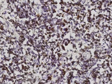
FasL positive immunohistochemical staining was detected in retinoblastoma (Figure 2). A FasL positive immunohistochemical staining expressed a brown colour in the cytoplasm and cell
membrane. CDK4 and Ki-67 positive immunohistochemical staining expressed most tumor cells and a dark brown colour in nuclear cells (Figure 3 and Figure 4). Statistical analysis of
0 20 40 60
33,3
16,6 13,3
36,8
0 53,3
16,6 30,1
10 36,7
53,3
E
X
P
R
E
S
S
ION
OF C
E
LLS
(
%
)
CRITERIA OF EXPRESSION
the results revealed statistically significant correlations of FasL, CDK4, and Ki-67. The expression of FasL was significantly related to
CDK4 (r: 0.363; p: 0.048). The CDK4 was also significantly related to Ki-67 expression (r: 0.601; p: 0.000).
Figure 2. FasL staining in retinoblastoma expressed in cytoplasm and membrane cells. Original magnification x400.
Figure 3. CDK4 staining was expressed many positive Cells with intact nuclei in retinoblastoma.Original magnification x400.
DISCUSSION
On this study FasL expressed 66,7% of cells and 36,8% expressed strong. This indicated the role of FasL in immune mediated process of cancer cells18. Fas/FasL system plays an important role in the event of apoptosis in cytotoxic reaction involving the role of cellular immunologic against tumor cells10,14,19. Associated of Fas and FasL activates its death domain and thus triggers a cascade of caspases that result in apoptosis cell. FasL is activator of extrinsic apoptosis in cancer cells. Apoptotic signal was sent by FasL-Fas associated to binding site of FADD protein and adapter enable Caspase-8 to Caspase-3 as apoptosis executor11,12. Expression of FasL accordance with expression on coutaneous squamous carcinoma18, bladder cancer20, cervical squamosa cancer 21, and colon cancer25.
Expression of CDK4 distribution showed that the majority of retinoblastoma is a weak and high expression. CDK4 is a key protein in G1 transition during cell-cycle progression. In complex with cyclin D, CDK4 phosphorylates G1-specific substrates, including the retinoblastoma protein (Rb). Rb phosphorylation in collaboration with cyclin D/CDK4 and cyclin E/CDK2 results in releasing of Rb from the E2F complex then the G1 phase of the cell enter S phase (DNA synthesis). If there was a mutation in the regulation of cyclin-D, then resulting in increased cell into S phase and the oncogenic transformation activities happen6.
Expression of Ki-67 showed that the majority of retinoblastoma is a high expression. The increasing Ki-67 proliferation marker indicate the aggressiveness of tumors that are characterized by an increase in the number of cells undergoing mitotic20. Ki-67 was expressed on G1, S, G2, and mitotic phase but not on G0 phase7,8. Ki-67 expression on retinoblastoma was accordance with expression on breast cancer7, colorectal carcinoma23, and gastric carcinoma24.
Our studies confirmed that expression of FasL
was significantly related to proliferation of retinoblastoma. FasL significantly correlated with CDK4 and Ki-67. The system of associated Fas and FasL was responsible for cytotoxic T cell- mediated apoptosis and played a role in the immunologic homeostasis. Increasing expression of FasL was related to carcinoma that metastatic, such as found on stomach and esophagus cancer9,18,25. Zhang et al., (2003) said that increased FasL expression on gastric cancer cells impact on immunologic response. A tumor cells having the immunity to kill a cytotoxic T cells. This mechanism called a Fas counterattack10,14.
Normally, cytotoxic T cells produced FasL to bind a tumor Fas and induce apoptotic process of tumor cells. In Fas counterattack mechanism, tumor cells produce a MMP-7 to shedding a FasL cytotoxic T cells. Shedding of MMP-7 producing a soluble FasL which will inhibit an associated Fas/FasL then inhibit a FADD protein to bind receptors of death domain then the death signal of Caspase-8 to Caspase-3 interrupted10,27-29.
In addition, some cancer cells have the ability to kill of cytotoxic T cells by producing FasL that binds Fas of cytotoxic T cells, then result T cell was apoptosis. The process of counterattack resulted in a increase a growth, invasion, and metastatic a tumor cell10,25,29. Based on our data was concluded that increased of FasL on the mechanism Fas counterattack induces proliferation of retinoblastoma cells.
Conflict of interest
The authors declare that there are no conflicts of interest.
REFERENCES
1. Divan J, Lawry IR, Dunsmore MA, Parsons JA. p53 and p21waf-1 Expression Correlates with Apoptosis or Cell Survival in Poorly Differentiated, but not Well-Differentiated, Retinoblastomas. Cancer Research. 2001;61:3157-63.
2. Khrisnakumar S, Kandalam M, Mohan A, Iyer A, Vankatesan N, Biswas J, Shanmugam MP. Expression of Fas Ligand in Retinoblastoma. American Cancer Society. 2004;101:1672-6.
3. Sehu KW. Ophthalmic Pathology. An Illustrated Guide for Clinican. British Library, Blackwell Publishing. 2005.
4. Sitorus RS, Gumay S, Der Valk PV. The apoptosis paradox in retinoblastoma. Natural compounds and their role in apoptotic cell signaling pathways. Ann.N.Y. Acad.Sci.2005;1171:77-86.
5. Ito Y, Matsuura N, Sakon M, Takeda T, Umeshita K, Nagano H, et al. Both cell proliferation and apoptosis significantly predict shortened disease-free survival in hepatocellular carcinoma. British Journal of Cancer. 1999;81:747–51.
6. Kumar V, and Stricker TP. Neoplasia in Robin dan Cotran, Pathologic Basic of Diasease, 8th. Ed. Saunders Elsevier. 2010;259-327.
7. Urruticoechea A, Smith IE, and Dowsett M. Proliferation Marker Ki-67 in Early Breast Cancer. J Clin Oncol. 2005;23:7212-20.
8. Colozza M, Azambuja E, Cardoso F, Sotiriou C, Larsimont D, Piccart MJ. Proliferative markers as
prognostic and predictive tools in early kanker payudara: where are we now? Ann Oncol. 2005;16:1723-27.
9. Bennett MW, O’Connell J, O’Sullivan GC, Roche D, Brady C, Kelly J, Collins JK, Shanahan F. Expression of Fas ligand by human gastric adenocarcinomas: a potential mechanism of immune escape in stomach cancer. Gut. 1999;44:156–62.
10. Maher S, Toomey D, Condron C, and Bouchier-Hayes D. Activation-induced cell death: The controversial role of Fas and Fas ligand in immune privilege and tumour counterattack. J.Imun.Cell.Biol. 2001;80:131-7.
11. Peng SL. Fas (CD95)-related apoptosis and rheumatoid arthritis. Rheumatology (Oxford). 2006;45:26-30
12. Lumongga F. Apoptosis. Departemen Patologi Anatomi Fakultas Kedokteran Universitas Sumatera Utara. USU Repository. 2008.
13. Tanaka M, Itai T, Adachi M, Nagata S. Downregulation of fas ligand by shedding. J.Nat. Med. 1998;4(1):31-6.
14. Houston A. and O’Connell J. The Fas signalling pathway and its role in the pathogenesis of cancer. Current Opinion in Pharmacology. 2004;4:321-6.
15. Tong Q, Zheng L, Tang S, Li S, Jiang G, Cai J, Liu Y, Ruan Q. Expression of Fas and FasL in human neuroblastoma and its clinical significance. World J Pediatr. 2007;3:209-13.
16. An H, Beckmann MW, Reifenberger G, Bender HG, and Niederacher D. Gene Amplification and Overexpression of CDK4 in Sporadic Breast Carcinomas Is Associated with High Tumor Cell Proliferation. American Journal of Pathology. 1999;154:113-8
17. Ben-Izhak, O, Bar-Chana M, Sussman L, Dobiner V, Sandbank J, Cagnano M, Cohen H, and Sabo E. Ki67 Antigen and PCNA proliferation markers predict survival in anorectal malignant melanoma. Histopathology. 2002;41:519-25.
19. Shimizu M, Kondo M, Ito Y, Kume H, Suzuki R, Yumaki K. Soluble fas and fas ligand provide new information on metastasis and response to chemotherapy in SCLC patients. cancer detection and prevention. Elsevier. 2004. doi:10.1016/j.cdp.2004.09.001.
20. Perabo F.G.E., Kamp S, Schmidt D, Lindner H, Steiner G, Mattes RH, et al. Bladder cancer cells acquire competent mechanisms to escape Fas-mediated apoptosis and immune surveillance in the course of malignant transformation. British Journal of Cancer. 2001;84:1330–8.
21. Zhou, JH, Chen HZ, Ye F, Lu WG, and Xie X. Fas-mediated pathway and apoptosis in normal cervix, cervical intraepithelial neoplasia and cervical squamous cancer. Oncology Reports. 2006;16:307-11.
22. Trihia H, Murray S, Price K, Gelbert RD, Golouh R, Goldhirsch A, et al. Ki-67 expression in breast carcinoma: its associations with grading systems, clinical parameters, and other prognostic factors– a surrogate marker? Cancer. 2003;97:1321-31
23. Nabi U, Nagi AH, Sami W. Ki-67 proliferating index and histological grade, type and stage of colorectal carcinoma. J.Ayub Med Coll Abbottabad. 2008;20:44-9.
24. Lazar D, Taban S, Sporea I, Dema A, Corianu M, Lazar E, Goldis A, Vernic C. Ki-67 expression in gastric cancer. Results from a prospective study with long-term follow-up. Romanian Journal of Morphology and Embryology. 2010;51:655–61
25. Pryczynicz, A, Guziñska-Ustymowicz K, and Kemona A. Fas/FasL expression in colorectal cancer. An immunohistochemical study. Folia Histochemica Et Cytobiologica. 2010;48:425-9.
26. Zhang W, Ding EX, Wang Q, et al. Fas Ligand expression in colon cancer: a possible mechanism of tumor immune privilege. World J Gastroenterol. 2005;11:3632-5.
27. Mitsiades N, Yu W, Poulaki V, Tsokos M, and Stamenkovic I. Matrix Metalloproteinase-7-mediated Cleavage of Fas Ligand Protects Tumor Cells from Chemotherapeutic Drug Cytotoxicity. Cancer Research. 2001;61:577–81.
28. Vargo-Gogola T, Crawford HC, Fingleton B, and Matrisian LM. Identification of novel matrix metalloproteinase-7(matrilysin) cleavage sites in murine and human Fas ligand. Archives of Biochemistry and Biophysics. 2002;408:155–61.
29. Webb SD, Sherratt JA, and Fish RG. Cells behaving badly: a theoretical model for the Fas/FasL system in tumour immunology. Mathematical Biosciences. 2002;179:113–29.
30. Schroter M, Peli J, Hahne M, Tschopp J, Reichmann E. Fas-dependent tissue turnover is implicated in tumor cell clearance. Oncogene. 2000;19:1794-1800.
31. Kurooka M, Nuovo GJ, Cagliuri MA, Nabel GJ. Cellular localization and function of Fas Ligand (CD95L) in tumors. Cancer Res. 2002;62:1261-5.
32. Zhu, Q, Liu JY, Xu HW, et al. Mechanism of counterattack of colorectal cancer cell by Fas/FasL system. World J Gastroenterol. 2005;11:6125-9.
33. Okada K, Komuta K, Hashimoto S, Matsuzaki S, Kanematsu T, Koji T. Frequency of apoptosis of tumor-infiltrating lymphocytes induced by fas counterattack in human colorectal carcinoma and its correlation with prognosis. Clin Cancer Res. 2000;6:3560-4.
Yazışma Adresi / Address for Correspondence:
Dr. Dr. Hendrian D. Soebagjo Medical Faculty of Airlangga University
Department of Ophthalmology Dr. Soetomo Hospital Jl. Mayjen. Prof. Dr. Moestopo no 6-8 Surabaya, East Java, INDONESIA
E-mail: [email protected]