I.J. Biotech, Vol. 20, No. 2
141
Arianingrum et al. I.J. Biotech.
Synergistic effects of
para-
hydroxy
meta-
methoxy chalcone (
p
H
m
MC)-
doxorubicin treatments on T47D breast cancer cells
Retno Arianingrum
1,2*, Retno Sunarminingsih
3, Edy Meiyanto
3, and Soia Mubarika
41 Doctor Candidate, Biotechnology Study Program, Universitas Gadjah Mada, Yogyakarta, Indonesia 2 Department of Chemistry Education, Faculty of Mathematics and Natural Science,
Universitas Negeri Yogyakarta, Yogyakarta, Indonesia
3 Faculty of Pharmaceutical, Universitas Gadjah Mada, Yogyakarta, Indonesia
4 Faculty of Medicine, Universitas Gadjah Mada, Yogyakarta, Indonesia
Abstract
Resistance to some cancer chemotherapeutic drugs has been identiied. One strategy to overcome that problem is by combining two or more of the drugs to get co-chemotherapeutic effects. A derivate chalcone, 3 - (4’-hydroxy-3’-methoxyphenyl)-1-phenyl-2-propene-1-on or para hydroxy meta methoxy chalcone (pHmMC), has been reported to have cytotoxic activity on some cancer cells through some pathways. The aim of this study was to investigate the effects of combinations of pHmMC and Doxorubicin (Dox) on the cytotoxicity, anti-proliferation, apoptosis, and the cell cycle of T47D (breast cancer cell-lines) in vitro. The cytotoxic and antiproliferative activity were determined by MTT (3-[4,5-dimethylthiazol-2-yl)-2,5 diphenyl tetrazolium bromide] assay. The combination index (CI) was used to determine the synergistic, additive or antagonistic effects of the combinations. Flowcytometry method was performed to determine the combination effects on the apoptosis and cell cycle.The results indicated that the combinations had a higher inhibitory effect on the cell growth compared to those of single treatments of pHmMC and Dox. All the doses used in the combinations were lower of the single doses at their IC50s. The results showed all the combinations gave synergistic (CI: 0.3 – 0.7) up to strong synergistic (CI: 0.1 – 0.3) effects. The synergistic effects of the combinations were due to increased apoptosis and induced cell cycle arrest in S and G2/M phases on the cancer cell lines.
Keywords: pHmMC, Dox, co-chemotherapy, T47D cells.
*Corresponding author : Retno Arianingrum
Doctor Candidate, Biotechnology Study Program, Universitas Gadjah Mada, Yogyakarta
Introduction
The American Cancer Society has reported that breast cancer is the second fatal cancer disease on women in the world (Anonymous, 2012). In Indonesia, breast cancer is the second most frequent one after cervix cancer and become the major caused on women mortality. The statistic data in 1991 reported that breast cancer cases reached 17.77% of all cancer cases in Indonesian women (Tjindarbumi, 2002). There are many cancer therapeutic strategies including surgery, chemotherapy, and
radiation (King, 2000), but some problems found in the treatment. The main problems are the low selectivity and resistance of the drug on the tumor cells (Wong et al., 2006). Accordingly, any efforts in developing compounds which selectively against cancer are still needed.
142
enhance the eficacy with a reduced toxicity
to normal tissue (Sharma et al., 2004; Tyagi et al., 2004).
C h a l c o n e s are α,β−unsaturated ketones which have the core structure of
1,3-difenilpropen-1-one. They have been
studied as anti-cancer agents on different cell and animal models (Sasayama et al., 2007). One of the previous studies has reported that most actions of chalcones as cancer chemotherapeutic agents are blocking the cell cycle progress and inducing apoptosis (Hsu et al., 2006). Several kinds of literature also revealed that chalcones act on different types of targets (Boumendjel et al., 2009) including inhibitions of tubulin assembling
(Ducki, 2009), angiogenesis (Mojzis et al., 2008), and kinases (Reddy et al., 2010); as well as modulating of ABC proteins which involve in multidrug resistance (Valdameri et al., 2012), inductions of anti-estrogenic effect (Ducki, 2007) and non-apoptotic cell death (Robinson, et al., 2012).
Shen et al.(2007) have proved that the core structure of chalcones inhibits the
activation of nuclear factor kappa (NF-κB),
a transcription factor that is very important in the development and progression of cancer. It regulates many genes involved in
inlammation, cell survival, cell proliferation,
invasion, angiogenesis, and metastasis
(Pahl, H.L., 1999). The inhibition of NF-κB
activation causes apoptosis induction, cell cycle inhibition, and reduction of Bcl-XL expression as the downstream targets of
NF-κB in T24 and HT-1376 bladder cancer cells, as well as MCF-7 and MDA -MB-231 breast
cells (Hsu et al., 2006; Guttridge et al.,1999;
Hinz et al., 1999).
Arty (2010) has been synthesizing
some chalcone-derivative compounds with a hydroxyl group at the para position. One derivate resulted which has structure
of (3-
(4’-hydroxy-3’-methoxyphenyl)-1-phenyl-2-propene-1-one) named as para hydroxy meta methoxy chalcone (pHmMC) (Figure 1). The compound had cytotoxic activity against HeLa, Raji, T47D, and
MCF-7 cancer cells. It was shown that it inhibited the proliferation of T47D and MCF-7 cancer cells by blocking the cell cycle progression. In MCF-7, pHmMC also induced the cell apoptosis (Arianingrum et al., 2012; Arianingrum et al., 2015). Interestingly, it did not have cytotoxic activity against Vero normal cell culture (Arianingrum et al., 2010). Based on these pHmMC’s anti-cancer potencies, this study is to investigate the probable co-chemotherapeutic effect of pHmMC in combination with Dox.
Figure 1. Structure of (3-
(4’-hydroxy-3’-methoxyphenyl) -1-phenyl-2-propene-1-one) or para hydroxy meta methoxy chalcone (pHmMC)
Materials and Methods
Materials
The pHmMC was obtained from Prof Indyah Sulistyo Arty, Faculty of Mathematics and Natural Sciences, Universitas Negeri Yogyakarta, Indonesia. The compound was used as a stock solution with a concentration
of 100 µΜ in dimethylsulfoxide (DMSO).
The final concentration of DMSO in the study wells was kept less than 0.1%. Chemotherapeutic agent doxorubicin was from Kalbe, Indonesia.
Cell Cultures
T47D breast cancer cells were obtained from the collection of the Laboratory of Parasitology, Faculty of Medicine, Universitas Gadjah Mada, Indonesia. The cells were grown in a medium culture that contained RPMI 1640 (Gibco), 10% FBS (Gibco), 0.5%
fungizone, and 1% penicillin-streptomycin (Gibco). The cells were developed at 37oC in
a humidiied atmosphere of 5% CO2/95% air.
Trypsin-EDTA 0.025% (Gibco) was used to
detach the cells on the lask.
HO
O O
I.J. Biotech, Vol. 20, No. 2
143
Arianingrum et al.
Cytotoxic and Anti-proliferation Assay
The cytotoxic and anti-proliferation tests were performed using MTT colorimetric assay. T47D cells were seeded at a density of 104 cells per well for the cytotoxic assay and 5x10³ cells per well for the anti-proliferative assay. The cells were grown for 24 hours in a
humidiied incubator at 37oC. After seeding, the cells were treated with solutions of pHmMC, Dox, and their combination. They were then incubated for 24 hours for the cytotoxic assay; and 0, 24, 48, and 72 hours for the antiproliferative assay. After incubations, the culture medium was removed and the cells were washed with 100 µL PBS (Sigma). The washed cells in the wells were added with
100 µL of MTT (Sigma) solution (0.5 mg/ml
diluted with RPMI medium) and incubated
for 4 hours at 37oC. The viable cells will
react with MTT to produce purple formazan
crystals. A 100 µL stopper reagent (10% SDS (Sigma) in 0.01M HCl) was added to dissolve
the formazan crystal. The cells were then
incubated for 12 hours (overnight) at room temperature and protected from light to determine the absorbance of the color formed due to the viable cells. The absorbance of each well was measured using ELISA reader
(Bio-Rad) at λ 595 nm; and it was converted
to a percentage of viable cells (Mosmann,
1983). The concentrations of pHmMC, Dox, and their combination at the IC50 were calculated. The co-chemotherapeutic effect
of the combination solutions was analyzed
using the Combination Index (CI) (Table 1)
(Doyle and Grifith, 2000).
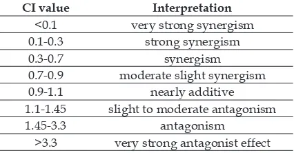
Table 1. Interpretation of Combination Index (CI) values
CI value Interpretation
<0.1 very strong synergism
0.1-0.3 strong synergism
0.3-0.7 synergism
0.7-0.9 moderate slight synergism
0.9-1.1 nearly additive
1.1-1.45 slight to moderate antagonism
1.45-3.3 antagonism
>3.3 very strong antagonist effect
The CI was calculated based on equation
below:
CI = D1/Dx1 + D2/Dx2
D1 : combination concentration of pHmMC D2 : combination concentration of Dox Dx1 : concentration of pHmMC in single
dose that could inhibit the T47D cells growth at the same point with combination concentration
Dx2 : concentration of Dox in single dose that could inhibit the T47D cells growth at the same point with combination concentration
(Reynold and Meurer, 2005)
The concentrations of pHmMC and Dox used in the combination were referred to the concentration of each single compound at its IC50. The IC50 value of pHmMC in the previous study was 48 µM (Arianingrum et al., 2012); while the IC50 value of Dox was measured before the CI determination. To get a linear regression of Dox treatment versus viable T47D cells, concentrations of Dox of 7.81,
15.63, 31.25, 62.5, 125, and 250 nM were used
for the treatment.
Analysis of the Apoptosis
T47D cells were seeded and put on a six tissue culture well-plate at 5 x 10⁵ cells per well then incubated for 24 hours. The cells then were treated with various combination concentrations of pHmMC and Dox with each around its own IC50. After 24 hours incubation, the cells were removed using 0.25% trypsin solution, then
were centrifuged at 2000 rpm for 3 minutes
and washed twice with cold PBS. The cells were re-suspended in 500 µl of Annexin V buffer (Roche), then were treated with Annexin V and propidium iodide (PI) for 10 minutes at room temperature and protected
from light, then were analyzed using low
cytometer. The flow cytometer resulted
144 marked in blue, indicated viable cells; the lower right (R2), marked in green, indicated
early apoptotic cells; the upper right (R3),
marked in orange, indicated late apoptotic cells; and the upper left (R4), marked in yellow, indicated necrotic cells.
Analysis of the Cell Cycle
The washed T47D cells obtained from the combination treatment of pHmMC-Dox plate above were re-suspended in 500 µl of cycletestTM Plus DNA Reagent Kit (BD
Biosciences). The Cellquest program, which
works based on the DNA content, was used to calculate the percentage of the distribution of cells in each stage of the cell cycles (G1, S and G2/M phases).
Results
Cytotoxic Effect of Doxorubicin
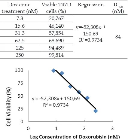
In this study, the IC50 of the pHmMC was got from the previous study of 48 µM (Arianingrum et al., 2012); while the IC50 of the Dox was got from the Dox treatment versus viable T47D cells. The linear regression of Dox-cytotoxic effect resulted (Table 2 and Figure 2) gave the IC50 of Dox of 84 nM. Dox treatments have also caused cells
morphology changing as presented in
Figure 3. Some cells appear became rounded and detached from the bottom lask with a
massive morphology. The rounded shape of cells indicated the mortality of T47D cells.
Figure 3. The T47D cells morphology: (A) with Dox-untreated, (B) treated with Dox at concentration of 7.8 nM, (C) 15.6 nM, (D) 31.3 nM, (E)125 nM, and (F)250 nM respectively. Observation of cell morphology was performed using inverted microscope with a magniication of 100x. The bold arrows indicate normal living cells, whereas
the dashed arrows indicate the cell morphological changes.
Figure 2. The linear regression of the Dox cytotoxic effect on the T47D cells viability.
Table 2. The cytotoxic effect of Doxorubicin on T47D cells
Dox conc. treatment (nM)
Viable T47D cells (%)
Regression IC50
(nM)
7.8 20,767
y=-52,308x +
150,69
R2=0.9734 84
15.6 46,140
31.3 57,854
62.5 68,690
125 94,489
250 99,814
B
E F
D
I.J. Biotech, Vol. 20, No. 2
145 Arianingrum et al.
Figure 4. The cytotoxic effect of pHmMC-Dox combination on T47D cell. Graph (A) showed that the combination of
pHmMC and Dox decreased cell viability compared to the single treatments. The combination treatment altered cell morphology: (B) untreated cells, (C) 18 µM pHmMC, (D) 31.5 nM Dox, and (E) combination of 18 µM pHmMC and
31.5 nM Dox. The bold arrows indicate normal living cells, whereas the dashed arrows indicate the cell morphological changes. The cell morphology was observed using an inverted microscope with 100x magniication.
The changes were more apparent in line with increasing concentrations of Dox.
Co-chemotherapeutic effect invitro of pHmMC-Doxorubicin on T47D cells
a. The cytotoxic effect of the pHmMC-Dox
treatment
Several combinations of pHmMC-Dox were constructed with concentrations which were lower than the IC50 values of the single compounds. IC50 value of pHmMC from the previous study was 48 µM (Arianingrum et al., 2012). The pHmMC concentrations
used in treatment were 12; 18; 24; and 36 µM, while the Dox concentrations were 21;
31,5; 42; and 63 nM. The cytotoxic effect of the combinations of pHmMC and Dox were presented in Figure 4. The data showed that the combinations treatment of pHmMC-Dox more decreased the viability cells compared to that of the single treatments of pHmMC and Dox. Treatment of 18 µM pHmMC on T47D cells caused the cell viability of 60.97%
and treatment of 31.5 nM Dox resulted in
67.08% cell viability. While the combination treatment at the same concentrations of the
(A)
146 single compounds resulted in cell viability
was 29.93%. The higher concentrations of
the single compounds in the combination gave decreasing cell viability. In this study, the lowest cell viability was obtained at combination concentrations of pHmMC and
Dox of 36 µM and 63 nM respectively.
b. The combination index of the pHm
MC-Dox treatment
The co-cytotoxic effects of the combination
treatments were conirmed by the CIs resulted (Table 3). It was indicated that the combination
treatments with concentrations of pHmMC and
Dox above respectively 18 µM and 31.5 nM gave
strong synergistic effect (CI=0.2). Combination treatments with lower concentrations of those showed weaker synergistic effects with CI
values of 0.3, 0.4 and 0.7. The combination treatments also caused some morphological changes of the cells. The changes were more obvious in the combination treatment compared to the single treatments. Such changes included
larger volume of the cells and cell nuclei, longer
cell shape, more transparent and lat, rounded cell shape and cell loating. The study showed that the pHmMC increased the Dox-cytotoxic effect on the T47D cells.
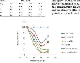
c. The anti-proliferation effect of the
pHmMC-Dox treatments
The Dox concentration used in the study was 21 nM, while the pHmMC concentrations were 18 and 24 µM. The observation results of the cell proliferative effect of the single and combination treatments are presented in Figure 5. The results showed that combination pHmMC-Dox treatments decreased the cell viability compared to that of the single treatments of pHmMC and Dox. The increase concentration of pHmMC or Dox in the combination treatment caused decreasing of the cell viability. All the treatments, both the single and the combination, decreased the cell viability until 48-hour incubation. After 48 hour incubation, the single treatments at lower concentration (18 µM pHmMC and 21 nM Dox 21) did not have antiproliferation effect so that the cells could proliferate and grow. While the single treatment with higher concentration (24 µM pHmMC) and the combination treatments still have an antiproliferative effect which inhibited the growth of the cells until 72-hour incubation.
Figure 5. The inhibiting effects of pHmMC, Dox, and pHmMC-Doc treatments on the T47D cell proliferation.
Table 3. The combination index of the pHmMC-Dox treatments of on T47D cells
pHmMC (µM)
Dox (nM)
21 31.5 42 63
12 0.7 0.4 0.4 0.3
18 0.4 0.2 0.2 0.2
24 0.3 0.2 0.2 0.2
I.J. Biotech, Vol. 20, No. 2
147 Arianingrum et al.
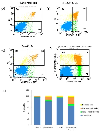
The pHmMC –Dox Combination Effect on Apoptosis
The concentrations of pHmMC and Dox used for the apoptotic effect analysis were half of the IC50 which were respectively 24 µM and 42 nM. The results showed that all the treatments induced apoptosis on T47D cells
at early and late phases as shown in Figure 6. After treatments with the pHmMC, Dox, and pHmMC-Dox, the percentage of late apoptotic cells increased by 12.05%; 2.21%; and 21.71% respectively from that of the control cells (2.05%). The percentage of the early apoptosis
also increased by 3.94%; 2.31%, and 6.87 %
Figure 6. The effect of pHmMC, Dox, and their combination on T47D cells apoptosis.
The lowcytometric proiles of (A), (B), (C), and (D) are the proiles of cell apoptosis after
the treatments with respectively 0 µM (control), 24 µM pHmMC, 42 nM Dox, and the
combination of 24 µM pHmMC-42 nM Dox. The Graph (E) shows the percentage of cell
viability after the treatments. The longer the blue bar indicates the more viable cells; the longer the green and brown bars indicate more induce the cell apoptosis.
–
R1 R2
R3 R4
R1 R2
R3 R4
R1 R2
R3 R4
R1 R2
R3 R4
T47D control cells
(A)
(E)
(D) (C)
(B)
pHmMC 24 M
148
The cytotoxic activity of the pHmMC, either as a single treatment or in combination with Dox, underwent the cell apoptosis and cell cycle arrest.
Studies on chalcones have been reported that apoptotic mechanism of the chalcones’ treatments are occurred through mitochondrial pathway and inhibiting of
NF-κB (Hsu et al., 2006; Shen et al., 2007; and Yadav et al., 2011); through increasing transcription of Bcl-2 that prevents the release of cytochrome c by Bax in mitochondria. The
inhibition of NF-κB also causes decreasing
of Bcl-2 and Bcl-XL expressions, two main anti-apoptotic proteins (Simstein et al., 2003). These mechanisms might be responsible for the apoptotic induction of pHmMC.
The single treatment of pHmMC
induced cell arrest at G2/M phase. This
result is consistent with the previous research (Arianingrum et al, 2012). While the Dox and pHmMC-Dox combination treatments induced
cell arrest at S and G2/M phase. Various
studies have shown that some chalcones inhibited cell cycle (Yadav et al., 2011). Some literature suggests that chalcones’ ability to inhibit the cell cycle by inhibiting cell division cycle25B (Cdc25B) phosphatase (Zhang et al., 2014 and Yuan et al., 2014). Cdc25B is a mitotic regulator that acts as a ‘starter phosphatase’ to initiate the positive feedback loop at the entry into M phase. The activity of Cdc25B appears during late S phase and peaks during G2 phase. Both in-vitro and in vivo cdc25B is activated through phosphorylation during S-phase. Inhibition of phosphatase results in inhibition
of cell cycle in G2/S-phase and mitotic arrest
occurs (Lammer et al., 1998; Nilsson et al., 2000, and Lindgvist et al., 2005). While some study about Dox mention that Dox damaged double stranded DNA by intercalation on DNA base pairs and inhibition topoisomerase IIa. DNA damage will activate kinase protein (ATM), and this will further activate Chk2. Activation of Chk2 will cause Cdc25 inactive which in turn inhibit as cdc2. Both proteins are needed in G and M phase of cell cycle. Inactivation of
these proteins will lead to G2/M arrest in the
respectively from the control cells (2.09%). Compared with the single treatments, the combination treatments more induced the cell apoptosis.
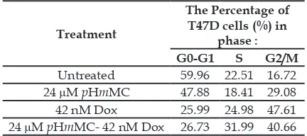
pHmMC –Dox Combination Effect on cell cycle progression
The concentrations of the pHmMC and
Dox used for analyzing the combination
treatment effect on the T47D cell cycle were the same as the doses for the apoptosis study (24 µM pHmMC, 42 nM Dox, and their combination). Table 4 shows the distribution cells into cell cycle phases of Go-G1, S, and
G2/M after the treatments. It shows that
the single treatments of pHmMC and Dox inhibited the cell cycle and caused the cells
arrest at G2/M, which were higher than
that of the cell control. The pHmMC-Dox combination caused cells accumulation in S
and G2/M phase.
Table 4. The inhibiting effects of pHmMC, Dox, and
pHmMC-Doc treatments on the T47D cell cycle
Treatment
The Percentage of T47D cells (%) in
phase :
G0-G1 S G2/M
Untreated 59.96 22.51 16.72
24 µM pHmMC 47.88 18.41 29.08
42 nM Dox 25.99 24.98 47.61
24 µM pHmMC- 42 nM Dox 26.73 31.99 40.66
Discussion
From the study results, it was concluded that the pHmMC increased the Dox eficacy in vitro on T47D breast cancer cells. The CI study showed that the combination treatment of pHmMC-Dox had a synergistic effect up to strong synergy; especially at the low concentrations (below IC50). The combination treatments also gave better anti-proliferative effect than that of the single ones. Those
above facts conirmed that combining Dox
with pHmMC could reduce the dosage of the
Dox. This will give the beneit of minimizing
I.J. Biotech, Vol. 20, No. 2
149 Arianingrum et al.
cell cycle (Drummond, 2007). Combination treatment of pHmMC and Dox caused cell cycle
arrest in S and G2/M. Combination treatment
also decreased the cell population. Based on the research, pHmMC could improve Dox cytotoxic effect by increasing the inhibition of cell cycle arrest from G2-M to S phase.
Conclusion
The combination treatments of the pHmMC-Dox resulted in a synergistic up to strong synergistic effect, an increase in apoptosis, and induction of the cell cycle
arrest in S and G2/M phase of T47D cancer
cells. The combination treatment of pHm MC-Dox is potential to be developed as a breast cancer therapy.
Acknowledgement
The author thanks for Prof. Dr. Indyah Sulistyo Arty, M.S for pHmMC sample. This study was supported by “Hibah Desertasi 2014” from The Directorate General of Higher Education Ministry of Education, Republik Indonesia.
References
Anonymous. 2012. Cancer facts and igures
2012. American Cancer Society Page 9. Arianingrum, R., Arty, I.S. and Atun, S. 2010.
Uji sitotoksisitas senyawa mono para hidroksi lalkon terhadap Cancer cell lines T47D.Saintek 16(2): 121 -132.
Arianingrum, R., Sunarminingsih, R., Meiyanto, E. and Mubarika, S. 2012. Potential of a chalcone derivate compound as cancer chemoprevention in breast
cancer IPCBEE 38: 41-45.
Arianingrum, R., Sunarminingsih, R., Meiyanto, E. and Mubarika, S. 2015. Cytotoxic effect of para hydroxyl chalcone (pHmMC) on MCF-7 breast cancer cells by inducing cell arrest and apoptosis. 2ndICRIEMS proceedings C-11:89-95.
Arty, I.S. 2010. Synthesize and cytotoxic
test of several compounds of mono para hydroxy Chalcones. Indo J. Chem. 10 (1): 110-115.
Boumendjel, A., Ronot, X. and Boutonnat. 2009. Chalcone derivatives acting as cell cycle blockers : potential anticancer drugs? J Curr Drug Targets. 10(4): 363-71.
Doyle, A. and Grifith, J.B. 2000. Cell and
tissue culture for medical research. New York: John Willey and Sons Ltd.
Drummond, C. 2007. The mechanismof anti-tumour activity of DNA binding agent SN 280409. Thesis. New Zealand-University of Auckland.
Ducki, S. 2007. The development of chalcones as promising anticancer agents. Drugs 10: 42–46.
Ducki, S. 2009. Antimitotic chalcones and related compounds as inhibitors of tubulin assembly. Anticancer Agents Med. Chem. 9: 336–347.
Ferreira, A.L.A., Matsubara, L.S. and Matsubara, B.B. 2008. Anthracycline-induced cardiotoxicity. Cardiovasc Hematol Agents Med Chem. 6: 278-281. Guttridge, D.C., Albanese, C., Reuther,
J.Y., Pestell, R.G. and Baldwin Jr., A.S.
1999. NF-κB controls cell growth and
differentiation throught transcriptional regulation of cyclin D1. Mol. Cell. Biol. 19: 5785 - 5799.
Hinz, M., Krappmann, D., Eichten, A.,
Heder, A., Scheidereit, C. and Strauss, M.
1999. NF-κB function in growth control:
regulation of cyclin D1 expression and G0/G1-to-S-phase transition. Mol. Cell. Biol. 19: 2690 - 2698.
Hsu, Y.L., Kuo, P.L., Tzeng, W.W. and
Lin, C.C. 2006. Chalcone inhibits the proliferation of human breast cancer cell by blocking cell cycle progression and inducing apoptosis. Food Chem Toxicol
44(5): 704-13
King, R. J. B. 2000. Cancer biology, Pearson Education, Second Edition, England.
p.1-7,228-231, 263-264.
Lammer, C., Wagerer, S., Saffrich, R., Mertens, D., Ansorge, W. and Hoffmann, I. 1998. The cdc25B phosphatase is essential for
the G2/M phase transition in human
150
Lindqvist, A., Kallstrom, H., Lundgren,
A., Barsoum, E. and Rosenthal, C.K. 2005. Cdc25B cooperates with Cdc25A
to induce mitosis but has a unique
role in activating cyclin B1-Cdk1 at the centrosome, J. Cell. Biol. 171(1): 35-45.
Mojzis, J., Varinska, L., Mojzisova, G.,
Kostova, I. and Mirossay, L. 2008.
Antiangiogenic effects of lavonoids and
chalcones. Pharmacol. Res. 57: 259–265.
Mosmann, T. 1983. Rapid colorimetric
assay for cellular growth and survival: a p p l i c a t i o n t o p r o l i f e r a t i o n a n d cytotoxicity assays. J. Immunol Methods
65: 55-63
Nilsson, I. and Hoffmann, I. 2000. Cell cycle regulation by the Cdc25 phosphatase family, Prog. Cell. Cycle Res. 4: 107-114. Pahl, H.L. 1999. Activators and target genes
of Rel/NF-κB transcription factors,
Oncogene 18: 6853–6866.
Reddy, M.V.R., Pallela, V.R., Cosenza, S.C.,
Mallireddigari, M.R., Patti, R., Bonagura, M., Truongcao, M., Akula, B., Jatiani, S.S., and Reddy, E.P. 2010. Design, synthesis
and evaluation of (E)-alpha-benzylthio
chalcones as novel inhibitors of BCR-ABL kinase. Bioorg. Med. Chem. 18: 2317–2326. Reynolds, C.P. and Maurer, B.J. 2005.
Evaluating response to antineoplastic drug combinations in tissue culture models. Methods Mol. Med. 110: 173-83. Robinson, M.W., Overmeyer, J.H., Young,
A.M., Erhardt, P.W. and Maltese, W.A. 2012. Synthesis and evaluation of indole-based chalcones as inducers of methuosis, a novel type of nonapoptotic cell death. J. Med. Chem. 55: 1940–1956.
Sasayama, T., Tanaka, K., Mizukawa, K.,
Kawamura, A., Kondoh, T., Hosoda, K. and Kohmura, E. 2007. Trans-4-lodo,4’-boranyl-chalcone induces antitumor activity against malignant glioma cell lines in vitro and in vivo. J. Neurooncol.
85: 123–132.
Sharma, G., Tyagi, A.K., Singh, R.P, Chan, D.C and Agarwal, R. 2004. Synergistic anti-cancer effects of grape seed extract and
conventional cytotoxic agent doxorubicin against human breast carcinoma cells. Breast Cancer Res Treat. 85: 1-12.
Shen, K.H, Chang, J.K., Hsu, Y.L and Kuo, P.L. 2007. Chalcone arrests cell cycle progression and induces apoptosis through induction mitochondrial pathway and inhibition of nuclear factor kappa B signaling in human bladder cancer cells. Basic Clin Pharmacol Toxicol. 101: 254-61.
Simstein, R., Burow, M., Parker, A., Weldon,
C. and Beckman, B. 2013. Apoptosis,
chemoresistance, and breast cancer: Insights from the MCF-7 cell model system. Exp Biol Med. 101:995-1003. Tjindarbumi, D. and Mangunkusumo, R.
2002. Cancer in Indonesia, present and future. Jpn. J. Clin. Oncol. 32 (Supplement 1): S17-21.
Tyagi, A.K., Agarwal, C., Chan, D.C.F. and Agarwal, R. 2004. Synergistic anti-cancer effects of silibinin with conventional cytotoxic agents Doxorubicin, Cisplatin and Carboplatin against human breast carcinoma MCF-7 and MDA-MB468 cells.Oncology Reports. 11: 493-499. Valdameri, G., Gauthier, C., Terreux, R.,
Kachadourian, R., Day, B.J., Winnischofer, S.M.B., Rocha, M.E.M., Frachet, V., Ronot, X., Di Pietro, A. and Boumendjel, A. 2012. Investigation of chalcones as selective inhibitors of the breast cancer resistance protein: critical role of methoxylation in both inhibition potency and cytotoxicity. J. Med. Chem. 55: 3193–3200.
Wong, H.L., Bendayan, R., Rauth, A.M., Xue, H.Y., Babakhanian, K. and Wu, X.Y. 2006. A mechanistic study of enhanced Doxorubicin uptake and retention in multidrug resistant breast cancer cells using a polymer-lipid hybrid nanoparticle system. The Journal of Pharmacology and Experimental Therapeutics 317 (3):
1372-1381.
NF-I.J. Biotech, Vol. 20, No. 2
151 Arianingrum et al.
kB-mediated inlammation and cancer.
International Immunopharmacology 11(3):
295-309.
Yuan, X., Li, T., Xiao, E., Zhao, H., Li, Y., Fu, S., Gand, L., Wang, Zheng, Q. and Wang, Z. 2014. Licochalcone B inhibits growth of bladder cancer cells by arresting cell cycle progression and inducing apoptosis. Food Chem. Toxicol. 65: 242-251.