Bioactivity of Proplis to the CD4 + and CD8+ T cells Producing IFN-γ Cytokines in BALB / C Mice
Yonna Ayundria 1), Muhaimin Rifa’I 2)
1,2 Jurusan Biologi, Fakultas Matematika dan Ilmu Pengetahuan Alam, Universitas Brawijaya
1
[email protected]2[email protected]
ABSTRACT
Propolis (bee glue) is a natural resinous product of honey bees which collected from exudates and plant buds, rich in biochemicals constituents including mostly flavonoids, phenols and various acids bond. These compound are believed to be responsible as immunomodulatory agents. The study aims to determine the immunomodulatory activity of ethanolic extract of propolis to the CD4+ and CD8+ T cells producing IFN-γ
cytokines and analyze the differences immune responses between control and treatment group by in vivo. Stages include animal acclimation for ± 1 week, preparation of Ethanolic Extracts of Propolis / EEP, Oral Administration with doses levels of 0 mg / kg BW; 50 mg / kg BW (DI); 100 mg / kg BW( DII); 200 mg / kg BW (DIII) for 2 weeks, isolation of lymphocyte cells from spleen, flowcytometry analysis to asses cell number and surface molecule expression. Data was analyzed using Kruskal Wallis Test with α = 0,05 and followed by Mann Whitney Test by SPSS 16.0 for windows with complete randomized design. The results showed that a dose of 50 mg / kg BW was increases the relative number of CD8+ T cells producing IFN-γ cytokines significantly (p <0.05) compared with controls. However, at the same dose the relative number of CD4+ T cells producing IFN-γ cytokines was decreased significantly (p<0,05). Based on this case, its dose supposedly that the ethanolic extract of propolis play role in maintaining the balance or homeostatic of IFN-γ cytokines production by T cell subsets. Dose of 100 mg/kg BW and 200 mg / kg BW could decrease the relative number of activated CD4+ T cells producing IFN-γ cytokine significantly compared to controls.
Keywords : CD4+ T cells, CD8+ T cells, ethanolic extract of propolis, IFN-γ cytokines, in vivo
INTRODUCTION
Indonesia has great Natural Resources. But, minimum research data for Indonesian natural resources is available yet. The use of herbal plants as health promoters are gaining increasing attention in both consumer and scientific because it has no side effects than drugs made from synthetic. One of Indonesia's natural resources promising as new source of herbal medicine is propolis.
Propolis is a natural resinous product of hooney bees which collected from exudates and plant buds, processed with enzymes secreted by bees and mixed with wax in the hive. Propolis
contains a variety of complex chemical
compounds, which composition varies depending on the plant source [1]. In general, composition of propolis in nature consisting of 30% wax, 50% resins and balsams, 10% essential and aromatic oils, 5% pollen and other substances [2]. The main compounds of propolis is a resin consisting of flavonoids, phenols and various acids bond [3]. Complex chemical compounds of propolis made
them have some benefits to health such as immunomodulatory agents [4].
Immunomodulator through natural or
synthetic substance that can modulate the function and activity of the immune system, enabling it to maintain balance (homeostasis) immune system [5]. Chemical compounds of propolis supposed as immunomodulatory agents are flavonoids and caffeic acid phenethyl ester (CAPE) [6].
Based on the previous research conducted
by Park et al. [7], oral administration of CAPE in
propolis at doses of 20 mg / kg BW could increase
the production of IL-2, IL-4 and IFN-γ cytokines
activation of Th1 cells and Tc [9]. IFN-γ cytokines
is a pro-inflammatory cytokine produced by
Jurnal Biotropika | Vol. 2 No. 2 | 2014 126
T cells and a small proportion of CD8+ T cells that
play role especially in the non-specific and specific immune system [9].
The last decade, interesting studies about immunomodulatory activity of propolis were performed, but no scientific data for the Indonesian product is available yet. Therefore, purpose of this study was to determine immunomodulator activity
of propolis to the CD4+and CD8+ T cells producing
IFN-γ cytokines and analyze the differences
immune responses between control and treatment groups by in vivo.
METHODS
Time and Research Place
The research was conducted on September 2013 to March 2014 in the Laboratory of Animal Physiology, Department of Biology, Faculty of
Science; Laboratory Biomedical and
Pharmacology, Faculty of Medicine, Brawijaya University, Malang.
Equipment and Materials
The equipment used are mice cages, husk, spatula, oral administration tool, erlenmeyer glass, surgical scissors & board, petri dish, syringe, wire, micropipette, propylene tubes, eppendorf tubes, yellow and blue tip, centrifugation, flowcytometry
cuvets and FACS CaliburTM flowcytometry, while
the materials used are BR 1 pellet, mineral water,
ethanolic extract of propolis, Na2CO3, distilled
water, alcohol 70%, Pbs sterile, cytofix, wasperm
and monoclonal antibody (rat anti-mouse IFN-γ).
Animal Studies and Experimental Design
Male BALB / c mice (Mus musculus), 8
weeks old, approximately 38 gram weight, and healthy condition. The animals were kept in groups of six per cage. Groups consist control and treatment groups (dose of EEP : 50 mg/kg BW, 100 mg/kg BW and 200 mg/kg BW). The animal were maintained on BR 1 pellet diet and mineral water ad libitum. The animal were acclimation 1 weeks before used in experiment.
Extraction of Propolis
The propolis sample was collected from
hives of the Trigona sp. bees of Lawang city, East
Java. The propolis has characteristics sticky, solid and black colour.
The propolis (200 g), added to 1 L of ethanol absolute and moderately shaken. The extract was filtered then solvent was evaporated at evaporator for ± 1.5-2 hours. The extract approximately 1:10 of the dry natural materials. The extract was filtered and evaporated in a heated dish in the oven. Crude extract of propolis, placed in vials and stored in a refrigerator with temperature of 4 ˚ C.
Oral Administration of Treatment Group with Ethanolic Extracts of Propolis / EEP
The need of extract each dose depending on the average weight of each groups. Extract dilution with distilled water (1:10). In the process of
dissolution was added with Na2CO3 to make it
easy to dissolved. EEP was orally administered to mice at dose levels of 50 mg/ kg BW, 100 mg/ kg BW and 200 mg/ kg BW. Oral administration carried out for 2 weeks.
Isolation of Lymphocyte Cells
Mice were killed by neck dislocation. Mice were dissected using surgical scissor on a surgical board in the dorsal part, then spleen was taken and washed with Pbs sterile twice.
Spleen was placed in a petri dish containing PBS sterile ± 2 ml, crushed with a syringe base, clockwise until organ was destroyed. The homogenate was filtered with wire and put into 15 ml propylene tube. Pbs was added to the tube until 10 ml. After that, suspension was centrifuged at 2500 rpm, 4 ° C, for 5 min. Supernatant was discarded and pellet was taken. Pellet was resuspended with 1 ml of PBS sterile. It was taken
70µl into eppendorf tube containing 500 mL PBS
Antibody Staining
Intracellular staining proc perforation of the cell membr
antibody staining. Pellet
centrifugation, resuspended wit incubation for 20-minutes in resuspended with 1ml washperm 2500 rpm, 4 ° C, for 5 min. The discarded & pellet ready to label specific antibody, pipeting, incub
and added 300µl Pbs, then
flowcytometry cuvet.
Flowcytometry Analysis
Suspension was prep
flowcytometry cuvet ready for a to the parameters that have flowcitometry tool. Flowcytome research is nozzleBD Biosciences
cytokines. Research using compl design. Data were analyzed statistics (Kruskal-Wallis test followed by Mann Whitney te using SPSS 16.0 for Windows.
RESULTS AND DISCU
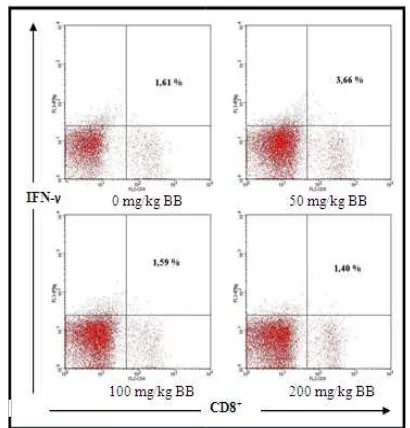
The relative number of
producing IFN-γ cytokines
Extract of Propolis with d BW gave significant different r
the activation of CD8+ T cells
cytokine (Fig 1). It have been sh
number of CD8+ T cells producin
by 3.66% compared to 1.61% fo 2). Meanwhile, doses of 100 mg mg / kg BW have no significa control (p>0,05). But, both significance different. Doses of and 200 mg/kg BW could dec
lysis using BD Cell
/ relative number of icant different with h of doses have of 100 mg/kg BW ecrease the relative
number of CD8+ T cells pr
by 1.59% and 1.40% (Fig 2
Figure 1. Differences the Tcells producing IFN-γ cytok
Figure 2. Profile the relative producing IFN-γ cytokines be
The increase in CD
IFN-γ cytokines at a dos
supposedly induced by propolis. This is suppo
conducted by Park et al. [
able to increase the produc
IFN-γ cytokines is a pro
produced by various cells
such as CD4+ T cells and
CD8+ T cells that play rol
specific and specific immu specific immunity, this
ab okines between groups.
ive number of CD8+ T cells between groups.
CD8+ T cells producing
ose of 50 mg / kg BW, y CAPE compounds in ported previous research . [7], CAPE in propolis is
uction of IFN-γ. cytokines.
ro-inflammatory cytokine lls of the immune system and a small proportion of role, especially in the
non-Jurnal Biotropika | Vol. 2 No. 2 | 20 cytokine of MAC (Macroph Cytokine), whereas in the specifi role in increasing activation of C cells [9]. Macrophages have fu specific defense that capture and antigens through phagocytosis ac it to T cells [10].
The decrease in the relative
T cells producing IFN-γ cyto
caused by multicompound prop which can act antagonists.
Active compounds in p immunostimulatory that enhan system or suppress the immune imunosupressor [5]. Simplicia in than one active compounds or m
multicompound, exist to
(reinforcing mutually) or a Propolis suppresses synthesis mediated by propolis compound and flavonoid groups quercetin hesperidin (flavonones) [12]. molecular mechanism of pro cytokine production by T c Extracts of Propolis with d BW, 100 mg / kg BW, and 200 m decrease the relative number o
producing IFN-γ cytokine con
These three different doses difference (p<0,05) by 2.20%; 1 compared to 3.24% for the contr However, there were no sig between treatment groups (Fig 3)
No. 2 | 2014
tive number of CD8+
tokines supposedly operties of propolis
plants can be as ance the immune une system, namely in plants have more r multicompound. In cells producing IFN-γ cytokin
Berdasarkan grafik
Figure 4. Profile the relative producing IFN-γ cytokines be
CD4+ T cells that w
differentiated into Th1 cytokines [13]. As descr cytokines production by C the specific immunity by
CD4+ it self/ autocrine ac
paracrine action [14,9]. Bu
cells producing IFN-γ
different trend with the n
producing IFN-γ cytokine,
mg/kg BW. Extract of p kines between groups.
fik perbedaan berat badan
ive number of CD4+ T cells between groups.
t were activated would be
1 that producing IFN--γ
scribed previously, IFN-γ
CD4+ T cells play role in
significantly. In this case, dose of 50 mg/kg BW supposedly play role to maintain homeostasis or
balance of IFN-γ cytokines in the body.
The existence of IFN-γ cytokines numbers
need to be controlled in order to maintain homeostasis or balance immunocompetent cells of the body. This is because cytokines are molecule mediators that plays a critical role in regulating lymphocyte cells, so the cells are maintained in number to keep it balanced. The unbalanced of the immune system components will lead to arising
cytokines significantly compared to controls.
However, the relative number of CD4+ T cells
producing cytokines IFN-γ were decrease
significantly. Based on this, the ethanol extract of propolis dose of 50 mg/kg BW supposedly play
role to maintain homeostasis or balance of IFN-γ
cytokines in the body. Dose of 100 mg/kg BW and 200 mg / kg BW could decrease the relative
number of activated CD4+ T cells producing IFN-γ
cytokine significantly compared to controls.
ACKNOWLEDGEMENTS
The author would like to thanks to Mr Muhaimin Rifa'i, S.Si., PhD., Med.Sc as supervisor, Mr. Dr.Ir.Moch.Sasmito Djati., MS and Drs. Aris Soewondo., MS as the reviewers, Dewi Satwika, S.Si., M. Si, Bambang, S.Si, Ririn, S.Si., M.Si and Animal Laboratory Physiology Team for the support in this research.
REFERENCES
[1] Bankova, V., Castro, S.L & Marcucci, M.C. 2000. Propolis:recent advances in chemistry
and plant origin. Apidologie.3:3–15.
[2] Burdock, G.A. 1998. Review of the
biological properties and toxicity of bee
propolis (propolis). Food and Chemical
Toxicology.36:347–363.
[3] Siregar, H.C.N., Asnanth M.F & Yuke, O.
2011. Propolis Madu Multikhasiat.
Penebar Swadaya. Jakarta.
[4] Girgin, G., Baydar, T., Ledochowski, M., Schennach, H., Bolukbasi, D.N., Sorkun, K., Salih, B., Sahin, G & Fuchs, D. 2009.
Immunomodulatory Effect of Turkish
Propolis :Changes in neopterin Release and
Tryptophan Degradation. Immunobiology.
214 (2):129-34.
[5] Nazir, N. 2013. Imunomodulatory Activity
of Isoflavones Isolated from Iris
Kashmiriana: Effect on T-Lymphocyte Proliferation and Cytokine Production in
Balb/C Mice. Journal of Biomedicine &
Preventive Nutrition. 3:151–157.
[6] Challem, J. 2004. Tuberculosis, Medical & H.Y. Oh. 2004. Immunomodulatory effect of caffeic acid phenethyl ester in Balb/c
mice. Intl.Immunopharmacol. 4: 429-436.
[8] Fatahinia, M., Khosravi, A.R & Shokri, H.
2012. Propolis efficacy on TNF-α, IFN-γ and
IL-2 cytokine production in old mice with
and without systemic candidiasis. Journal de
Mycologie Medicale. 22,237-242.
[9] Wahab, Shadma & Hussain, Arshad. 2013.
Cytokines As Targets For
Immunomodulation. International Journal of
Pharmacy and Pharmaceutical Sciences. Vol 5, Suppl 3.
[10] Baratawidjaja, K.G dan Rengganis, Iris. 2010. Imunologi Dasar Edisi Ke-9. Balai Penerbit, Fakultas Kedokteran Universitas Indonesia.
[11] Robinson, T. 1995. Kandungan Organik
Tumbuhan Tinggi. ITB. Bandung.
[12] Ansorge, S., Reinhold, D., & Lendeckel U. 2003. Propolis and some of its constituents
down-regulate DNA synthesis and
inflammatory cytokine production but induce TGF-beta production of human immune
cells. Zeitschrift fur Naturforschung.
58c:580—9.
[13] Rifa’i, M., Z. Shi, S.Y. Zhang, Y.H. Lee, H. Shiku, K. Isobe, and H. Suzuki. 2008. CD8+CD12+ regulatory T cells recognize activated T cells via conventional
MHC class I–αβTCR interaction and
become IL-10-producing active regulatory
cells. International immunology. 20.
Jurnal Biotropika | Vol. 2 No. 2 | 2014 130
[14] Cruse, J.M dan Lewis, R.E. 2010. Atlas of
Immunology, Second Edition. CRC Press. United States.
[15] Santamaria, Pere. 2002. Cytokines and
Chemokines in Autoimmune Disease.
Advances in Experimental Medicine and Biology. Vol 520.
[16] Nelson, Brad.H. 2004. IL-2, Regulatory T
Cells and Tolerance. J Immunol
.172:3983-3988.
[17] Rifa’i,M., Y Kawamoto, I Nakashima, H
Suzuki . 2004. Essential roles of