ISSN : 2356-3303
Research and Development on
Nanotechnology in Indonesia
Vol.2, No.1, 2015
Selected papers from
Symposium Nanotechnology & Biotechnology ITB
November 2014
Editors :
H. K. Dipojono, B. S. Purwasasmita, H. Kasai
Center for Advanced Sciences
KATA PENGANTAR
Assalamu’alaikum wr. wb
Perkembangan sains dan teknologi dalam skala berdimensi nano, membawa dampak perubahan yang besar dalam industri dan kehidupan sehari-hari, kemajuan teknologi material ini tentunya harus dapat di tangkap dan di implementasikan di Indonesia agar dapat bersaing dengan produk dari luar negeri. Kekayaan alam dan kekayaan hayati Indonesia yang banyak ini haruslah dapat dimanfaatkan untuk kepentingan bangsa Indonesia seutuhnya. Keberadaan lembaga penelitian menjadi salah satu upaya peningkatan kualitas produk material berbasis sumberdaya Indonesia ini,
Kebutuhan akan ajang tukar informasi dan pertemuan karya ilmiah para peneliti di Indonesia dalam bidang nanosains dan nanoteknologi, perlu diwadahi melalui publikasi ilmiah yang dapat menjadi cermin kemajuan pencapaian karya anak bangsa dalam bidangnya. Untuk itu Research and Development on Nanotechnology in Indonesia
(RDNI) diterbitkan, yang direncanakan akan terbit rutin tiap tahun. Sesuai dengan agenda kegiatan workshop, seminar atau symposium dalam bidang nanosains dan nanoteknologi.
Institut Teknologi Bandung, sebagai salah satu penghela bidang sains dan teknologi di Indonesia sudah seyogyanya memainkan peranan penting dalam teknologi ini. Sesuai dengan rencana akademik ITB 2010 – 2015 serta bantuan dana JICA untuk pengembangan ITB, yang mana salah satu tujuannya adalah pengembangan nanoteknologi dalam bentuk kegiatan riset dan akademik. Penelitian dalam bidang
Nano science dan nanoteknologi di ITB telah dimulai sejak sekitar tahun 2000, dan di lakukan secara terpisah-pisah di beberapa fakultas, misalnya FMIPA, FTI, SF dan SITH. Keterbatasan anggaran dan peralatan mengakibatkan sulitnya pertumbuhan riset dalam bidang ini, sedangkan upaya untuk mengintegrasikan riset dalam bentuk kerjasama riset antar fakultas perlu difasilitasi.
Symposium Nanotechnology dan Biotechnology yang telah diselenggarakan di tahun 2014 ini adalah kegiatan pengembangan pusat riset bidang nanoteknologi yang terangkum dalam agenda kegiatan pengembangan Center for Advances Sciences (CAS)
pada Proyek Pengembangan ITB (III) dengan bantuan dana dari JICA. Makalah-makalah terpilih dari aktivitas workshop tersebut menjadi bahan utama dalam RDNI volume 2.
Besar harapan kami melalui RDNI ini, kerjasama riset dalam bidang nanosains dan nanoteknologi akan lebih memacu pertumbuhan riset antar instansi sehingga pemanfaatan hasil-hasil riset dapat lebih cepat terasa untuk masyarakat.
EDITORS
Prof. Ir. Hermawan Kresno Dipojono, MSEE. Ph.D. (ITB)
Prof. Dr. Bambang Sunendar Purwasasmita, M.Eng. (ITB)
Research and Development on
Nanotechnology in Indonesia
Vol .2, No.1, 2015
Hak cipta © CAS 2015
Hak publikasi pada penerbit CAS
Dilarang memperbanyak, menyalin, merekam sebagian atau seluruh
bagian buku ini dalam bahasa atau bentuk apapun tanpa izin tertulis dari
penerbit.
DAFTAR ISI
Daftar Isi i
1. Synthesis, Characterization and Functionalization of Bio-based Nano Filler
Made from Ash Resulted from Wood Waste Burning to Reduce Formaldehyde Emission of Bio-Composite Products
Sutrisno, Bambang Sunendar Purwasasmita, Eka Mulya Alamsyah, dan Tati Suryati
Syamsudin
1
2. Natural Product Utilization As Antioxidant Formulated in Nanoemulsion
Rachmat Mauludin, Irda Fidrianny, Dita Sasri Primaviri, Okti Alifiana, Rino R. Mukti
20
3. Study on Electronic and Magnetic Properties of xMnO3 by DFT+U
Approximation
Nurul Ikhsan, Suprijadi, Ely Aprilia
28
4. Self- Nanoemulsion Containing Combination of Curcumin and Silymarin :
Formulation and Characterization
Utami Wulandari Rachmadi, Dahlia Permatasari, Annisa Rahma, Heni Rachmawati
37
5. Effect of Deviation in Image Quantification for Internal Dosimetry
Assessment in Radionuclide Therapy
Nur Rahmah Hidayati, Deni Hardiansyah, Gerhard Glatting
49
Informasi Symposium Nanotechnology & Biotechnology ITB 2014
ii
ii | CAS – Center for Advanced Sciences
Symposium Nanotechnology & Biotechnolog
ITB 2014
Date : 12 -14 November 2014
Venue : Institut Teknologi Bandung, Indonesia
Auditorium IPTEKS, East Campus Center
Organized by :
Center for Advanced Sciences (CAS)
http://nrcn.itb.ac.id/
Institut Teknologi Bandung (ITB)
Jln. Ganesa No.10 Bandung
STEERING COMMITTEE
Dr. Aditianto Ramelan Dr. Sony Suhandono Dr. Suprijadi
ORGANIZING COMMITTEE
Dr. Triati Dewi Kencana Wungu (Chairman) Dr. Heni RachmawatiDr. Ferry Iskandar Dr. Veinardi Suendo Dr. Brian Yuliarto
Dr. Mohammad Kemal Agusta
Symposium & Workshop
Nanotechnology & Biotechnology
Computational Material Design
(CMD)& Bio-Informatika
12 – 14 Nopember 2014
Rabu, 12 Nopember 2014
Auditorium
Campus Center
Timur
08.00 – 08.30 Registrasi Peserta
08.30 – 09.00 Pembukaan Wakil Rektor Bidang Riset dan Inovasi (WRRI) ITB Sambutan Kepala UIP Pengembangan ITB (III) - JICA
Moderator : Dr. Freddy Haryanto
09.00 – 09.30 Keynote speaker: Prof. Dr. rer. nat. Gerhard Glatting -
Heidelberg University, German
Molecular Radiotherapy using physiologically based
pharmacokinetic (PBPK) modeling
09.30 – 09.45 Photo Session & Coffee Break
09.45 – 10.15 Keynote speaker: Prof. Dr. Bambang Sunendar Purwasasmita, M.Eng - ITB
“Membangun Kerangka Riset Nano Kolaboratif Berbasis Agenda Riset Nasional”
10.15 – 10.45 Invited Speaker: Leenawaty Limantara, Ph.D– Ma Chung University
“Chlorophylls And Carotenoids In Foods, Healths, and Future Energy Sectors: an Outlook For Nanotechnology Applications”
10.45 – 11.00 Dr. Veinardi Suendo, S.Si., M.Eng
“Fabrikasi DSSC (Dye Sensitized Solar Cell) Berbasis Elektrolit Padat Polianilina”
11.00 – 11.15 Dr.rer.nat. Freddy Haryanto
”Kajian In-vitro eksperimen dan simulasi Monte Carlo pada sel dalam Karakterisasi Gadolinium sebagai agen kontras”
11.15 – 11.30 Nurul Ikhsan
iv | CAS – Center for Advanced Sciences
11. 30 – 11.45 Nur Rahmah Hidayati, M.Sc
“Effect of Deviation in Image Quantification for Internal Dosimetry Assessment in Radionuclide Therapy”
11.45 – 12.00 Raafqi Ranasasmita, M.Biomed
“Halal Production of Mycelle-Encapsulated Calcitrol as
Nanotechnology-Based Product”
12.00 – 13.00 ISHOMA
13.00 – 13.30 Keynote speaker: Dr. Ir. Usman Pasarai, M.Eng - LEMIGAS
“Peptida untuk Surfactant EOR”
13.30- 14.00 Keynote speaker: Prof. Yoshitada Morikawa - Osaka University
14.00 – 14.15 Utami Wulandari Rachmadi, S. Farm
“Self- Nanoemulsion Containing Combination of Curcumin and Silymarin : Formulation and Characterization”
14.15 – 14.30 Dr. Heni Rachmawati
“Pengembangan Formula dan Karakterisasi In Vitro Tablet Multipartikulat Lepas Lambat di Kolon Mengandung Fraksi Bioaktif dari Cacing Tanah (Lumbricusrubellus)”
14.30 – 14.45 Dr. Rachmat Mauludin, MSi, Apt
“Pemanfaatan bahan alam sebagai antioksidan yang diformulasikan dalam bentuk nanoemulsi”
14.45 – 15.15 Coffee Break
15.15 – 15.30 Brian Yuliarto, Ph.D
“Pengembangan Material Komposit Multiwalled Carbon Nanotubes dan Zinc Oxide Berstruktur Nano untuk Aplikasi Sensor Gas Methane”
15.30 – 15.45 Ir. Sutrisno, M.Si
“Sintesis, Karakterisasi dan Fungsionalisasi Bio-Nano Filler dari Abu Pembakaran Limbah Kayu pada Boiler Industri Kayu Lapis untuk Menurunkan Emisi Formaldehida Produk Bio-Komposit” 15.45 – 16.00 Dr. rer. nat. Rino Rakhmata Mukti
“Synthesis of Zeolite Catalyst at Low Temperature in a Non-Hydrothermal Method”
16.00 – 16.15 Nina Siti Aminah, M.Si
“Fabrikasi Nanopatterned Sensing Layer Berbasis Polimer Hibrid Untuk Pengawasan Pencemaran Lingkungan”
“Studi Efek Nanopartikel Kitosan Terhadap Proses Pematangan Buah untuk Aplikasi Pasca Panen Buah Pisang Raja Bulu”
16.30 – 16.45 Sony Suhandono, Ph.D
“Karakterisasi Kandidat Nano-Vaksin VLP (Virus Like Particle) HPV-16 Rekombinan”
16.45 – 17.00 Oktira Roka Aji
“A New Whole Cell Biocatalyst To Degrade Poly-Ethylene Terephthalate (Pet)”
17.00 – 17.15 Rahmat Azhari Kemal
vi | CAS – Center for Advanced Sciences
Kamis, 13 Nopember 2014
Auditorium
Campus Center
Timur
08.00 – 08.30 Registrasi
08.30 – 08.45 Annistia Rahmadian Ulfah
“Virus Like Particles (VPLs) Production in Plant Transient System”
08.45 – 09.00 Sparisoma Viridi
Simulasi Deposisi Berdasrkan Penumbuhan Butiran Menggunakan Dinamika Molekular
09.00 – 09.15 Agil Nawa Irawan Putro, ST
“Application of Fe3+-Doped Tio2/Sno2nanoparticles for
Bacterial Disinfectant and as Inhibitor of The Fruit Ripeningrate”
09.15 – 09.30 Adi Surya Pradipta
“Influence of Starch and Chitosan on Silica Nanorod Formation for Hydrophobic Textiles Application”
09.30 – 10.00 Dr. Mohammad Kemal Agusta – FTI ITB
10.00 -10.15 Photo session – Coffee break
10.15 – 10.45 Prof. Hiroshi Nakanishi –Osaka University 10.45 – 11.15 Dr. Florian Stieler
“Modern Radiation Treatment Techniques”
11.15 – 11.30 Hayyu Widiatma Sakya, S.T
“Synthesis of Sio2nanosphere and Nanoporous As Filler In Tire Rubber From Na2sio3 And Chitosan Coupling Agent”
11.30 – 11.45 Danang Crysnanto
“Encapsulation dsRNA GIH (Gonad Inhibiting Hormone) Using Chitosan Nanoparticle With Several Physiochemical Optization”
11.45 – 12.00 Nuke Ayu Febryana, S.Si
“Study Characterization of Aflatoxin-Whole Cell Biosensor Based on Co-transformed Escherichia coli BL21 (DE3) with pKCYP and pSOSGFP”
12.00 – 12.15 Sri Indah Ihsani, ST
“Encapsulation of Fe3o4 Superparamagnetic Nanoparticles Using Chitosan and Alginate, Impregnated With Mangosteen and Modification of Fe3o4 Morphology Using Chitosan and Starch”
Kamis, 13 Nopember 2014
Workshop Computational Material Design
Lab. Komputer, Gedung TP. Rahmat, Lt.4
13.30 – 15.00 Hands on Computational Material Design – CMD
(Quantum Espresso)
15.00 –15.15 Coffee Break
15.15 – 17.00 Hands on NANIWA
Workshop Biotechnology
Lab. Komputer, Gedung TP. Rahmat, Lt.4
13.30 – 15.00 Hands on Bioinformatika
15.00 –15.15 Coffee Break
15.15 – 17.00 Hands on Bioinformatika
Jum’at, 14 Nopember 2014
Workshop CMD
Lab. Komputer, Gedung TP. Rahmat, Lt.4
08.30 – 09.00 Registrasi peserta Workshop CMD
Research and Development on Nanotechnology in Indonesia, Vol.2, No.1, 2015, pp. 49-57 ISSN : 2356-3303
49 | CAS – Center for Advanced Sciences
Effect of Deviation in Image Quantification for Inter nal
Dosimetr y Assessment in Radionuclide Ther apy
Nur Rahmah Hidayati1), Deni Hardiansyah2),Gerhard Glatting
1) Pusat Teknologi Keselamatan dan Metrologi Radiasi–
2)
Badan Tenaga Nuklir Nasional, Indonesia
2) Medical Radiation Physics/Radiation Protection, Medical Faculty Mannheim of Heidelberg University, Germany
* e-mail: [email protected]
Received : 14 January 2015 Accepted : 16 February 2015
ABSTRACT
There is a raising interest regarding internal dosimetry assessment for radionuclide therapy. One focus of interest lies on the importance of patient specific dosimetry for the efficacy of therapy, which needs a step of imaging quantification from gamma camera scans for a particular time interval. The quantification must be conducted according to Pamphlet No. 16 from Medical Internal Radiation Dosimetry Committee (MIRD) with a conjugate view method to determine the time-integrated activity coefficients (TIACs), which are necessary for absorbed dose prediction in the patients. The region of interest (ROI) in a gamma camera image reflects the accumulated activity of radionuclides in the respective organ. Hence, it is important to define the region of interest for target and source organs to find the amount of cumulated activity in each organ. The objective of this study is to observe the effect of deviation in the drawing process of organ ROI on the TIACs. Five patients with neuroendocrine tumors (NET) were intravenously
injected with111In-labelled DTPAOC (OctreoscanTM) as bolus for (120 ±
Hidayati et al., RDNI, Vol. 2, No.1, 2015, pp. 49-57
corresponding relative change of the time-integrated activity coefficients can be approximated by a linear relationship. Linear regression yielded for
the slope s and the y-intercept y0 of the organs the following values:
kidneys: s = (0.61 ± 0.05), y0 = (0.37 ± 0.05), liver: s = (0.58 ± 0.10), y0 =
(0.44 ± 0.10), and spleen: s = (0.51 ± 0.04), y0
Radionuclide therapy has been acknowledged as a loco regional treatment for killing cancer cells in which radiation energy has been delivered selectively to diseased cells or tissues, while minimizing the damage on surrounding normal tissues. Moreover, radionuclide therapy has been proved as therapy with revolutionary approach, especially for the cancer type which is inoperable
= (0.49 ± 0.04). To conclude, the step of drawing ROIs is a crucial step in the process of calculation of time-integrated activity coefficients. Hence, the enlargement and reduction of a ROI will affect the obtained TIAC. In general, the relative error in the TIACs is in the same order of magnitude as the errors in the organ ROI drawing process.
Keywords: patient specific dosimetry, gamma camera, image quantification, ULMDOS software
INTRODUCTION
[1]
With regard to internal dosimetry assessment for patients in the nuclear medicine department, the importance of patient specific dosimetryhas been stressed for achieving the efficacy of therapy
.
[2-4]
. As initial step, an imaging quantification from gamma camera scans in a particular timing scale should
be done as a treatment planning study prior the therapy[ ]4
The latest studies have reported that performing patient specific dosimetry assessment prior radionuclide therapy will reduce the damage to the organ at risks and maximize the drug delivery to the target tissues
.
[2, 3, 5, 6] . There are few ways to perform patient specific dosimetry using either a simple planar gamma camera, or advanced imaging equipments such as SPECT/CT
or PET/CT, and a computational tool such as Monte Carlo code[7-10]
Patient specific dosimetry is developed by a conventional internal dosimetry method. One of available and the most favorable is a method from Medical Internal Radiation Dosimetry (MIRD) Committee in United States Nuclear Medicine community, which has been reported in the MIRD pamphlet no. 16
.
[9]
Hidayati et al., RDNI, Vol. 2, No.1, 2015, pp. 49-57
51 | CAS – Center for Advanced Sciences
ULMDOS is a software for facilitating internal dosimetry studies for
radionuclide therapy based on the MIRD pamphlet 16 method[11]. The
ULMDOS evaluates a pair of anterior and posterior images from gamma camera scans and will produce time-integrated activity coefficients (TIACs) as the output. These outputs are needed in further calculation steps,
especially when software such as OLINDA/EXM[12] will be used for
predicting the organ doses .
In ULMDOS the delineation of source and target organ region of interest (ROI) is performed, which reflects the accumulated activity of radionuclides in the organs. Since in the determination of accumulated activity by using image quantification method, need a precise delineation of target organ, hence the objective of this study is to observe the effect of error in the drawing process of target organ ROI on the TIACs.
MATERIAL AND METHODS
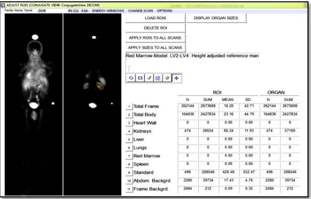
To perform image quantification for patient specific dosimetry, patients need to be scanned in a series of gamma camera acquisitions. The acquired images have been subjected to investigate the response of radiopharmaceutical administration on either cancer cells or relevant organs. The responses which have been presented in the images can be quantified as cumulated activity of radiopharmaceutical by drawing the region of interest for relevant organs. Figure 1 shows the sample of display menu in ULMDOS which represent the number of cumulated activity in the organs.
In this study, five patients with neuron endocrine tumors (NETs) were
intravenously injected with 11In-labelled DTPAOC (OctreoscanTM) as bolus
Hidayati et al., RDNI, Vol. 2, No.1, 2015, pp. 49-57
Figure 1.Display menu in ULMDOS for creating ROIs on the organs.
Hidayati et al., RDNI, Vol. 2, No.1, 2015, pp. 49-57
53 | CAS – Center for Advanced Sciences
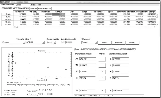
Figure 2.PLOT&FIT menu - ULMDOS software
RESULTS AND DISCUSSION
Hidayati et al., RDNI, Vol. 2, No.1, 2015, pp. 49-57
Figure 3.The relationship between relative size of the ROI and the
corresponding relative change of the TIAC (τ) forkidneys.
Figure 4.The relationship between relative size of the ROI and the
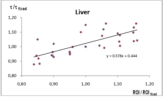
corresponding relative change of the TIAC (τ) for liver.
y = 0.611x + 0.369
0,80 0,90 1,00 1,10 1,20
0,80 0,90 1,00 1,10 1,20 τ/τfixed
ROI / ROI fixed
Kidneys
y = 0.578x + 0.444
0,80 0,90 1,00 1,10 1,20
0,80 0,90 1,00 1,10 1,20
Hidayati et al., RDNI, Vol. 2, No.1, 2015, pp. 49-57
55 | CAS – Center for Advanced Sciences
Figure 5.The relationship between relative size of the ROI and the
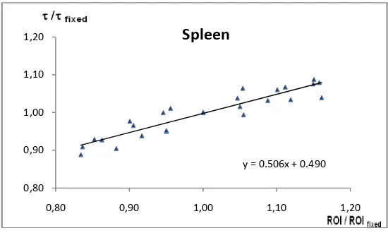
corresponding relative change of the TIAC (τ) for spleen.
From the plot analysis, when the ROIs have been set to be decreased and increased, the effect onTIACs will be either larger or smaller respectively. For example, 10% smaller or larger error will produce the similar error at 5 % error of TIAC in spleen, while in kidneys and liver, the relationship is on the contrary, for example the smaller ROI in kidneys produce larger error in TIAC, but the larger ROI in kidneys produces the smaller error of TIAC.
During the ROI drawing process, there were a few challenges in defining ROI for the livers in some patients since it seems that they have metastases in the liver. As a result a relationship which is less accurate can be seen in Figure 3. This is one of the particular problems in image quantification using planar images from gamma camera, for example a similar problem exists in the area where organs overlap, which can reduce the accuracy of ROI definition. To solve the problem, some correction need to be performed, for example the metastasis area needs to be subtracted from the total gross of ROI, but in this study, the correction has not been done, and
the metastasis has been disregarded. It has been suggested[2] that this
correction will not be necessary for image quantification for PET images, since it represents a three dimensional volume. Moreover, a calibrated PET image can also be used to eliminate the effect of overlapping organs.
y = 0.506x + 0.490
0,80 0,90 1,00 1,10 1,20
Hidayati et al., RDNI, Vol. 2, No.1, 2015, pp. 49-57
CONCLUSION
The step of drawing ROIs is a crucial step in the process of calculation of time-integrated activity coefficients. Hence, the enlargement and reduction of a ROI will affect the obtained time-integrated activity coefficient. In general, the relative error in the TIACs is in the same order of magnitude as the errors in the organ ROI drawing process.
ACKNOWLEDGMENT
The first author gratefully acknowledges a grant by The Non-Degree Program for Research Innovation in Science and Technology Project (RISET-Pro) for the financial support to visit Medical Faculty Mannheim of Heidelberg University, Germany. We also are grateful for a DIKTI Scholarship which is awarded to the second author for his PhD study.
[1] M. Sathekge, "Targeted Radionuclide Therapy, Continuing
Medical Education," 31, vol. 8, pp. 289-294, 2013.
Both grant and Scholarship are funded by the Ministry for Research, Technology and Higher Education, Republic Indonesia.
REFERENCES
[2] M. B, "Nuclear Medicine radiation Dosimetry – advanced
Theoretical Principles," pp. 455-457, 2010.
[3] M. Lyra, N. Lagopati, P. Charalambatou, and I. Vamvakas,
"Patient-specific dosimetry in radionuclide therapy," Radiat Prot Dosimetry, vol. 147, pp. 258-63, 2011.
[4] S. R. Thomas, "Options for radionuclide therapy: from fixed
activity to patient-specific treatment planning," Cancer Biother Radiopharm, vol. 17, pp. 71-82, 2002.
[5] M. Cremonesi, M. Ferrari, L. Bodei, G. Tosi, and G. Paganelli,
"Dosimetry in Peptide radionuclide receptor therapy: a review," J Nucl Med, vol. 47, pp. 1467-75, 2006.
[6] E. J. Rolleman, M. Melis, R. Valkema, O. C. Boerman, E. P.
Krenning, and M. de Jong, "Kidney protection during peptide receptor radionuclide therapy with somatostatin analogues," Eur J Nucl Med Mol Imaging, vol. 37, pp. 1018-31, 2010.
[7] T. Das and M. R. Pillai, "Options to meet the future global demand
of radionuclides for radionuclide therapy," Nucl Med Biol, vol. 40, pp. 23-32, 2013.
[8] Z. Fan, "Theranostic nano medicine for cancer detection and
Hidayati et al., RDNI, Vol. 2, No.1, 2015, pp. 49-57
57 | CAS – Center for Advanced Sciences
[9] G. Glatting and M. Lassmann, "Nuclear medicine dosimetry:
quantitative imaging and dose calculations," Z Med Phys, vol. 21, pp. 246-7, 2011.
[10] E. B. Silberstein, "Radioiodine: The Classic Theranostic Agent,
Seminars in Nuclear Medicine," vol. 42, pp. 164-170, 2012.
[11] G. Glatting, M. Landmann, T. Kull, A. Wunderlich, N. M.
Blumstein, A. K. Buck, et al., "Internal radionuclide therapy: the ULMDOS software for treatment planning," Med Phys, vol. 32, pp. 2399-405, 2005.
[12] M. G. Stabin, R. B. Sparks, and E. Crowe, "OLINDA/EXM: the