Association between
Helicobacter pylori
Infection
and Graves’ Disease: A Meta-Analysis
Guntur Darmawan*, Marcellus Simadibrata**, Indah Suci Widyahening***
*Department of Internal Medicine, Faculty of Medicine, University of Padjadjaran, Bandung **Division of Gastroenterology, Department of Internal Medicine, Faculty of Medicine, Universitas
Indonesia/Dr. Cipto Mangunkusumo General National Hospital, Jakarta
***Department of Community Medicine, Faculty of Medicine, Universitas Indonesia, Jakarta
Corresponding author:
Marcellus Simadibrata. Division of Gastroenterology, Department of Internal Medicine, Dr. Cipto Mangunkusumo General National Hospital. Jl Diponegoro No.71 Jakarta Indonesia. Phone: 3153957; facsimile: +62-21-3142454. E-mail: [email protected]
ABSTRACT
Background:Helicobacter pylori (H. pylori) infection is proposed to be related with autoimmune diseases, such as Graves’ disease. This study aimed to assess the association between H. pylori infection and Graves’ disease.
Method: A systematic literature review was conducted using Pubmed and Cochrane library. The quality of enrolled studies was assessed by the Critical Appraisal Skills Program Oxford. A fixed-effect model approach was used if there was no heterogeneity; otherwise, a random-effect model was used. Heterogeneity was assessed using I2. Publication bias was assessed by funnel plot. All data were analyzed using REVIEW MANAGER 5.3.
Results: Six studies from Europe and Asia involving 983 patients were included. Overall H. pylori infection was significantly associated with Graves’ disease (OR = 2.7; 95% CI: 1.47-4.99; p < 0.001). In subgroup analysis of 3 studies using non-serological diagnostic method, the prevalence rate of H. pylori infection was higher in Graves’ disease group (78.26% vs. 42.42%) with significant relationship (OR = 4.93; 95% CI: 3.16-7.69; p < 0.00001; I2 = 0%). The Cytotoxin associated gene A (CagA) antibody prevalence was significantly higher in Graves’ disease group (46.57% vs. 20.29%; OR = 4.41; 95% CI: 2.65-7.33; p < 0.00001; I2 = 56%). No publication bias was observed.
Conclusion:Our study showed association between H. pylori infection and Graves’ disease. It might suggest the need of H. pylori examination in Graves’ disease patients and the impact of H. pylori eradication in the treatment of Graves’ disease.
Keywords:Helicobacter pylori, Graves’ disease, meta-analysis
ABSTRAK
Latar belakang: Beberapa penelitian melaporkan infeksi Helicobacter pylori (H. pylori) berkaitan dengan penyakit otoimun, antara lain Graves’ disease. Penelitian ini bertujuan untuk mengevaluasi hubungan antara infeksi H. pylori dengan Graves’ disease.
Metode: Telaah literatur dilakukan dengan menggunakan Pubmed dan Cochrane library. Kualitas studi dinilai dengan Critical Appraisal Skills Program Oxford. Pendekatan fixed-effect model digunakan jika tidak terdapat heterogenitas dan random-effect model digunakan jika terdapat heterogenitas. Heterogenitas dievaluasi dengan I2. Bias publikasi dikaji dengan funnel plot. Semua data dianalisis dengan program REVIEW MANAGER 5.3.
1.47-4.99; p < 0.001). Pada analisis subgrup dari 3 studi yang menggunakan metoda diagnostik non-serologik, didapatkan prevalensi infeksi H. pylori yang lebih tinggi secara bermakna pada grup Graves’ disease (78.26% vs. 42.42%; OR = 4.93; 95% CI: 3.16-7.69; p < 0.00001; I2 = 0%). Prevalensi antibodi CagA secara signifikan lebih tinggi pada grup Graves’ disease (46.57% vs. 20.29%; OR = 4.41; 95% CI: 2.65-7.33; p < 0.00001; I2 = 56%). Tidak ditemukan adanya bias publikasi.
Simpulan: Penelitian ini menunjukkan hubungan antara infeksi H. pylori dengan Graves’ disease. Hal ini dapat mendasari perlunya pemeriksaan H. pylori pada pasien Graves’ disease dan dampak eradikasi H. pylori dalam terapi Graves’ disease.
Kata kunci: Helicobacter pylori, Graves’ disease, meta-analisis
references cited by the original published studies and relevant review articles. Article selection and assessment were done by reviewers. We contacted the authors via email to obtain the required information when relevant information was not available in the published article.
Studies were included for analysis based on the following inclusion criteria: (i) observational study having control group; (ii) the outcome was Graves’ disease; (iii) Graves’ disease was diagnosed by the presence of hyperthyroidism (suppressed TSH, elevated FT3, elevated FT4), diffuse goiter with positive antibody titers (TSI, TPOAb, TgAb), and, in some cases, opthalmopathy; (iv) the exposure was H. pylori infection; (v) the diagnosis of H. pylori infection was based on serological tests (antigen-specific enzyme-linked immunosorbent assay (ELISA) and Western blotting) or non-serological tests (rapid urease test, stool antigen test (SAT), 13C-urea breath test (UBT)); (vi) studies had extractable data and sufficient information on the association between H. pylori infection and Graves’ disease.
We also recorded the CagA serology examination result if it was done in the study. Since all parameter observed were objectively measured and having a written record, neither recall bias nor observer bias were occur in each study. We assessed the quality of each study by using the criteria from the critical appraisal skills program (CASP) Oxford United Kingdom consisting of 11 systematic questions for appraising case control study. The quality levels then were graded as good, fair, and poor.10 Only studies with good quality were included in our final analysis review. Discrepancies and disagreements were resolved by consensus.
Study characteristics were taken as follows: first author; year of publication; study design; country; H. pylori test method; subjects characteristics (case subjects, age in mean or median, sex, and matched INTRODUCTION
Graves’ disease, having thyrotoxicosis as clinical hallmark, is characterized by formation of autoantibody thyroid stimulating immunoglobulins (TSI) to the thyroid stimulating hormone receptor (TSH-R). Other antibodies, thyroid peroxidase antibody (TPOAb) and thyroglobulin antibody (TgAb), might also present in Graves’ disease. The etiology of Graves’ disease is considered as a complex combination of genetic and environment factors.1,2
The discovery of Helicobacter pylori (H. pylori) by Marshall and Warren in 1982 has contributed significantly in understanding pathogenesis of diseases. Globally, the prevalence of H. pylori infection is more than 50%. The impact of this microaerophilic, gram negative curved bacillus is not just limited at the gastrointestinal; moreover, it has been proposed to have extra-gastrointestinal manifestations, such as coronary heart disease, diabetes mellitus, autoimmune diseases.3-7 The presence of seropositivity to the cytotoxin-associated gene A (Cag-A) is commonly used to identify the virulence of H. pylori.6,7
The interaction of H. pylori as infectious environmental exposure and genetic susceptibility resulting in autoimmune disorder such as Graves’ disease has become an appealing issue.1,3,4,8 The present study was performed to evaluate the association between H. pylori infection and Graves’ disease through review of existing studies.
METHOD
between two groups); CagA test availability; number of subjects with positive H. pylori test in each outcome group; number of subjects with positive CagA test in each outcome group.
We calculated the odds ratios (OR) with 95% confidence interval (CI) for H. pylori positivity. The Mantel-Haenszel method was used to weight the studies included. A fixed-effect model approach was use if there was no heterogeneity; otherwise, a random-effect model was used. Heterogeneity was assessed using I2. Negative value of I2 was put equal to 0. I2 values ranged from 0% (no observed heterogeneity) to 100%, and interpreted according to Cochrane Consumers and Communication Review Group.11 For sub-analysis, we calculated the OR of CagA seropositivity. Moreover, to evaluate the possible bias due to difference in diagnostic method, we did sub-analysis ofthe OR of H. pylori positivity by serological diagnostic method and the OR of H. pylori positivity by non-serological diagnostic method (study using both serological and non-serological diagnostic method will be included here). Publication bias was assessed by funnel plot. All statistical analysis was performed using Review Manager 5.3.
RESULTS
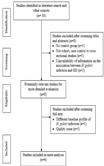
Our literature search identified 16 studies, all published in English. We excluded studies which were not cohort, or case control studies (n = 5), had no available information on the association between H. pylori infection and Graves’ disease (n = 2), had no control group (n = 1). After final-text screening, we decided to exclude 1 study due to different baseline profile of H. pylori infection and 1 study due to quality issue. Finally, a total of 6 studies involving 983 patients met our criteria. The flowchart showed the process of studies selection (Figure 1).
The studies were published between 1998 and 2013, and the characteristics of which are summarized in Table. Four studies were performed in Europe, and 2 studies were performed in Asia.7,8,12-15 Four studies had Graves’ disease subjects as a subgroup of the case
group (subjects with autoimmune thyroid disease) and 2 other studies had Graves’ disease subjects as the whole subjects of case group.7,8,12,15 All studies had greater number of female than male subjects in both Graves’ disease and control groups. None of the studies performed endoscopy due to young age subjects and had no alarm symptoms. Three studies used serology method, 2 studies used SAT method, and 1 study used combination of serology and UBT method12 to assess the infection status of H. pylori. Four studies examined CagA antibody to identify infection with H. pylori strain possessing CagA.7,8,13,14,15
Figure 1. Flow diagram of literature selection
Table 1. Characteristic of studies included in meta-analysis
Author Year of publication
Study
design Country HP test method Case subjects
Age
(mean or median)
De Luis12 1998 CC Spain UBT & ELISA ATD (Graves’ disease as subgroup) patients 33.9 ± 1.2
Larizza14 2006 CC Italy ELISA ATD (Graves’ disease as subgroup) patients 11.7
Bassi7 2010 CC Italy SAT Graves’ disease patients 42.8 ± 8.8
Soveid15 2012 CC Iran ELISA ATD (Graves’ disease as subgroup) patients 36.3 ± 7.6
Bassi13 2012 CC Italy SAT ATD (Graves’ disease as subgroup) patients 48.8 ± 3.9
Wang8 2013 CC China ELISA ATD (Graves’ disease as subgroup) patients 28.0 ± 5.4
Table 3. Characteristic of studies included in meta-analysis
HP: Helicobacter pylori; CagA: cytotoxin associated gene A
Prevalance rate of H. pylori positivity showed statistically significant different between Graves’ disease and control group in 4 studies.7,8,13,14 One study showed higher prevalence of H. pylori positivity in Graves’ disease group although it was not statistically significant.12 One study, in contrary, showed higher prevalence of H. pylori positivity in control group but not statistically significant.15 The overall prevalence rate of H. pylori positivity was 69.87% (327 of 468) in Graves’ disease group and 47.96% (247 of 515) in control group. Positivity of H. pylori infection was significantly associated with Graves’ disease (pooled OR = 2.7; 95% CI: 1.47-4.99; test for overall effect Z = 3.18; p < 0.001). However, there was a substantial heterogeneity (I2 = 74%).
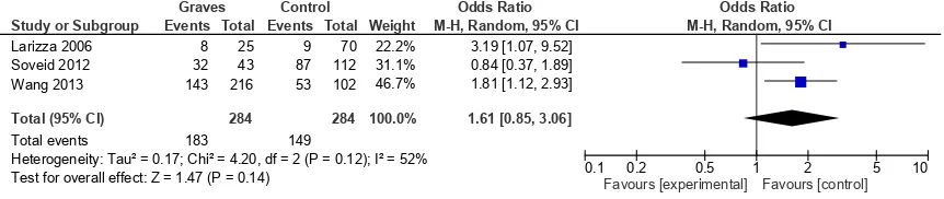
We performed subgroup analysis of study using different method of diagnostic test. In the 3 studies using serological diagnostic method, the prevalence rate of H. pylori infection was higher in Graves’ disease than in control group (64.43% [183 of 284] vs. 52.46% [149 of 284]). However, the association was not significant (pooled OR = 1.61 [95% CI: 0.85-3.06, test for overall effect Z = 1.47, p 0.14]) with moderate heterogeneity (I2 = 52%) (Figure 2).
In the 3 studies using non-serological diagnostic method, the prevalence rate of H. pylori infection was also higher in Graves’ disease group than in control group (78.26% [144 of 184] vs. 42.42% [98 of 231]). Moreover, the relationship was statistically significant (pooled OR = 4.93 [95% CI: 3.16-7.69, test for overall effect Z = 7.03, p < 0.00001]). There was no heterogeneity existed (I2 = 0%) (Figure 3).
The CagA antibody prevalence in the pooled 4 studies was higher in Graves’ disease group (46.57% [197 of 423]) than in control group (20.29% [84 of 414]) with significant OR = 4.41 (95% CI: 2.65-7.33), test for overall effect Z = 5.70, p < 0.00001). The heterogenity was moderate (I2 = 56%) (Figure 4).
Table 2. Characteristic of studies included in meta-analysis
Author Sex (F/M)
Graves’ disease
Sex (F/M) Control
Matched between case & control
CagA test
Age Sex S SE Area
De Luis12 13/7 21/10 Yes Yes Yes NA Yes NA
Larizza14 23/2 55/15 Yes Yes NA NA Yes NA
Bassi7 98/14 87/13 Yes Yes Yes Yes Yes A
Soveid15 38/5 89/23 Yes Yes NA Yes Yes A
Bassi13 48/4 90/10 Yes Yes Yes Yes Yes A
Wang8 122/94 59/43 Yes Yes Yes NA Yes A
HP: Helicobacter pylori; F: female; M: male; A: available; NA: not available; S: smoking; SE: socioeconomic; ELISA: enzyme-linked
immunosorbent assay; UBT: 13C-urea breath test; SAT: stool antigen test; CagA: cytotoxin associated gene A
Figure 2. Association between H. pylori infection and Graves’ disease in studies using serological diagnostic method Study or Subgroup
M-H, Random, 95% CI
3.19 [1.07, 9.52] 0.84 [0.37, 1.89] 1.81 [1.12, 2.93]
1.61 [0.85, 3.06]
Graves Control Odds Ratio Odds Ratio
M-H, Random, 95% CI
0.1 0.2 0.5 1 2 5 10
Favours [experimental] Favours [control]
Figure 3. Association between H. pylori infection and Graves’ disease in studies using non-serological diagnostic method Study or Subgroup Test for overall effect: Z = 7.03 (P < 0.00001)
Events
Graves Control Odds Ratio Odds Ratio
M-H, Fixed, 95% CI
0.1 0.2 0.5 1 2 5 10
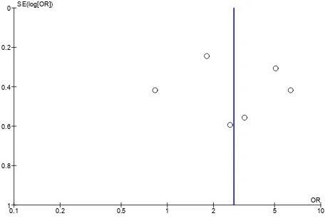
For the overall 6 studies, no evidence of publication bias was observed in the funnel plot (Figure 5).
H. pylori cannot differentiate either past or current infection. Moreover, the background of H. pylori prevalence influences the positive predictive value (PPV) of this method, as discussed in the study by Soveid in Iran.15-22 We found no significant association between H. pylori seropositivity and Graves’ disease in this subgroup, as shown by the pooled OR of 3 studies using serology method, and moderate heterogeneity. In contrary, the association was statistically significant in the non-serology method subgroup with 0% of heterogeneity. This finding may underline the importance of ensuring the infection status (current infection) since the antibody itself can persist even after eradication therapy. Therefore, either UBT or SAT method is considered as the preferred non-invasive diagnostic method.
For the subgroup analysis of CagA seropositivity, overall studies showed that infection by the more virulent strain (CagA seropositive) group was significantly correlated with Graves’ disease. The possession of CagA plays role in the development of Graves’ disease through “antigenic mimicry” and cytokines expression.17,19 Different diagnostic kit used in each study may contribute to the heterogeneity.
In this study, we had heterogeneous race of subjects, consisting of Caucasian (4 studies), Asian Chinese (1 study), and Asian Iranian (1 study). Genetic factors, which may be clustered more in people depending on their race might play role in the development of Graves’ disease. However, there were just 2 studies specifically assessed the genetic factor (HLA alleles), one in Caucasian and one in Asian Chinese. Unfortunately, the result of both studies was not supporting each other.8,14 Additional studies need to be done to confirm the specific alleles preventing or increasing risk developing of Graves’ disease in correlation with H. pylori infection.
To our knowledge, this is the first meta-analysis study to investigate the association between H. pylori positivity and Graves’ disease. This association might give an idea of the need H. pylori examination in
Study or Subgroup Test for overall effect: Z = 5.70 (P < 0.00001)
Events
M-H, Random, 95% CI
5.74 [3.09, 10.65] 8.46 [3.96, 18.11] 2.61 [1.25, 5.45] 3.09 [1.67, 5.71]
4.41 [2.65, 7.33]
Graves Control Odds Ratio Odds Ratio
M-H, Random, 95% CI
0.1 0.2 0.5 1 2 5 10
Favours [control] Favours [Graves]
Figure 4. Association between CagA seropositivity and Graves’ disease
0.1 0.2 0.5 1 2 5 10
Figure 5. Funnel plot analysis of H. pylori infection and Graves’ disease
DISCUSSION
Based on meta-analysis of 6 studies, there was a significant association between H. pylori positivity and Graves’ disease. Graves’ disease, a member of autoimmune thyroid disease, arises from the complex interaction of genetic susceptibility and environmental factor. Infectious agent is one of the environmental factors receiving particular attention. Among infectious agents, H. pylori infection has been proposed to trigger the Graves’ disease by several mechanisms. Earlier studies showed that the lipopolysaccharide (LPS) of H. pylori has a similar structure with human cell surface glycoconjugates Lewis X, Lewis Y. The “antigenic mimicry” expression of Lewis determinant in LPS is associated with CagA status of the H. pylori strain.16-18 This “antigenic mimicry” might induce autoimmune response which influences the thyroid gland. Interestingly, H. pylori might play role through modulation of dendritic cells, T helper cells, resulting in expression of proinflammatory cytokines such as interferon (IFN)-gamma, interleukin (IL)-10, IL-17.19-21
Graves’ disease patients and further investigation whether the treatment of H. pylori will positively influence the treatment of Graves’ disease. However, this study has some potential limitations. First, since all studies collected the data of exposure and outcome in the same time, temporal relationship could not be clearly defined. Second, there were just six studies met the criteria and were included the meta-analysis.
CONCLUSION
Our study showed association between H. pylori infection and Graves’ disease. We realize that more longitudinal studies with uniform diagnostic method, larger sample sizes, and multiethnic are needed for better understanding of H. pylori and Graves’ disease relationship.
REFERENCES
1. Effraimidis G, Wiersinga WM. Mechanisms in endocrinology: Autoimmune thyroid disease: Old and new players. Eur J Endocrinol 2014;170:R241-52.
2. Prabhakar BS, Bahn RS, Smith TJ. Current perspective on the pathogenesis of Graves’ disease and ophthalmopathy. Endocr
Rev 2003;24:802–35.
3. Smyk DS, Koutsoumpas AL, Mytilinaiou MG, Rigopoulou EI, Sakkas LI, Bogdanos DP. Helicobacter pylori and autoimmune disease: Cause or bystander. World J Gastroenterol 2014;20:613–29.
4. Franceschi F, Tortora A, Gasbarrini G, Gasbarrini A. Helicobacter pylori and extragastric diseases. Helicobacter 2014;19:52–8.
5. Hasni S, Ippolito A, Illei GG. Helicobacter pylori and autoimmune diseases. Oral Dis 2013;17:621–7.
6. Mayr M, Kiechl S, Mendall M a, Willeit J, Wick G, Xu Q. Increased risk of atherosclerosis is confined to CagA-positive
Helicobacter pylori strains: Prospective results from the
bruneck study. Stroke 2003;34:610–5.
7. Bassi V, Santinelli C, Iengo A, Romano C. Identification of a
correlation between Helicobacter pylori infection and Graves ’ disease. Helicobacter 2010;15:558–62.
8. Wang Y, Zhu S, Xu Y, Wang X, Zhu Y. Interaction between
gene A-positive Helicobacter pylori and human leukocyte
antigen II alleles increase the risk of Graves disease in Chinese
Han population: An association study. Gene 2013; 531:84–9.
9. Moher D, Liberati A, Tetzlaff J, Altman DG. Preferred
reporting items for systematic reviews and meta-analyses:
The PRISMA statement. PLoS Med 2009;6:e1000097. 10. Critical Appraisal Skill Program. Eleven questions to make
you make sense of case control study [serial online] [cited 2015 July]. Available from:URL:http://www.casp-uk.net/.
11. Cochrane Consumers and Communication Review Group. Heterogeneity and subroup analyses in Cochrane Consumers and Communication Review Group reviews: Planning the analysis at protocol stage [cited 2015 July]. Available from:
http://www.cccrg.cochrane.org/.
12. De Luis D, Varela C, de La Calle H, Canton R, de Argila
C, San Roman A. Helicobacter pylori infection is markedly increased in patients with autoimmune atrophic thyroiditis. J Clin Gastroenterol 1998;26:259–63.
13. Bassi V, Marino G, Iengo A, Fattoruso O, Santinelli C. Autoimmune thyroid diseases and Helicobacter pylori: The correlation is present only in Graves’s disease. World J Gastroenterol 2012;18:1093–7.
14. Larizza D, Calcaterra V, Martinetti M, Negrini R, De
Silvestri A, Cisternino M, et al. Helicobacter pylori infection and autoimmune thyroid disease in young patients: The
disadvantage of carrying the human leukocyte antigen-DRB1*0301 allele. J Clin Endocrinol Metab 2006;91:176–9.
15. Soveid M, Asl KH, Omrani GR. Infection by Cag A positive strains of Helicobacter pylori is associated with autoimmune thyroid disease in Iranian patients. Iran J Immunol 2012;9:48–52.
16. Appelmelk BENJ, Simoons-smit INA, Negrini R, Moran
AP, Aspinall GO, Forte JG, et al. Potential role of molecular mimicry between Helicobacter pylori lipopolysaccharide and
host Lewis blood group antigens in autoimmunity. Infection
and immunity 1996;64:2031–40.
17. Wirth H, Yang M, Karita M, Blaser MJ. Expression of the human cell surface glycoconjugates Lewis X and Lewis Y by
Helicobacter pylori isolates is related to cagA status. Infection and immunity 1996;64:4598–605.
18. Censini S, Lange C, Xiang ZY, Crabtree JE, Ghiara P, Borodovsky M, et al. Cag, a pathogenicity island of
Helicobacter pylori, encodes type I-specific and disease-associated virulence factors. Proc. Natl Acad Sci USA 1996, 93:14648–53.
19. Oghumu S, Satoskar A. The emerging role of dendritic cells in
the host immune response against Helicobacter pylori. Front Microbiol 2014;5:1–2.
20. Nagayama Y, Saitoh O, McLachlan SM, Rapoport B, Kano H, Kumazawa Y. TSH receptor-adenovirus-induced Graves’
hyperthyroidism is attenuated in both interferon-gamma and
interleukin-4 knockout mice; implications for the Th1/Th2 paradigm. Clin Exp Immunol 2004;138:417–22.
21. Park S, Kim J, TS H, JI S. The Role of Interferon-gamma and interleukin 17 between Helicobacter pylori infection and Graves ’ disease. Helicobacter 2011; 15:338.
22. Chey WD, Wong BCY. American College of Gastroenterology