16
O R I G I N A L A R T I C L E
CORRELATION BETWEEN C-REACTIVE PROTEIN AND LEFT
VENTRICULAR EJECTION FRACTION IN ANTERIOR ST ELEVATION
MYOCARDIAL INFARCTION
1 2 3
Rosa Priambodo , Bambang Irawan , Siti Nurdjanah
1. Division of Internal Medicine, Faculty of Medicine, Gadjah Mada University/Dr Sardjito Hospital, Yogyakarta 2. Sub Division of Cardiology, Internal Medicine Dr Sardjito Hospital/ Faculty of Medicine Gadjah Mada University
Yogyakarta
3. Sub Division of Gastro-Entero-Hepatology, Intenal Medicine Dr Sardjito Hospital/ Faculty of Medicine Gadjah Mada University Yogyakarta
ABSTRACT
Background: C-Reactive Protein (CRP) levels were found increase in ST elevation myocardial infarction (STEMI) patients, before and after STEMI. Increase of CRP levels were able to activate complement pathway to induce inflammation by attracting neutrophil and macrophage to enter to infarcted myocardial. Infarct expansion followed by remodeling process, led to left ventricular dysfunction and decrease of ejection fraction.
Aim: This study aimed to investigate a correlation between CRP plasma levels and left ventricular ejection fraction (LVEF) in anterior STEMI patients.
Subject and method: This study was conducted cross-sectionally. Subjects were new anterior STEMI patients, with maximum onset of 48 hours. Exclusion criteria included evidence of infection, inflammation, history of surgery or stroke in last three months, malignancy, congestive heart failure and inferior STEMI. There were 30 subjects who met the eligible criteria. CRP blood samples were collected at least 48 hours after onset. LVEF measurement was done during hospitalization. The correlation between CRP levels and LVEF was analyzed by Spearman rank correlation test.
Result: CRP levels in female subjects were higher than males [CRP median 23.4 mg/l (5.73 – 61.4 mg/l) vs. 12. 2 mg/l (5.6 – 66.5 mg/l)], but with no significance (P= 0.297). The thrombolytic therapy group had lower CRP levels than non thrombolytic therapy group [10. 7 mg/l (5.6 – 44.5 mg/l) vs. 14.7 mg/l (8.7 – 66.5 mg/l), also with no significance (P= 0.178). There was a
non-significantly negative correlation between CRP level and LVEF (r = - 0.100, P= 0.597).
Conclusion: There was no correlation between CRP level and LVEF in anterior STEMI.
Keywords: STEMI – C-reactive protein – inflammation – left ventricular ejection fraction.
INTRODUCTION
ST elevation myocardial infarction (STEMI) was a spectrum of acute coronary s y n d r o m e ( A C S ) , t h e r u p t u r e s t a g e o f atherosclerotic plaque. Inflammatory process involved in all of atherosclerotis stages, since fatty
1
streak formation to plaque rupture . In myocardial infarction patients, inflammatory process also related to the expansion of myocardial necrosis that affect short and long term outcome in post STEMI
2.3
and non NSTEMI patients .
Acute phase protein was a non specific protein produced by hepatocyte as a response to inflammatory cytokines i.e. interleukin-1 (1). IL-6 and tumor necrosis factor-α (TNF-α) released by tissue damage, infectious conditions, inflammatory
4
disorder and malignancy . C-reactive protein (CRP) as a positive acute phase protein play a role in the development of post myocardial infarction complications, such as ventricular wall rupture, aneurysm, papillary muscles rupture, infarct expansion, remodeling and ventricular
2.3
dysfunction . C-reactive protein was able to activate complement system that could lead neutrophil and macrophage enter myocardium
5.6
leading to necrosis expansion .
17 Study Protocol
Acta Interna - The Journal of Internal Medicine
18
Rosa Priambodo, et al
Table 1. Baseline characteristics of study subjects (n=30) Characteristics mean±SD
(Proportion)
CI 95% Median Minimum
–
Maximum
Age (years) 58.46±10.46 54.31-62.62
Sex:
? Male 23 (76.7%)
? Female 7 (23.3%)
Onset of Pain (hour) 5.5 2-30
CRP time (hour) 8 3-30
Diabetic:
? Yes 2(6.7%)
? No 28(93.3%)
Hypertension:
? Yes 16(53.3%)
? No 14(46.7%)
Smoking
? Yes 16(53.3%)
? No 14(46.7%)
Dislipidemia
? Yes 3(10%)
? No 27(90%)
Blood pressure (mmHg)
? Systolic 123.73±26.48 113.04-134.42
? Diastolic 80 56-105
Laboratory
? Leucocyte (103 /mm3) 13.25±3.64 11.78-14.72
? CKMB (IU/I) 34.6 11-208
? LDH (IU/I) 740.5 378-2675
? AST (IU/I) 65 24-502.8
? CRP (IU/I) 13.5 5.6-66.5
? Total Cholesterol (mg/dl) 216.31±44.3 198.41-234.2
? LDL (mg/dl) 134.23±51.54 113.41-155.05
? HDL (mg/dl) 43.7±9.86 39.72-47.68
? TG (mg/dl) 128.5 55-587
? Glucose level (mg/dl) 165 96-429
ST Elevation (mm) 3 2-6
Ejection fraction (%) Trombolysis:
53.23±11.95 48.4-58.06
? Yes 17(56.7%)
? No 13(43.3%)
Note: AST = aspartate aminotransferase, CKMB = creatin kinase MB, CRP = C-Reactive Protein, HDL = high density lipoprotein, CI 95% = confidential interval 95%, LDH = lactic dehydrogenase, LDL = low density lipoprotein, TG = trigliseride.
19 Table2. Distribution of C-reactive protein levels (mg/L) by sex (n= 300)
Table 3. Distribution of C-reactive protein levels (mg/L) by thrombolytic therapy (n=30)
Sex Mean±SD Median
Minimum-Maximum
P S/NS
Male(N= 22) 18.49±15.58 12.2 5.6–66.5 0.297 NS
Female (N= 7) 26.18±20.47 23.4 5.73–61.4
Thrombolytic Mean±SD Median Minimum– P
Therapy Maximum
Done(N = 17) 16.64±13.42 10.7 5.6–44.5 0.178
Not done (N = 13) 25.05±19.93 14.7 8.7 – 66.5
Note: SD = standard deviation
Note: SD = standard deviation
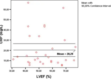
We found negative correlation between CRP levels and LVEF (r = - 0.100 and P = 0.597) (figure 1).
Figure 1. Correlation between C-reactive protein levels and left ventricular ejection fraction
Acta Interna - The Journal of Internal Medicine
20
DISCUSSION
In this study, CRP levels of females were non-significantly higher than the ones of males (median 23.4 mg/l versus 12.2 mg/l) (P= 0.297).
11
Podrid reported that female has higher CRP level than male and the increasing of CRP levels could be used to predict cardiovascular disease as in male. Female with increased CRP level more than 3 mg/l. had an 8-fold mortality risk compared to female
12
having CRP levels less than or equal to 1 mg/l . Female with cardiovascular disease had CRP levels higher than female without cardiovascular disease
13
(0.42 mg/l vs. 0.28 mg/l) . Post menopause female that used hormonal replacement treatment (HRT) had CRP levels twice as much as female that did not
13
use it . Other study reported on 493 post menopause healthy female that the HRT user had CRP levels
14
twice as much as the non user .
In this study there was difference of CRP levels between thrombolytic group and non thrombolytic group, even though it did not reach significance. A different result was reported by
2
Pietila et al cit. Anzai et al that found that CRP levels of open infarct- related coronary artery were less than levels of closed infarct-related coronary artery or when compared to control group (non thrombolytic therapy). In control group, they found closed relationship between infarct size and CRP levels (r= 0.58; P< 0.001). This correlation was particularly found in closed infarct-related coronary artery (r= 0.62; P< 0.001). In open infarct-related coronary artery, the correlation between infarct size and CRP levels were weak (r=30;P<0.01).
Left venticular ejection fraction had inverse correlation with CRP levels, in control group and
15
reperfusion group. Zairis et al found that in 319 STEMI patients subjects with CRP levels at high tertile had lower ST segment resolution to normal limit, lower thrombolysis in myocardial infarction score and higher mortality caused by cardiac survival.
The correlation between CRP levels and LVEF in this study was very weak, with coefficient correlation (r) = - 0.100 and P = 0.597. However,
16
Pandian et al found a closer relationship with r = - 0. 67 and P = 0.011. Many explanations of this result are as follows: First, this study only included anterior STEMI patients resembling the inclusion
7
done by Antman & Braunwald who found that anterior infarct related to the declining of cardiac output, congestive heart failure and cardiogenic
9
shock. DeMaria & Blanchard reported that left ventricular remodeling may occur in ten days after STEMI and related to worse prognosis caused by decreasing of left ventricular function. In ventricular inferior infarction, increasing of right cardiac filling pressure (increasing of central venous pressure, right atrium and right ventricular diastolic pressure) can be found, but left ventricular
7
filling pressure is still in normal level . Pandian et
16
al did not discriminate infarct location in their study population. Second, this study included only patients with new STEMI and had no congestive history of heart failure to avoid any interference to
16
LVEF measurement. Pandian et al did not mention congestive heart failure as an exclusion criteria. Third, in this study, the total time for CRP sampling were varied, ranged from 3 hours to 30 hours after the onset of pain (mean±SD = 7.46±6.98 hours).
3
Suleiman et al found that CRP levels after 12–24 hours of STEMI onset were independent markers of
17
30 days mortality and heart failure. Tomassi et al
who collected CRP samples eight hours after onset of pain found correlation between CRP levels and cardiac events one year afterward. The limiting time for CRP sampling in the present study was set at admission until 48 hours after STEMI. This was
4
based on observation by Hirschfield & Pepys reported that CRP was produced by hepatocytes immediately after onset. This is induced by releasing of IL-6 from infarct tissue and reached peak levels at 48 hours when it then gradually decline to normal limit. Fourth, CRP levels that increased immediately after STEMI onset had relationship to less infarct size and better left ventricular function at post reperfusion therapy in anterior STEMI patients. This protective effect of high CRP levels was silent myocardial ischemia which lead to ischemic preconditioning effect at myocardium. Inflammatory process induced an increase expression of angiogenic growth factor. This results in decreasing of infarct size and increasing of endogenous nitric oxide production to
18
keep myocardium off ischemia . The correlation between CRP levels and LVEF in this study was very weak. This can be caused by time of CRP levels Rosa Priambodo, et al
sampling that might provide protective effect of high CRP levels.
This study has some limitations. The design of cross sectional did not allow us to take conclusion since exposure and outcome variables were measured at the same time. This study did not defined CRP sampling based on thrombolysis therapy. Left ventricular diastolic function was not measured despite the fact that prognosis evaluation in post STEMI patients is not LVEF alone. Besides, the examiner of LVEF was not only one person. Finally, LEFV technical measurement could be better if Simpson method is used.
CONCLUSION AND SUGGESTIONS
This study showed that there was no correlation between plasma CRP levels and left venticular ejection fraction at anterior STEMI patients. Further study needs to perform a prospective design and consider the status of thrombolytic therapy. CRP sampling at peak level (48 hours) may discover the real inflammation condition. Left ventricular diastolic function also needs to be measured beside LVEF. The LVEF can be assessed using Simpson method that also c o n s i d e r i n t r a a n d i n t e r o b s e r v e r i n echocardiography examination.
REFERENCES
1. Fruchart, J.C. 2003. Atherosclerosis: an Inflammatory Disease? International Task Force for Prevention of Coronary Heart Disease. Task Force Symposium
2. Anzai, T., Yoshikawa, T., Shiraki, H., Asakura, Y., Akaishi, M., Mitamura, H., Ogawa, S. 1997. C-Reactive Protein as a Predictor of Infarct Expansion and Cardiac Rupture After a First Q-Wave Acute Myocardial Infarction. Circulation. 96: 778-784 3. Suleiman, M., Aronson, D., Reisner, S.A.,
Kapeliovich, M.R., Markiewicz, W., Levy, Y., Hammerman, H. 2003. Admission C-Reactive Protein Levels and 30- Day Mortality in Patients with Acute Myocardial Infarction. Am. J. Med. 115:695-701
4. Hirschfield, G.M., Pepys, M.B., 2003. C-Reactive Protein and Cardiovascular Disease: New Insight from an Old Molecule. Q. L. Med.. 96: 793 – 807
Volume 2, Number 1, June 2012 Correlation Between C-Reactive Protein and Left Ventricular Ejection
21 5. Griselli, M., Herbert, J., Hutchinson, W.L., Taylor,
K.M., Sohail, M., Krausz, T., Pepys, M.B. 1999. C-Reactive Protein and Complement are Important Mediators of Tissue Damage in Acute Myocardial Infarction. J Exp Med. 190: 1733-1739
6. Lagrand, W.K., Niessen, H.W.M., Wolbink, G.J., Jaspars, L.H., Visser, C.A., Verheugt, F.W.A, Meijer, C.J.L.M., Hack, C.E. 1997. C-Reactive Protein Colocalizes With Complement in Human Hearts During Acute Myocardial Infarction. Circulation. 95: 97-103
7. Antman, E.M., Braunwald, E.1997. Acute Myocardial Infraction. in: E. Braunwald (editor) Heart Disease. A Textbook of Cardiovascular
th
Medicine. 5 edition. W. B. Saunders Company. page: 1233-1241.
8. McMurray, J.V., McDonagh, T.A., Davie, A.P.1998. Should We Screen for Asymptomatic Left Ventricular Dysfunction to Prevent Heart Failure ? Eur Heart J. 19: 842-846.
9. DeMaria, A.N., Blanchard, D.G.1998. The Echocardiogram. in: R.W. Alexander. R.C. Schlant. V. Fuster. R.A. O'Rourke. R. Roberts. E.H. Sonnenblick (editor) Hurst's The Heart. Arteries and
th
Veins. vol. 1. 9 edition. Mc Graw-Hill. page: 415-517.
10. Galasko, G.I.W., Basu, S., Lahiri, A., Senior, R. 2004. Is Echocardiography a Valid Tool to Screen for Left Ventricular Systolic Dysfunction in Chronic Survivors of Acute Myocardial Infarction? A Comparison With Radionuclide Ventriculography. Heart. 90: 1422-1426.
11. P o d r i d , P. 2 0 0 4 . C - R e a c t i v e P r o t e i n i n Cardiovascular Disease-I. in: B.D. Rose (editor). UptoDate 12.2 edition. UptoDate. Wellesley. M.A 12. Tice, J.A., Browner, W., Tracy, R.P., Cummings, Inflammation and Coronary Artery Disease. Cleveland Clinic Journal of Medicine. Vol. 68. No. 6: 521-534
Acta Interna - The Journal of Internal Medicine Rosa Priambodo, et al
22
15. Zairis, M.N., Manousakis, S.J., Stefanidis, A.S., Papadaki, O.A., Andrikopulos, G.K., Olympios, C.D., Hadjissavas, J.J., Argyrakis, S.K., Foussas, S.G. 2002. C- Reactive Protein Levels on Admission Are Associated With Response to Thrombolysis and Prognosis After ST-segment Elevation Acute Myocardial Infarction. Am Heart J . 144: 782-789.
16. Pandian, S., Amuthan, V., Sukumar, P., Janarthanan, R.A., Murugan, S., Palanichamy, S., Subramaniam, G., Annamatai, M. 2005. Plasma CRP Level Predicts Left Ventricular Function and Exercise Capacity in Patients With Acute Myocardial Infarction. Indian Heart Journal. 57: 54-57.
17. Tommasi, S., Carluccio, E., Bentivoglio, M., Buccolieri, M., Mariotti, M., Politano, M., Corea, L.1999. C-Reactive Protein as a Marker for Cardiac Ischemic Events in the Year After a First. Uncomplicated Myocardial Infarction. Am J Cardiol. 83:1595-1599.