Natural regulatory anticoagulants proteins among
Sudanese patients with dengue virus infection
Introduction
Infection with certain viruses can tilt the hemostasis toward bleeding and cause viral hemorrhage fever (VHF).1 Among VHFs, dengue virus (DENV) is the most important and prevalent.2 More than 2.5 billion people or half of the world’s population in tropical and subtropical is at risk of DENV infection.3 DENV is a mosquito-borne Flavivirus that is transmitted by mosquitoes such as Aedes aegypti or
Aedes albopictus. DENV are a positive-stranded RNA with envelope.2 Based on the antigenic difference of E protein, DENV can be divided into ive different serotypes, DENV 1-5. DENV infection might lead to inluenza-like illness, which is called dengue fever (DF) or cause more severe dengue hemorrhage fever (DHF) or dengue shock syndrome (DSS).4
Thrombocytopenia is common in DF and constant inding in DHF. The mechanism of thrombocytopenia is poorly understood.5 The platelet (PLT) counts usually drops to below 100,000/mm3 1-2 days before defervescence and remain low for 3-5 days in most cases. The levels then increase rapidly to normal during convalescence. The PLT counts in shock cases are frequently below (50,000/mm3).6 The coagulation system appears to be abnormal during infection manifesting as decreased ibrinogen (FB) levels, increase levels of ibrin degradation products, prolonged partial thromboplastin time (PTT) and prothrombin time (PT), low levels of coagulation factors VIII and XII.7 The in vivo existence of natural anticoagulant systems is essential to prevent thrombosis. These natural anticoagulant systems include antithrombin (AT), heparin cofactor II (HC-II), and protein C (PC) and its
The Coagulation System: Abnormalities in natural physiologic anticoagulants are observed in dengue infection. Laboratory values such as protein C (PC), protein S (PS), and antithrombin (AT) indicate this problem on the coagulation system in dengue. Recently, an interrelationship between dengue and the levels of natural anticoagulants has been observed.
Objective: The study conducted to ind out the effect of dengue on the natural
anticoagulant proteins.
Methods: Acase–control study was conducted in Port Sudan Teaching Hospital from February 2013 to June 2014 for 334 cases of dengue caused by dengue virus, 217 (65%) males and 117 (35%) were females along with 101 cases of control 64 (63.4%) males and 37 (36.6%) were females. Laboratory-positive dengue cases were conirmed by immunoglobulin (Ig) M and IgG immune chromatography rapid test and the WHO criteria were used for classifying the dengue severity. Platelet count (PLT), plasma prothrombin time (PT), partial thromboplastin time (PTT), ibrinogen, D-dimer (DD), aspartate transaminase, alanine transaminase, PC, PS, and AT were performed.
Results: Of 334, 289 patients had dengue fever (DF) and 45 patients had dengue hemorrhagic fever (DHF). Thrombocytopenia was present in 279 (83.5%). PLT was found to be signiicantly low in the case of dengue (P < 0.000). There was a highly signiicant difference between the prolongations of PT and PTT in DF (P < 0.000). Prolongations of PT and PTT were signiicantly higher (90% and 76.2%, respectively) in DF than DHF patients (10% and 23.8%, respectively). PC and PS were signiicantly higher in DHF 100% and 80% than DF 89% and 57%, respectively.
Conclusion: Theindings of this study suggest that lower levels of these proteins in patients with dengue are attributed to disseminated intravascular coagulation.
Keywords: Antithrombin, D-dimer, dengue, natural anticoagulants, protein C, protein S, Sudan
Bashir Abdrhman Bashir
1,
Osman Khalaf Allah Saeed
21Department of Hematology, Medical Laboratory
Sciences Division, Port Sudan Ahlia College, Port Sudan, Sudan, 2Department of Internal
Medicine, Faculty of Medicine, University of
Gezira, Wad Madani, Sudan
Address for correspondence:
Dr. Bashir Abdrhman Bashir,Department of Hematology, Medical Laboratory Sciences Division, Port Sudan Ahlia College, Port Sudan, Sudan. Tel.: 00249912358772.
Fax: 00249 3118 26537.
E-mail: bashirbashir17@hotmail.com
WEBSITE: ijhs.org.sa
ISSN: 1658-3639
PUBLISHER: Qassim University
cofactor protein S (PS). AT is a serine protease glycoprotein inhibitor that is synthesized in the liver. PC is a vitamin K-dependent synthesized in the liver, when activated, capable of degrading activated factors V (Va) and VIII (VIIIa) in the presence of the cofactor PS.8 PS is a vitamin K-dependent plasma glycoprotein synthesized in the liver, endothelial cells, and megakaryocytes. It has two forms, free form (active) and bound form (inactive) complexed to complement 4b binding protein. The activity of natural anticoagulants may be important in determining the thrombin formation, also may alter in some cases of dengue.9 Increase the level of plasma thrombomodulin (TM) observed in patients with dengue could contribute to the activation of this natural anticoagulation.10 Unfortunately, due to short facilities, TM level and HC II were not assessed in this study. The current case study aimed to ind out the varying levels of the natural anticoagulant proteins and establish predictors for dengue infection using sophisticated tests such as PC, S, and AT.
Methods
This prospective study was conducted during the recent outbreak of dengue in Port Sudan Teaching Hospital, Red Sea State, Sudan, from February 2013 to June 2014. This study comprised 334 randomly selected patients positive with dengue infection. The inclusion criteria were all patients with clinical features and serologically positive dengue infection. No hemostatic agents were administrated to the patients. The exclusion criteria include patients with serologically negative dengue or any other disease. A hundred and one, apparently healthy normal individuals with no any clinical sign of dengue infection were selected randomly to be the control group. Blood samples were collected from all of the studied population. 3 ml blood was placed in tri-potassium ethylene diamine tetra acetic acid, 3 ml treated with sodium citrated buffer and 3 ml in lithium heparin.
Patient’s indicators
Patient characteristics of interest included: (1) Demographic: Sex, age, residence, tribe, and occupation; (2) Hematological: PLT count was used semi-automated hematology analyzer (Sysmex KX-21N, B 7151, and MF 9/2008 Japan); (3) Coagulation tests: PT, PTT, FB, PC, PS, and AT were examined within 4 h of collection using a semi-automated blood coagulation analyzer (bio bas-1 manufactured by RAL for SPINREACT, SN 536, Spain-European Community). (4) Chemical tests: Aspartate transaminase (AST) and alanine transaminase (ALT) were determined by semi-automated chemistry analyzer (WP21B Tough Biochemistry Analyzer, Mindary, China) and Biosystem reagents. Coagulation tests were determined by Biomed Diagnostic Reagent, Germany for PT/PTT. Reagents of Tulip Diagnostic, India were used in the established in the laboratory for FB, PC, PS and AT; (5) D-dimer (DD) was determined by NycoCard® method using NycoCard® READER II (SN 67498, Axis- Shield PoC AS, Oslo, Norway).
Criteria for dengue severity
Patients were classiied as DF, DHF or DSS according to the WHO guidelines and laboratory diagnosis of dengue was established by demonstration of immunoglobulin (Ig) M and IgG immune chromatography rapid strip test (BioTracer/ BioFocus, REF: 17112, Exp.12/2015, Korea) sensitivity 95.6 and 96 speciicity.
Criteria for DIC
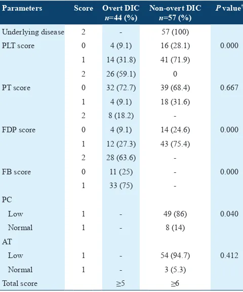
DENV infection was the underlying disease referred to the DIC scoring system for this study. The DIC scoring system used adopted from Taylor et al.11 The DIC scoring system evaluates the following parameters: The underlying disease, PLT, FB, PT, PC, AT, and DD.
Statistical analysis
Differences in laboratory data between patients with DF, DHF, and coagulation tests were tested by compare mean and Chi-square test whichever was appropriate. A P < 0.05 were considered statistically signiicant. The Statistical Package for Social Sciences (SPSS 20.0 version, IBN. Chicago, IL, USA) was used for data analysis.
Ethical considerations
This study was approved by the Regional Ethical Review Committee and written informed consent was obtained from all the patients.
Results
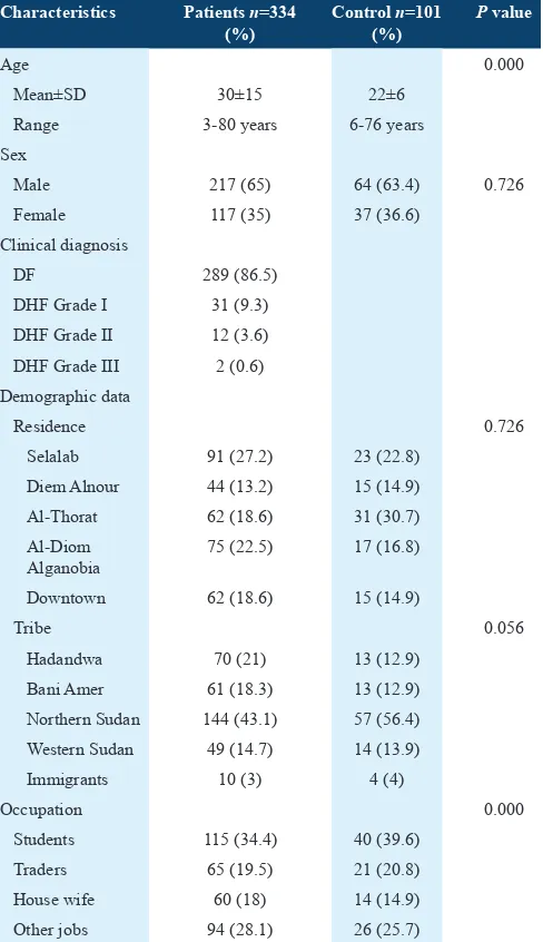
This is a case-control analytical study conducted in Port Sudan Teaching Hospital, Red Sea State, Sudan. The total number of the conirmed diagnosed dengue patients was 334. The age of the patients in this study was between 3 and 80 years (mean age 30 ± 15 years). The control individual aged between 6 and 76 years (mean age 22 ± 6 years). Of the 334 clinical patients, 65% (217) were males and 35% (117) were female. In control group, 63.4% (64) were males and 36.6% (37) were females.
dengue patients. Bleeding manifestations included hematuria in 19 (54.3%) cases, hematemesis in 1 (2.9%), hemoptysis in 1 (2.9%), epistaxis in 5 (14.2%) cases, and gum bleeds in 9 (25.7%) cases.
All of the patients 334 presented were screened for PLT, PT, PTT, and FB. The normal and altered PT, PTT, and FB are shown in Table 2. PLT count was lower in patients than in control (P
< 0.000). Thrombocytopenia was present in 279/334 (83.5%), 234 (81%) in DF cases and 45 (100%) DHF cases. Median PTT was 32.4” (mean ± standard deviation [SD] - 33.5 ± 9.7) (range 15.0-80.7”) and median PT was 13.8” (mean ± SD - 14.1 ± 2.1) (range 10.0-20.4”) (Table 3). PT and PTT were signiicantly higher in the DF (90%) and (72.7%) than DHF (10%) and (27.3%) of patients, respectively. Prolongations of PT, PTT, and low FB were observed in DF patients (P < 0.000) (Table 2). The difference between the patients and the control were found to be signiicant in PLT, PT, PTT, and FB (Table 3).
Out of 334 patients’ positive dengue infection, 101 (30.2%) had abnormal coagulation results. 81 (80.2%) patients with DF and 20 (19.8%) patients with DHF were screened for
special parameters summarized in Table 4. Of 101 patients
72 correction tests for PT and PTT were performed. Corrected results which are indicate a deicient coagulation factor. 72 mixing experimental studies for PT and PTT revealed many coagulation factor deiciencies as in Table 5. This is suggested increased consumption of these factors.
PC was lower in DF 72 (89%) cases and 20 (100%) cases with DHF. No signiicant difference between PC and DENV infection (P < 0.295). So, PS was lower in DF 46 (56.7%) cases and 16 (80%) cases with DHF. No signiicant between dengue infection and PS (P < 0.158). AT was lower in DF patients 79/81 (97.5%) and 18/20 (90%) with DHF. No signiicant correlation between AT and dengue infection (P < 0.226). DD was high in DF patients 70 (86.4%) and 17 (85%) with DHF. No signiicant correlation between DD and dengue infection (P < 0.287). The transaminase liver enzymes were
relatively higher in DHF than DF. 44(43.6%) patients had an elevated level AST, whereas 22 (21.8%) patients had an elevated level ALT. No signiicant correlation between AST/ ALT and the DENV infection (P < 0.978, 0.617, respectively) (Table 6).
A total of 101 (30.2%) had disseminated intravascular coagulation (DIC). DENV infection was the underlying disease referred to the DIC scoring system for this study. The
DIC scoring system used are summarized in Table 7. Nearly
43.6% diagnosed as overt DIC (classic) score ≥5, and 56.4% diagnosed as non-overt DIC score ≥6. The non-overt DIC was signiicantly higher in patients in the study. Interestingly, DIC was signiicantly higher in DHF 20 (19.8%) patients than the DF 81 (80.2%) patients.
Table 1: Characteristics of patients and control in the study
Characteristics Patients n=334
(%)
Range 3-80 years 6-76 years
Sex
Male 217 (65) 64 (63.4) 0.726
Female 117 (35) 37 (36.6)
Clinical diagnosis
Selalab 91 (27.2) 23 (22.8)
Diem Alnour 44 (13.2) 15 (14.9)
Al-Thorat 62 (18.6) 31 (30.7)
Al-Diom Alganobia
75 (22.5) 17 (16.8)
Downtown 62 (18.6) 15 (14.9)
Tribe 0.056
Hadandwa 70 (21) 13 (12.9)
Bani Amer 61 (18.3) 13 (12.9)
Northern Sudan 144 (43.1) 57 (56.4)
Western Sudan 49 (14.7) 14 (13.9)
Immigrants 10 (3) 4 (4)
Occupation 0.000
Students 115 (34.4) 40 (39.6)
Traders 65 (19.5) 21 (20.8)
House wife 60 (18) 14 (14.9)
Other jobs 94 (28.1) 26 (25.7)
DHF: Dengue hemorrhagic fever, DF: Dengue fever, SD: Standard deviation
Table 2: PTT, PT and FB results in comparison to DF/DHF
Parameters DF (%) DHF (%) Total (%) P value PTT
Prolong 32 (76.2) 10 (23.8) 42 (12.6) 0.000
Short 18 (100) 0 18 (5.4)
Normal 242 (88.3) 32 (11.7) 274 (82.0)
Total 292 (87.4) 42 (12.6) 334 (100)
PT
Prolong 27 (90.0) 3 (10.0) 30 (9.0) 0.000
Normal 265 (87.2) 39 (12.8) 304 (91.0)
Total 292 (86.8) 42 (12.6) 334 (100)
FB
Low 37 (60.7) 24 (39.3) 61 (18.3) 0.000
High 42 (87.3) 6 (12.5) 48 (14.4)
Normal 210 (93.3) 15 (6.7) 225 (67.3)
Total 289 (86.5) 45 (13.5) 334 (100)
Discussion
The defects in DF/DHF are multifactorial mechanisms that include thrombopathy, coagulopathy, and vasculopathy. Thrombocytopenia is common in DF, and is a constant finding in DHF.11 The present study increasing the hypothesis that thrombocytopenia is not only a laboratory changing in DF but also may be an important cause of bleeding. Many factors can contribute to the onset of thrombocytopenia in DF from a reactive immune response
against PLTs to decreased PLT production.12 However, in this outbreak the majority of patients (279/334; 83.5%) had thrombocytopenia a inding similar to Karoli et al.; Chairufatah et al.13,14
Hottz et al., studying the dengue induce PLT activation in
Brazil, reported DENV infection induces PLT consumption due to DIC, PLT destruction due to increased apoptosis, lysis by the complement system and by the involvement of anti PLT antibodies.15 Eventually, we suggest the cause of thrombocytopenia in the current study is related to the DIC.
Coagulopathy is also found in DF and most DHF cases, the PTT and FB are more frequently abnormal than PT.16 In this study, PT was demonstrated to be abnormal in some (30/334, 9.0%) but not all patients with dengue infection, these results
are in agreement with previous indings,16-19 whereas PTT
prolongation was observed in 42/334, 12.6% of patients which is similar to results stated by Wills et al., Krishnamurti et al., Lin et al., and Chuang et al.20-22 On the other hand, prolongation of PT and PTT indicates the coagulation in patients with dengue was impaired. Mairuhu et al. and Orsi et al. concluded that the relationship between dengue and activation of coagulation pathway is controversial. However, the causes of this coagulation disorder remain speculative.7,23 Accordingly, this study hypothesized that the coagulation and ibrinolysis are both activated; we attribute this to the presence of DENV. Recently, found nonstructural protein-1 (NS1) of DENV can bind to both thrombin and prothrombin. The thrombin activity is not altered when NS1 bind to thrombin, the binding of NS1 to prothrombin can inhibit its activation, which may contribute to the prolongation of PTT in dengue patients.10 This may explain why PTT abnormality occurs within the irst week of fever onset when antibodies are still underdeveloped.18 In addition, NS1 may also contribute to plasma leakage by mechanism without antibody involved. These results suggest that DENV secreted NS1 plays a direct and important role in vascular leakage and hemorrhage in DHF/DSS.22 Regardless of the causes of the coagulopathy, our study showed that the coagulation activity may be impaired during dengue infection and that this disorder may cause bleeding.
With respect to plasma levels of the naturally occurring anticoagulants PC, PS and AT were signiicantly reduced in the present study. PC, PS and AT are predominantly synthesized in the liver and lower circulating levels probably relect capillary
Table 3: The difference between test and control in studied parameters
Parameters Mean±SD Median test Median control Range test Range control P value
Test group Control group
PLT count×109/L 95.691±57 219.099±59 95 214 3-443 93-509 0.000
PT seconds 14.1±2.1 13.3±1.6 13.8 13.5 10-20.4 10-16.5 0.000
PTT seconds 33.5±9.7 29.6±4.5 32.4 28.9 15-80.7 20-39 0.000
FB g/dl 4.15±6.99 3.26±1.44 2.60 2.90 0.5-63.96 1.37-10.23 0.000
PT: Prothrombin time, PTT: Partial thromboplastin time, FB: Fibrinogen, SD: Standard deviation, PLT: Platelet
Table 4: Laboratory finding of specific parameters in the study
Parameters Median Mean±SD Range of test Normal range
PC 47% 48±22 8-140 70-140%
PS 60% 76±50 4-273 65-140%
AT 2.1 mg/dl 5.1±6.6 0.23-30 17-30 mg/dl
DD 1.3 mg/dl 3.9±5.9 0.10-22.5 <0.3 mg/dl
AST 34 U/l 54±69 5-480 Up to 40 U/L
ALT 22 U/l 34±43 2-279 Up to 41 U/L
PC: Protein C, PS: Protein S, AT: Antithrombin, DD: D-dimer, AST: Aspartate transaminase, ALT: Alanine transaminase, SD: Standard deviation
Table 5: Summary of factors deficient by mixing experiment study
Blood factor deficient Number of patients n=72
Frequency (%)
X 14 19
V 7 10
II 9 12
VIII 25 35
IX 7 10
XII 10 14
Total 72 100
Table 6: Special parameters among DF and DHF patients
Parameters DF n=81 (%) DHF n=20 (%) P value
Low PC 72 (89) 20 (100) 0.295
Low PS 46 (56.7) 16 (80) 0.158
Low AT 79 (97.5) 18 (90) 0.226
High DD 70 (86.4) 17 (85) 0.287
High AST 35 (43.2) 9 (45) 0.978
High ALT 17 (21) 5 (25) 0.617
leakage alone. An increase in the rate of consumption of these proteins may be a contributing factor in the severity of disease.20 Cabello-Gutiérrez et al. studying the modiication of the
cytoprotective PC pathway during dengue infection of human endothelial vascular cells in Mexico, reported an increased level of soluble TM in the plasma of patients with DHF/DSS, and they present a participation of DENV in down-regulation of TM-thrombin-PC complex formation at the endothelial surface with a reduction in activated PC.24 Chuang et al. proposed that the expression of several other PC activations related molecules such as epithelial cell receptor and PS are also increased together with TM in DENV infection,25 a result relatively similar to our inding regarding PS. During severe infection, AT may become very low due to consumption, decreased synthesis, and degradation by elastase resulting from activated neutrophils. Glycosaminoglycan could cause reduction of AT function because glycosaminoglycan has a role as a co-factor like physiologic heparin for AT. Whereas AT reduction in DHF mainly caused by capillary leakage, and may be also by the dilution effect of the administered intravenous luid and elevated consumption, so that AT is related to the severity degree of DHF.26 Low levels of the anticoagulant proteins are usually associated with the development of thrombotic complications or signiicant laboratory evidence of DIC.27
On the other hand, excessive ibrinolysis was demonstrated in our patients with dengue infection, since plasma levels DD were signiicantly increased in those patients. In a similar way, previous studies have demonstrated that patients with DHF may
also present with excessive ibrinolysis.9,11,20 Chuang et al. in a study by Institute of Basic Medical Sciences, Taiwan, reported data suggest dengue-induced plasminogen cross-reactive antibodies may enhance plasminogen conversion to plasmin, which would be expected to contribute to hyperibrinolysis.28 This study showed signiicantly higher DD level in 86.4% of DF patients compared with 85% DHF with the sensitivity of DD in predicting DF of 80%. These results are strongly consistent with Setrkraising et al. and Orsi et al.23,29 DD was also found to be positively correlated with DIC (P < 0.000). Detection of DD in patients with dengue infection may be beneicial for predicting the clinical course of the disease. This helps the clinicians in predicting the disease severity before the patients’ progress into the toxic stage. Detection of DD suggests that DIC and activation of the ibrinolytic system occur early in patients with DENV infection before the onset of severe hemorrhagic manifestations.
Aminotransferase levels are useful in predicting the occurrence of hepatic dysfunction and spontaneous bleeding. Liver enzyme elevation is a common feature in dengue infection.30 However, Wong and Shen were reported that AST abnormality was predominantly high as compared to ALT; 91% and 72%, respectively.31 In this study, AST levels were equal to or greater than those of ALT levels in most of the infected patients, indings that have also been reported earlier by Ali and Elgasim; Shukla and Chandr.32,33
A correlation between the levels of AST and PTT shows a strong association between AST elevation and PTT prolonged time in dengue infection in our patients (P < 0.003), this might seem to relate to the process of hepatic parenchymal damage than the biliary tract obstruction. Ultimately, our indings suggest that lower levels of these proteins in conjunction with high rates of the liver enzyme are due to the presence of DIC. To the best of our knowledge, this is the irst study in our area to investigate the association between dengue infection and natural physiologic anticoagulants. Limitations of this study are that the data were collected manually and some information was missing. Some factors inluencing natural physiologic anticoagulants such as chronic inlammatory state. Another limitation is that the sophisticated laboratory values such as PC, PS, and AT were performed only for the patients had abnormal coagulation tests, this needs to be addressed in the further studies.
Conclusion
This study demonstrated and conirmed the evidence that the coagulation in patients with dengue was abnormal. Moreover, the causes of this abnormality are attributed to DIC.
References
1. Chen JP, Cosgriff TM. Hemorrhagic fever virus-induced changes in hemostasis and vascular biology. Blood Coagul Fibrinolysis 2000;11:461-83.
Table 7: Summary of DIC score results
Parameters Score Overt DIC
n=44 (%)
Non‑overt DIC
n=57 (%)
P value†
Underlying disease 2 - 57 (100)
PLT score 0 4 (9.1) 16 (28.1) 0.000
†P<0.05. DIC: Disseminated intravascular coagulation, PT: Prothrombin time, FDP: Fibrin
2. Henchal EA, Putnak JR. The dengue viruses. Clin Microbiol Rev 1990;3:376-96.
3. Chang SF, Huang JH, Shu PY. Characteristics of dengue epidemics in Taiwan. J Formos Med Assoc 2012;111:297-9.
4. Mustafa MS, Rasotgi V, Jain S, Gupta V. Discovery of ifth serotype
of dengue virus (DENV-5): A new public health dilemma in dengue control. Med J Armed Forces India 2015;71:67-70.
5. Schexneider KI, Reedy EA. Thrombocytopenia in dengue fever. Curr Hematol Rep 2005;4:145-8.
6. Jameel T, Mehmood K, Mujtaba G, Choudhry N, Afzal N, Paul RF.
Changing haematological parameters in dengue viral infections. J Ayub Med Coll Abbottabad 2012;24:3-6.
7. Mairuhu AT, Mac Gillavry MR, Setiati TE, Soemantri A, ten Cate H, Brandjes DP, et al. Is clinical outcome of dengue-virus infections
inluenced by coagulation and ibrinolysis? A critical review of the
evidence. Lancet Infect Dis 2003;3:33-41.
8. Turgeon ML. Clinical Hematology: Theory and Procedures. 5th ed.
China: Wolters Kluwer Health, Lippincott Williams and Wilkins; 2011. p. 420-3.
9. Sosothikul D, Seksarn P, Pongsewalak S, Thisyakorn U, Lusher J.
Activation of endothelial cells, coagulation and ibrinolysis in children
with Dengue virus infection. Thromb Haemost 2007;97:627-34.
10. Lin SW, Chuang YC, Lin YS, Lei HY, Liu HS, Yeh TM. Dengue virus nonstructural protein NS1 binds to prothrombin/thrombin and inhibits prothrombin activation. J Infect 2012;64:325-34.
11. Taylore FB, Toh CH, Hoots WK, Wada H, Levi M. Toward deinition,
clinical and laboratory criteria and a scoring system for disseminated intravascular coagulation. Thrombo Haemost 2001;86(5):1327-30.
12. Saito M, Oishi K, Inoue S, Dimaano EM, Alera MT, Robles AM, et al.
Association of increased platelet-associated immunoglobulins with thrombocytopenia and the severity of disease in secondary dengue virus infections. Clin Exp Immunol 2004;138:299-303.
13. Karoli R, Fatima J, Siddiqi Z, Kazmi KI, Sultania AR. Clinical proile
of dengue infection at a teaching hospital in North India. J Infect Dev Ctries 2012;6:551-4.
14. Chairufatah A, Setiabudi D, Agoes R, Colebunder R. Thrombocytopenia and platelet transfusions in dengue haemorrhagic fever and dengue shock syndrome. Dengue Bull 2003;27:138-43.
15. Hottz ED, Oliveira MF, Nunes PC, Nogueira RM, Valls-de-Souza R,
Da Poian AT, et al. Dengue induces platelet activation, mitochondrial dysfunction and cell death through mechanisms that involve DC-SIGN and caspases. J Thromb Haemost 2013;11:951-62.
16. Sellahewa KH. Pathogenesis of dengue haemorrhagic fever and its impact on case management. ISRN Infect Dis 2013;2013:6.
17. Nimmamnitya S. Dengue Hemorrhagic Fever: Disorders of Hemostasis. IX Congress of the International Society of Haematology
Asia-Paciic Division. Bangkok, Thailand; 1999. p. 185-7.
18. Wills B, Tran VN, Nguyen TH, Truong TT, Tran TN, Nguyen MD,
et al. Hemostatic changes in Vietnamese children with mild dengue correlate with the severity of vascular leakage rather than bleeding. Am
J Trop Med Hyg 2009;81:638-44.
19. Díaz-Quijano FA, Villar-Centeno LA, Martínez-Vega RA. Early
indicators of dengue infection in children. An Pediatr (Barc) 2006;64:523-9.
20. Wills BA, Oragui EE, Stephens AC, Daramola OA, Dung NM, Loan HT, et al. Coagulation abnormalities in dengue hemorrhagic fever: Serial investigation in 167 Vietnamese children with dengue shock syndrome. Clin Infect Dis 2002;35:277-85.
21. Krishnamurti C, Kalayanarooj S, Cutting MA, Peat RA, Rothwell SW, Reid TJ, et al. Mechanisms of hemorrhage in dengue without circulatory collapse. Am J Trop Med Hyg 2001;65:840-7.
22. Chuang YC, Lin YS, Liu HS, Wang JR, Yeh TM. Antibodies against thrombin in dengue patients contain both anti-thrombotic and
pro-ibrinolytic activities. Thromb Haemost 2013;110:358-65.
23. Orsi FA, Angerami RN, Mazetto BM, Quaino SK, Santiago-Bassora F,
Castro V, et al. Reduced thrombin formation and excessive ibrinolysis
are associated with bleeding complications in patients with dengue fever: A case-control study comparing dengue fever patients with and without bleeding manifestations. BMC Infect Dis 2013;13:350.
24. Cabello-Gutiérrez C, Manjarrez-Zavala ME, Huerta-Zepeda A, Cime-Castillo J, Monroy-Martínez V, Correa BB, et al. Modiication
of the cytoprotective protein C pathway during dengue virus infection of human endothelial vascular cells. Thromb Haemost 2009;101:916-28.
25. Chuang YC, Lin YS, Liu CC, Liu HS, Liao SH, Shi MD, et al. Factors
contributing to the disturbance of coagulation and ibrinolysis in
dengue virus infection. J Formos Med Assoc 2013;112:12-7.
26. Jong JB, Pohan HT, Zulkarnain I, Tambunan KL, Panggabean MM, Setiabudy RD, et al. The correlation between coagulation test and albumin with antithrombin III in dengue hemorrhagic fever. Acta Med Indones 2004;36:57-61.
27. Vervloet MG, Thijs LG, Hack CE. Derangements of coagulation and
ibrinolysis in critically ill patients with sepsis and septic shock. Semin
Thromb Hemost 1998;24:33-44.
28. Chuang YC, Lei HY, Liu HS, Lin YS, Fu TF, Yeh TM. Macrophage migration inhibitory factor induced by dengue virus infection increases vascular permeability. Cytokine 2011;54:222-31.
29. Setrkraising K, Bongsebandhu PC, Voraphani N, Pancharoen C, Thisyakorn U, Thisyakorn C. D-dimer as an indicator of dengue severity. Asian Biomed 2007;1:53-7.
30. Parkash O, Almas A, Jafri SM, Hamid S, Akhtar J, Alishah H. Severity of acute hepatitis and its outcome in patients with dengue fever in a tertiary care hospital Karachi, Pakistan (South Asia). BMC Gastroenterol 2010;10:43.
31. Wong M, Shen E. The utility of liver function tests in dengue. Ann Acad Med Singapore 2008;37:82-3.
32. Ali KA, Elgasim SA. A correlation study between clinical manifestation of dengue fever and degree of liver injury. J Microbiol Antimicrob 2012;4:45-8.