L
Journal of Experimental Marine Biology and Ecology 249 (2000) 1–12
www.elsevier.nl / locate / jembe
The ionic basis of cardiac activity in the bivalve mollusc
Perna perna
* ˜ ˆ
Rosangela Pereira Ferreira, Luiz C. Salomao
Department of Physiology, University of Sao Paulo, Sao Paulo, Brazil
Received 25 October 1999; received in revised form 7 December 1999; accepted 24 January 2000
Abstract
The ionic basis of cardiac activity and aspects of excitation–contraction (E–C) coupling were investigated in the isolated heart of the bivalve mollusc Perna perna, using the sucrose-gap technique. The role of the principal ions was established employing artificial seawater, in which specific ion concentrations were modified, and ion channel blockers. The mean membrane resting potential (MP) and the action potential (AP) were 23360.7 mV (n589) and 1360.3 mV
1
(n571), respectively. The MP potential was primarily dependent on K ions. Three types of cardiac APs were identified: fast, slow and spike-plateau potentials. Cardiac activity was
1 21 21
maintained in Na - or Ca -free salines but ceased when either Cd or EDTA was added to
21
these salines. Other Ca channel blockers reduced the amplitude and increased duration of the cardiac APs. Tetrodotoxin (TTX) and procaine did not alter the AP. The data showed that the
21
depolarizing phase of the AP was dependent on Ca influx while the plateau phase, when
1 21
present, resulted from Na influx that was modulated by Ca . The mechanical responses were
21
more sensitive to changes in extracellular Ca concentration than were the electrical responses. 2000 Elsevier Science B.V. All rights reserved.
Keywords: Perna perna; Membrane potentials; Heart; Mollusc; Sucrose-gap technique
1. Introduction
The mollusc heart is myogenic in nature and its pacemaker cells are modified muscle fibers. However, efforts to identify the pacemaker have failed because this system is believed to be a diffuse structure (Irisawa, 1978). One of the advantages of studying
˜ ˜
ˆ
*Corresponding author. Instituto de Biociencias, Universidade de Sao Paulo, Rua do Matao, Trav. 14[101, ˜
´
CEP 05508-900, Cidade Universitaria, Sao Paulo, Brazil. Tel.: 155-11-818-7518; fax: 155-11-211-5028. ˜
E-mail address: [email protected] (L.C. Salomao)
the marine bivalve mollusc Perna perna (Mytilidae) and to examine the role of the identified ions in the process of excitation–contraction (E–C) coupling.
2. Materials and methods
´
Specimens of the intertidal bivalve Perna perna (Linne, 1758) were collected from banks off the coast of Sao Paulo State (238499S; 458279W), Brazil, and transported to the Department of Physiology of the University of Sao Paulo. They were maintained in 100-l aerated aquaria in sea water of 34‰ salinity.
The sucrose-gap apparatus consisted of two cylindrical lucite hemi-chambers, each 13.5 mm long and 17 mm in external diameter, with a central orifice of 3.3 mm diameter. Each chamber also had three 2-mm diameter orifices arranged radially. In the upper chamber, these orifices allowed the inflow and outflow the test solutions, and the insertion of the test electrode, respectively. In the lower chamber, the orifices allowed perfusion with a depolarizing solution (0.5 M KCl) and insertion of the reference electrode. A rubber disk with the same external and internal diameters of the hemi-chambers, covered with thin latex membranes, interposed between them, formed the sucrose chamber. The sucrose chamber was perfused with a 1.0-M sucrose solution through needles inserted radially into the rubber disk.
Test solutions were obtained by modifying Pantin (1948) artificial seawater (ASW) which contains the following salts per liter solution: NaCl (23.43 g), KCl (0.73 g),
CaCl (1.12 g), MgCl (5.01 g), NaHCO (0.21 g), Na (SO) (3.95 g) and NaBr (0.062 2 3 2 4
g). The osmolality of each solution was measured with a Fiske osmometer. Where specific ion-free solutions were used, the solution was osmotically adjusted with
21 2
choline-chloride, potassium aspartate or sodium aspartate. Ca and Cl concentrations
1 1
were measured by microtitration (Beckman-Spinco Model 150) and Na and K
concentrations by flame photometry (Zeiss PM Q II). The ionic concentrations of the
1 1 21 2
control saline were (in mM): Na (460), K (10), Ca (10), Cl (540).
The following ion channel blockers were added directly to the perfusion solutions:
1
tetrodotoxin (TTX) or procaine for Na channels; cobalt chloride, manganese chloride,
cadmium iodide and verapamil for calcium channels; tetraethylammonium (TEA),
1
4-aminopyridine (4-AP) and cesium chloride for K channels.
Although the composition of the control ASW was identical in all experiments, inexplicable variability was present in the cardiac activity of each preparation. For this reason, the respective control values were previously established in each experimental
condition. All experiments were performed at room temperature (24618C).
Data were given as the mean6standard error of the mean (S.E.M.). Statistical analysis was performed using ANOVA followed by the Student–Newman–Keuls (SNK) test, or
a paired t-test. Minimum significance was set at P50.05.
3. Results
3.1. Characterization of membrane potentials
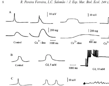
Three types of cardiac APs (fast, spike-plateau and slow) were identified in P. perna ventricles perfused with ASW of 34‰ salinity by using the sucrose-gap (Fig. 1). The mean values obtained for the resting potential (RP) and AP amplitude (measured from
the RP to the AP peak) were 23360.7 (n589) and 1360.3 mV (n571), respectively.
The depolarizing phase was never positive.
3.2. Effects of extracellular potassium and chloride ions
1
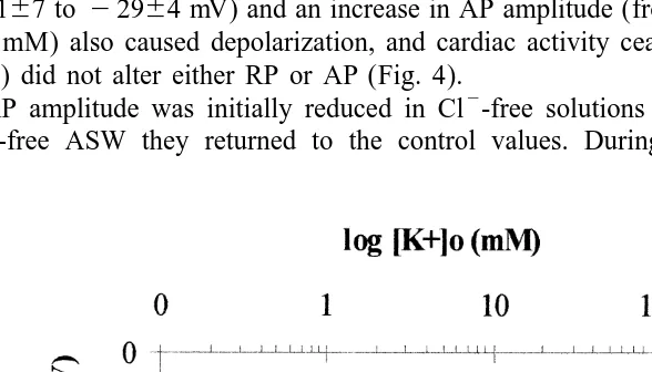
An increase in extracellular K concentration from 10 to 100 mM caused significant
21
depolarizations at a rate of 18 mV decade rather than the 58 mV predicted from the
1
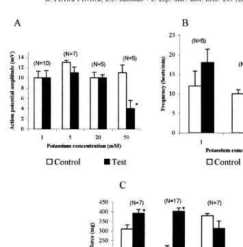
Nernst equation for a perfect K selective electrode (Fig. 2). The amplitude of APs
recorded in salines containing 1, 5, 10 and 20 mM KCl was not changed, however, in 50 mM KCl, the amplitude was markedly reduced. Reductions in external KCl from 10 to 5 or 10 to 1 mM caused positive inotropic response. Conversely, an increase from 10 to 20 mM KCl induced negative chronotropic and inotropic responses, however not significant by t-test (Fig. 3). At KCl concentrations of 50 mM or greater cardiac activity was reduced and eventually ceased. However, rhythmicity returned when the hearts were perfused with the control ASW.
1
Fig. 1. Different types of cardiac action potential obtained by the sucrose-gap method from the isolated heart of Perna perna perfused with artificial seawater of 34‰ salinity (control). Fast (A), spike-plateau (B), and slow action potentials (C).
24167 to 22964 mV) and an increase in AP amplitude (from 1364 to 2166). 4-AP
(20 mM) also caused depolarization, and cardiac activity ceased. Cesium chloride (50 mM) did not alter either RP or AP (Fig. 4).
2
AP amplitude was initially reduced in Cl -free solutions although after 30 min in
2
Cl -free ASW they returned to the control values. During this time, the RP was
Fig. 2. Relationship between the resting potential and extracellular potassium concentration in isolated heart of
Perna perna. The asterisks indicate significant differences compared to the control concentration. Data is a
Fig. 3. Action potential amplitude (A), cardiac frequency (B), and systolic force (C) obtained from isolated
1
hearts of Perna perna in salines containing different K concentrations. Data are the mean6S.E.M.; asterisks indicate significant differences between the control and test salines.
depolarized and returned to initial values only when the hearts were perfused with control saline.
3.3. Effects of sodium ions
1
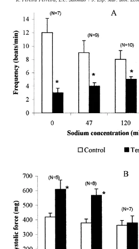
A reduction in external Na concentration caused hyperpolarization of the RP and
1
increased AP amplitude. In external Na concentration of 47 mM the RP changed from
23662.5 to 24162 mV and the amplitude increased from 1060.8 to 1360.6 mV.
1
Fig. 4. Action potentials recorded from isolated hearts of Perna perna perfused initially with artificial seawater of 34‰ salinity (control saline), then either TEA (50 mM) (A), 4-AP (20 mM) (B) or CsCl (50 mM) (C). 4-AP was added from a stock solution at the time indicate by the arrow.
21 1
Cd (10 mM) (Fig. 5). The Na channel blockers TTX or procaine did not alter RP or
1 1
AP. Na concentrations lower than that of the control saline and Na -free saline caused
a negative chronotropic response coupled to increased systolic force (Fig. 6).
3.4. Effects of calcium ions
21
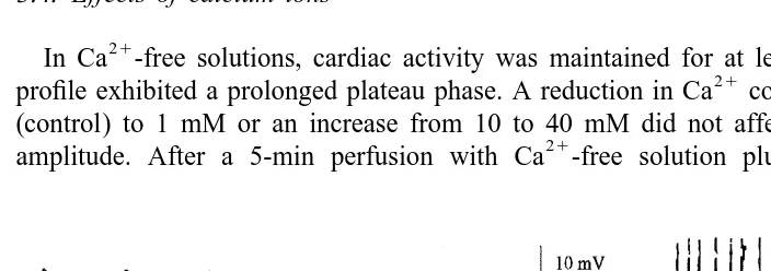
In Ca -free solutions, cardiac activity was maintained for at least 30 min; the AP
21
profile exhibited a prolonged plateau phase. A reduction in Ca concentration from 10
(control) to 1 mM or an increase from 10 to 40 mM did not affect either RP or AP
21
amplitude. After a 5-min perfusion with Ca -free solution plus EDTA (2 mM),
Fig. 5. Action potentials (upper trace) and respective cardiograms (lower trace) recorded from isolated hearts
1
of Perna perna when perfused with artificial seawater of 34‰ salinity (control saline), followed by a Na -free
1
Fig. 6. Frequency (A) and systolic force (B) recorded from the isolated heart of Perna perna perfused with
1
different Na concentrations. The data are the mean6S.E.M.; asterisks indicate significant differences between
1
Fig. 7. Action potentials (upper trace) and the respective cardiogram (lower trace) recorded from isolated 21
hearts of Perna perna in control saline, in Ca -free saline and in Ca-free saline plus EDTA (2 mM) (A).
21 21 21
Action potentials were altered by Cd (5 and 10 mM) (B), Co (20–30 mM) (C) and Mn (30–40 mM) (D).
depolarization occurred and rhythmicity was gradually lost; the APs exhibited an atypical profile with a prolonged repolarization period (about 4 min) (Fig. 7A). In all cases, the hearts recovered their normal activity after 40-min perfusion with control saline.
21
Typical response to Ca channel blockers are shown in Fig. 7B–D. Cadmium, cobalt
or manganese increased AP duration and finally abolished cardiac activity at con-centrations of 10, 30 and 40 mM, respectively. Similar results were obtained with L-type channel blocker verapamil (0.65 mM).
21
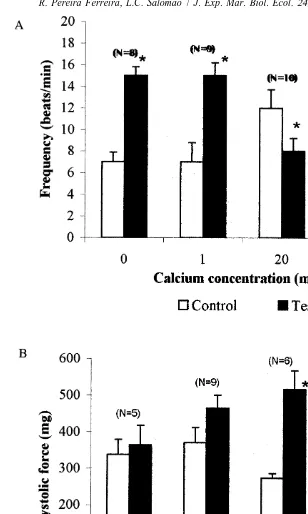
Reductions in Ca concentration caused a positive chronotropic effect while
21
conversely Ca increases caused a negative chronotropic effect. Systolic force was
21
unaffected by calcium concentrations lower than normal, but at 20 mM Ca , systolic
force increased twofold (Fig. 8).
4. Discussion
Fig. 8. Frequency (A) and systolic force (B) recorded from isolated hearts of Perna perna, perfused with 21
21
blockers and to the L-type channel blocker verapamil. In Ca -free solutions there was a
conspicuous occurrence of a plateau phase, the duration of which increased with
21
perfusion time. The same effect was observed when the Ca -chelator EDTA was added
to the saline.
1
Although AP amplitude did not change in a Na -free solution, amplitude was
1 1
enhanced when the Na concentration was reduced ten-fold suggesting that Na ions
1
play little or no role in the depolarizing phase. The Na channel blockers, TTX or
1
procaine did not change the AP, suggesting that the Na channel present in the heart of
P. perna was TTX-insensitive as described in the hearts of other mollusc species (M.
edulis, Irisawa et al., 1967; an oyster species, Irisawa et al. 1968; M. demissus, Wilkens, 1972b; Spisula sachalinensis, Filippov et al. 1988).Given these results, we suggest that
21 1
the depolarising phase was due to an inward Ca current, and the plateau to Na influx.
1 21
The putative Na channels were sensitive to Cd since cardiac activity that persisted in
1 21
Na -free saline was abolished by the addition of Cd . The plateau appeared to be
1 21
Na -dependent and modulated by Ca since the duration of the plateau seen in
21 21
Ca -free solution was reduced by the addition of Ca . Such modulation does not
1
necessarily imply the existence of a Na -specific channel but rather, the presence of a
1 21
non-selective cation channel unable to discriminate between Na and Ca (Hess and
21 21
Tsien, 1984). We possibly identified a Ca channel that, at low extracellular Ca
1
concentrations, was permeant to Na , as that observed in guinea pig cardiac myocytes (Hess and Tsien, 1984). Wilkens (1972b) and Devlin (1993) have suggested that the
21 1
extracellular Ca concentration modulated the transient Na current responsible for the
AP plateau in Modiolus and Mercenaria. Wilkens (1972b) suggested that such a plateau was characteristic of species exposed to the reduced and variable salinities found in intertidal and estuarine regions. However, species living in stable environments in which
21
Ca concentration was fairly stable, did not show the AP plateau. Perna perna is found
in intertidal regions and was tolerant to a broad range of salinities.
The contractile mechanism in P. perna was more sensitive to a reduction in
21
extracellular Ca concentration than was the electrical response. During perfusion with
21
a Ca -free saline plus EDTA, mechanical activity ceased before electrical activity.
Curiously, an initial increase in systolic force was observed concomitant with
extracellu-21
1
An increase in systolic force was seen when hearts were perfused with either low Na
1
salines or a Na -free solution.This may be related to a rise in cytosolic calcium due to:
1
(i) an increase in calcium influx facilitated by absence of Na , assuming that the same
1 21
channel was shared by these two ions; (ii) a reduction in Na -Ca antiporter activity as
1
a consequence of the lack of or low Na concentrations in the extracellular medium.
Deaton and Greenberg (1980), have proposed that the ionic sensitivity of the bivalve myocardium might be distributed by taxon. Bivalves of the subclass Pteriomorphia are
21 1
sensitive to Ca but not to Na removal, while the opposite is seen in the Heterodonta
and Paleoheterodonta. Of 25 species studied, Deaton and Greenberg (1980) found that
1
hearts from the Heterodonta and Paleoheterodonta did not beat in Na -free seawater, but
21 1
that activity was maintained in Ca -free medium, suggesting a critical role for Na in
cardiac excitability. Conversely, hearts from pteriomorphia bivalves maintained their
1 21
spontaneous activity in the absence of Na but not in Ca -free medium. The present
results for the pteriomorphia mytilid P. perna clearly support this hypothesis, although
21
the Ca requirement seems to be very low.
Acknowledgements
We thank to Centro de Biologia Marinha–CEBIMAR, for supplying the mussels. This work was partially supported by CNPq–Conselho Nacional de Pesquisa, Brazil. [SS]
References
Brezden, B.L., Gardner, D.R., 1984. The ionic basis of the resting potential in a cross-striated muscle of the aquatic snail Lymnaea stagnalis. J. Exp. Biol 108, 305–314.
Deaton, L.E., Greenberg, M.J., 1980. The ionic dependence on the cardiac action potential in bivalve molluscs: systematic distribution. Comp. Biochem. Physiol. 67A, 155–161.
Devlin, C.L., 1993. An analysis of control of the ventricle of the mollusc Mercenaria mercenaria. I. The ionic basis of autorhythmicity. J. Exp. Biol. 179, 47–61.
Dorset, D.A., Evans, C.G., 1989. The ionic basis of the resting potential of molluscan unstriated muscle. J. Comp. Physiol. B 159 (3), 305–312.
Filippov, A.K., Porotikov, V.I., Novolesova, V.D., Bessonov, B.I., Butsuk, V., 1988. Action potential and ionic currents of myocardial fibers in the mollusc Spisula sachalinensis. Comp. Biochem. Physiol. 89A, 1–5. Hess, P., Tsien, R.W., 1984. Mechanism of ion permeation through calcium channels. Nature (Lond.) 309,
453–456.
Irisawa, H., 1978. Comparative physiology of the cardiac pacemaker mechanisms. Physiol. Rev. 58 (2), 461–498.
Irisawa, H., Kobayashi, M., 1964. On the spontaneous activities of oyster myocardium caused by several inorganic ions in sucrose solution. Jpn. J. Physiol. 14, 165–176.
1 21
Irisawa, H., Shigeto, N., Otani, M., 1967. Effect of Na and Ca on the excitation of the Mytilus (Bivalve) heart muscle. Comp. Biochem. Physiol. 23 (1), 199–212.
Irisawa, H., Noma, A., Ueda, R., 1968. Effect of calcium on the spontaneous activities of the oyster myocardium in sodium free solution. Jpn. J. Physiol. 18, 157–168.
Jones, H.D., 1983. The circulatory systems of gastropods and bivalves. In: Wilbur, K.M. (Ed.), The Mollusca, Academic Press, New York, pp. 189–238.
´