www.elsevier.com / locate / bres
Interactive report
1
Activation of trigeminovascular neurons by glyceryl trinitrate
*
Geoffrey Andrew Lambert , Cathy Donaldson, Peter Michael Boers,
Alessandro Stefano Zagami
Institute of Neurological Sciences, The Prince Henry and Prince of Wales Hospitals, University of New South Wales, Little Bay NSW 2036, Australia
Accepted 12 September 2000
Abstract
The effect of intra-carotid arterial infusions of glyceryl trinitrate (GTN), a substance known to precipitate headache, including migraine, upon the spontaneous activity of trigeminal neurons with craniovascular input was studied in cats. Second-order craniovascular neurons which received sensory input from the superior sagittal sinus were recorded in the trigeminal nucleus caudalis. Infusions of GTN were administered via a catheter inserted retrogradely into the common carotid artery through the lingual artery. Infusions of GTN (100mg
21 21 21
kg min in a volume of 2 ml min ) increased the mean basal discharge rate of all second-order neurons to 239647% of control. GTN produced a fall in mean blood pressure, but there was no correlation between this fall and the changes in discharge rate. GTN infusions sensitised neurons to the effects of electrical stimulation of the superior sagittal sinus, but not to subsequent GTN infusions. Infusions of similar volumes of vehicle did not alter the discharge rate of neurons. We conclude that GTN activates craniovascular sensory pathways at a site at, or peripheral to, the second-order neuron and that such an action may account for at least the acute-onset headache induced by GTN. 2000 Elsevier Science B.V. All rights reserved.
Theme: Sensory systems
Topic: Pain modulation: pharmacology
Keywords: Glyceryl trinitrate; Trigeminal; Vascular headache
1. Introduction likely that they are due to release of nitric oxide (NO)
from the GTN donor molecules.
Glyceryl trinitrate (GTN, nitroglycerin) was first syn- Glyceryl trinitrate administration leads to an immediate thesised in 1846 by Sobrero who immediately observed throbbing headache in nearly all subjects [12,19]. In that it produced a ‘migraine’ when he tasted it [29]. It was susceptible subjects, GTN reliably triggers a cluster head-used as a homeopathic remedy for headache two years ache like pain [4]. In migraineurs, it also produces a later [7] and for angina and hypertension later in the delayed headache highly reminiscent of the spontaneous nineteenth century [19]. Alfred Nobel observed that work- migraine headaches which these people suffer. These ers in dynamite factories where the active principal, effects have been attributed to a selective action of GTN nitroglycerin, was incorporated into the explosive, suffered on extracranial and intracranial blood vessels, principally from chronic migraine-like headaches at work and angina- the dural arteries and large penetrating cerebral arteries, like problems on their vacations [19]. The therapeutic and mainly through the formation of NO [22]. It is tempting to side effects were thought to be due to the nitrate [20], as assume that the immediate headache is a consequence of they were shared by other organic nitrates [23], but it is the vasodilator action of GTN, but both it and the delayed headache might be due to an effect of GTN or NO on neuronal function [32]. In 1993, Olesen and co-workers demonstrated that the immediate headache induced by 1
Published on the World Wide Web on 7 November 2000. GTN could be ameliorated by prior subcutaneous injection *Corresponding author. Room 136, Clinical Sciences Building, Prince
of the antimigraine drug sumatriptan, which also reduced Henry Hospital, Little Bay NSW 2036, Australia. Tel.:161-2-9382-5728;
the frequency of the delayed headache [11]. In rats, fax:161-2-9382-5127.
E-mail address: [email protected] (G.A. Lambert). intravenous administration of GTN leads to increased
expression of c-fos, a marker for neuronal activation, in the supramaximal square-wave shocks (#150 V, 250 ms trigeminal nucleus caudalis and several other brain nuclei duration, 0.3 Hz) with a Grass S88 stimulator.
[31]. This activation was suppressed by the intraperitoneal The central tungsten wire of a glass-coated tungsten administration of the non-steroidal anti-inflammatory drug electrode [18] was used to record single unit activity in the indomethacin [33], which is effective in migraine [2]. trigeminal nucleus caudalis. The electrode was placed on GTN-induced expression of c-fos in the caudal trigemi- the dorsal surface of the brainstem, 1.5–3 mm caudal to nal nucleus does not however demonstrate unequivocally the obex and 2.5–4.5 mm lateral to the midline, and whether GTN specifically activates those neurons respon- advanced to a depth of up to 2500mm below the surface sible for dural or cerebrovascular pain processing, synaptic by means of a piezoelectric microdrive. Single unit activity activation is only inferred. The experiments we describe was amplified, filtered and displayed on an oscilloscope. here were carried out to determine whether GTN adminis- Peri-stimulus and post-stimulus histograms were compiled tered into the carotid artery of cats can produce activation and used for post-experiment analysis of the latency and of neurons which also process sensory information from firing frequency of single units.
the dura. Neurons were located first by the presence of a response
to the search stimulus-electrical stimulation of skin or superior sagittal sinus. Some units were tested for the presence of a cutaneous receptive field. The skin and hair
2. Materials and methods of the face were examined systematically with a variety of
stimuli (brush, light touch, heavy pressure and pinch), and Seven male or female cats (mass 2.760.5 kg; the cells classified according to response. Low threshold mean6standard deviation) were used in these experiments. mechanoreceptive (LTM) units responded to light touch or They were anesthetised with halothane 1.5%, and then brush, and did not increase firing rate with noxious stimuli. with intraperitoneal injections of a-chloralose (60 mg Nociceptive specific (NS) units responded only to heavy
21
kg ). The femoral artery and vein were cannulated to pressure or pinch, and wide dynamic range (WDR) units measure blood pressure and heart rate and to administer responded to non-noxious stimuli, but had an increased intravenous drugs and fluids respectively. Animals were firing rate in response to noxious stimuli [9]. Congruence intubated and ventilated with 30% oxygen in air to keep of action potential shape between SSS-induced responses end-expiratory CO in the range 3.5–4.0%. Throughout the2 and RF-induced responses was assessed visually or via an experiment, the animal was immobilised with intermittent averaging program to ensure that both modes of
stimula-21
intravenous gallamine triethiodide, 20 mg kg . The depth tion were activating the same neuron.
of anesthesia during immobilisation was assessed period- One to three post-stimulus histograms, each consisting ically during the experiment by testing for sympathetic of 50 successive recordings of discharges in response to responses (pupillary dilatation, tachycardia, raised blood stimulation, were recorded for each neuron under control pressure) to noxious stimulation, and regularly by allowing conditions. Histograms were acquired in pairs, with sagittal the effects of gallamine to wear off and testing for sinus and receptive field stimuli alternating in each acquisi-withdrawal reflexes to pinching a hindpaw. Supplementary tion run. The spontaneous discharge rate of the neurons doses of either chloralose or gallamine were given when was monitored by recording peri-event histograms in necessary. Rectal temperature was monitored throughout which each bin recorded the number of discharges re-the experiment with a re-thermistor, and was maintained at corded over a sample time of 4 s. Control discharge rates 37–388C by means of a servo-controlled heating blanket. were recorded for 15–20 min. Glyceryl trinitrate (David
21
The lingual artery on one side was catheterised re- Bull Laboratories) 5 mg ml in 30% (v / v) ethanol, 30% trogradely with a small diameter catheter (SV8 polyvinyl (v / v) propylene glycol was diluted 1:100 in normal saline. tubing, Critchley Electrics), advanced until its tip lay in the Drug-free vehicle was made from ethanol (30%) and common carotid artery. Heparinised normal saline (50 I.U. propylene glycol (30%) in distilled water, similarly diluted
21
ml ) was continuously infused through the catheter at 5 and infused as a control substance. Drug or vehicle was
21 21 21
ml h to maintain its patency. infused at a rate of 100 mg kg min or equivalent
21 21
The cat was mounted in a David Kopf stereotaxic frame volumetric rate (2 ml kg min ) from a glass syringe, and the lower brainstem was exposed through a C1 with a Braun Perfusor. Infusions were carried out for 15 laminectomy and occipital craniotomy. The superior sagit- min or until neuron discharge rates peaked and began to tal sinus was exposed by making parallel incisions in the decline again, whichever came first.
dura and the falx; a plastic sheet was then passed through Selected recording sites were marked by producing an the incision in the falx [16]. Current spread to the cortex electrolytic lesion with a cathodal DC current (5 mA) was further prevented by constructing a paraffin-filled well passed through the tungsten wire for 5–10 s. At the end of around the craniotomy site. The superior sagittal sinus was the experiment, the cat was deeply anesthetised with
21
phosphate-buf-fered formalin. The lower brainstem / upper cord was (2, division not decidable) and were classified as WDR removed and stored in phosphate-buffered formalin. The (7 / 8) or LTM (1 / 8). The locations of 10 lesioned record-brainstem was later sectioned on a freezing microtome (50 ing sites out of 12 could be found on sections of the
msections) and stained with cresyl violet. Recording sites brainstems of 6 cats. Three sites were in laminae I / II of were reconstructed from a combination of electrolytic the trigeminal nucleus caudalis (mean depth 1.4 mm) and 2 lesions or track marks and microdrive readings. were in laminae III / IV (mean depth 2.2 mm); all of these All group data are presented as mean6standard error neurons responded to GTN with accelerated discharge (S.E.M.), except where indicated. Neurons were classified rates following GTN infusion. Two sites were in the neck as displaying an altered discharge rate by means of the of the nucleus caudalis (mean depth 1.7 mm); neither of critical ratio test [24]. A variance ratio test and a two-tailed the neurons recorded from these sites responded to GTN Kolmogorov–Smirnov test [28] were used to test whether with acceleration. The other three sites were in the adjacent the pre-GTN and post-GTN discharge rate frequency ventrolateral medulla or nucleus retroambigualis (mean histograms could have been drawn from the same popula- depth 2.5 mm); 2 of the neurons recorded from these sites tion. The variance ratio test was used to compare more responded to GTN infusions with an increase in discharge than two populations of neuronal discharge rates. rate.
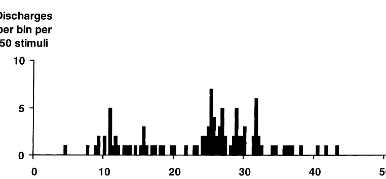
All experiments described in this report were approved Fig. 1 shows a post-stimulus histogram obtained from a by this university’s Animal Care & Ethics Committee and neuron in the trigeminal nucleus caudalis activated with conformed to its guidelines. A2dfibre latency by electrical stimulation of the superior
sagittal sinus.
Infusion of GTN through the indwelling lingual artery
3. Results catheter resulted in a rapid increase in the discharge rate of
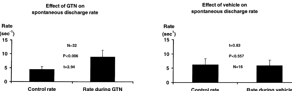
neurons, commencing immediately and reaching a maxi-Twenty-seven neurons in the trigeminal nucleus caudalis mum 3–4 min after the start of infusion. Discharge rates of 7 cats responded to electrical stimulation of the superior rose to about 400% of control at this peak and fell back to sagittal sinus and were chosen for further study. The average about 170% of control for the remaining time of latencies of the response to stimulation were in the A2d infusion. The average duration of infusions was 8.960.9 fiber range (mean latency to earliest discharge510.561.2 min. The mean rate of discharge during infusion was ms). These latencies are similar to those recorded in our 239647% of control over the duration of the infusion in previous experiments [16]. Neurons discharged 1 to 15 all 32 tests. In 23 out of 32 tests (20 out of 27 neurons), times after a single supramaximal shock (mean number of this increase in rate was significant at the 0.05 level, discharges 3.660.6). All 27 neurons discharged in the according to the criteria of the critical ratio test.
absence of impressed electrical stimulation with a basal Two neurons in the neck of the trigeminal nucleus did
21 21
rate of 4.461.0 s (range 0.012–23 s ). Thirteen of the not respond to GTN with an accelerated discharge rate but 27 neurons were tested for cutaneous receptive fields and otherwise there appeared to be no preferential location in all 13 were found to have them on the face: in the first the nucleus for neurons which fulfilled the criteria of the (N55), second (N55) or third (N51) trigeminal divisions test, nor was there a significant difference in the
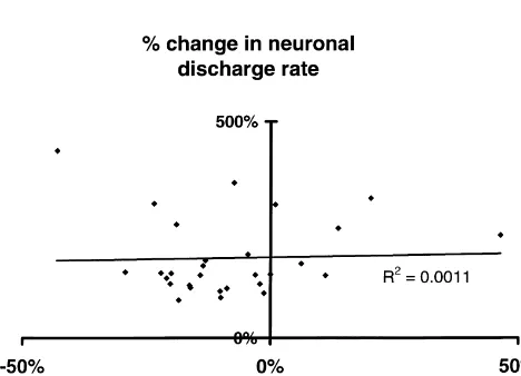
infusion discharge rate between those neurons which of changes in blood pressure, heart rate and expired CO is2 responded (N523 tests) and those which did not (N59 shown in Fig. 2. However, there was no correlation tests) (t51.48, P NS). The Kolmogorof–Smirnoff test between the size of the changes in blood pressure and the showed a significant change in population characteristics size of the changes in neuronal discharge rate. Fig. 4 for all 32 infusions. The only neuron with LTM input was shows a scattergram of the percentage changes in neuronal
also accelerated. firing rate induced by GTN plotted against the percentage
A significant increase in discharge rate persisted after changes in mean blood pressure occurring at the same cessation of infusion; the mean was still 150% of control time.
30 min after the infusion (Fig. 3). Furthermore, in some Infusion of the vehicle alone in 5 cats during the cats the same neuron was tested more than once for its monitoring of 16 neurons did not produce any significant responses to GTN infusions and, in most cats, more than change in the discharge rates of the neurons (Fig. 5) nor in one neuron was tested for its response. Two to six hours mean blood pressure (t51.07, NS) or heart rate (t50.01, separated these successive tests. The mean basal discharge NS). Based on average carotid flow rates (usually about 25
21 ¨
rates of ‘non-naıve’ neurons recorded prior to the Nth ml min ), vehicle or GTN infusion should have produced infusion were higher than the mean basal discharge rates an average ethanol concentration in the carotid blood of recorded for the (N21)th infusion, but there was no 0.02% (0.2 mg / kg) and, at the end of an average infusion, difference in the percentage increase in discharge rate a body load of about 0.002% (0.02 mg / kg), not allowing induced by GTN in successive infusions. However, there for removal mechanisms.
was some evidence that infusions of GTN sensitised neurons to the effects of electrical stimulation of the
sagittal sinus: mean responses to SSS stimulation in 4. Discussion
discharges per stimulus increased according to whether the
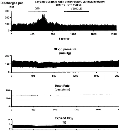
neurons had experienced one or more previous GTN These results support the idea that glyceryl trinitrate infusions. These results, with the results of an analysis of (nitroglycerin, GTN), which produces headache in humans variance for each measure, are shown in Table 1. [34], activates the trigeminovascular sensory system in Fig. 2 shows the ongoing activity of a neuron which cats, and could conceivably also do so in humans. The responded to electrical stimulation of the superior sagittal results show that GTN increases the discharge rate of sinus and was then tested with GTN and vehicle. The trigeminovascular second-order neurons immediately, but neuron was spontaneously active, with a discharge rate that also that it leads to an overall increase in the discharge rate varied from 5 to 8 per second. Fig. 3 shows the time- of all craniovascular neurons, beyond the time of infusion. averaged mean response from 32 tests in 27 neurons These two effects might correlate with the immediate [12]
subjected to GTN infusions. and delayed [11] effect of GTN in producing human
Infusion of GTN led to a fall in mean blood pressure, a headache.
rise in pulse pressure and a rise in heart rate in most tests, Experiments in animals over the last 20 years have but there was a rise of mean blood pressure in five tests. demonstrated that there is a pool of neurons in the Before infusion, the value of the mean arterial blood trigeminal nucleus caudalis and upper cervical spinal cord, pressure was 10363 mmHg and the mean value of heart which are activated by electrical, mechanical or chemical rate was 25165 beats per minute. At the trough of the stimuli of the dural blood vessels [3,14,16,30]. Neurons in blood pressure fall during infusion, mean arterial blood this trigeminovascular pool are potential mediators of pressure was 9263 mmHg and the mean heart rate, headache in humans, because the same types of stimuli in measured at the time of maximum changes was 26365 migraineurs effectively trigger their typical headaches beats per min. The changes were significant (t52.89, [5,26,27].
N530, P,0.01 for blood pressure; t54.1, N528, P, Neurons which responded to GTN infusions with an 0.0005 for heart rate; Student’s paired t-test). An example accelerated discharge rate were found in the trigeminal
Table 1
Effects of successive infusions of GTN on ‘basal’ discharge rates of trigeminovascular neurons in the same cat prior to the Nth infusion, on mean responses a
to the Nth infusion of GTN and on mean responses to stimulation of the sagittal sinus prior to the Nth infusion
1st 2nd 3rd 4th F P
Fig. 2. Top: Peri-event histogram of the same neuron shown in Fig. 1 to intravenous infusions of glyceryl trinitrate (‘GTN’) and of vehicle at a rate of 100
21 21 21
mg kg min (first and second bars, gray). The volume rate of both infusions was 2 ml min . There was no change of discharge rate in response to vehicle infusion, but the rate of discharge increased to 2.5 times control at the peak of the response to GTN. Vertical axis5discharges per bin of 4 s. 2nd, 3rd and 4th panels: Blood pressure, heart rate and expired CO recorded simultaneously with the cell discharge changes shown in the top panel. There was2 a small fall in blood pressure during the infusion, but no significant changes in heart rate or CO . Horizontal axis on all panels2 5time in s, 1 sampling bin54 s.
nucleus caudalis itself and in the sub-adjacent ventrolateral activation, the expression of c-fos in the trigeminal nucleus medulla and nucleus retroambigualis. Two neurons found caudalis [31]. It is not possible, however, to say whether to be in the neck of the nucleus caudalis did not respond to neurons activated in this way had any connection with GTN. GTN responsive neurons in this study were found in dural sensation. The usual infusion rate for GTN in human the same locations as fos-expressing neurons seen after cardiovascular disease is 5–20 mg / min (approximately
21 21
GTN infusions in rats [31] and also after SSS stimulation 0.07–0.28mg kg min ) and, according to the product
in cats [13]. literature, this will produce headache in only about 2% of
The experiments of Olesen et al. examined the headache patients, but, under these circumstances, headache may not produced by GTN [25] and demonstrated that it could be be a symptom to which such patients pay attention, and relieved by administration of the anti-migraine drug sumat- they may also be receiving other drugs which may prevent riptan [11]. The same group also showed that spontaneous or ameliorate headaches. Previous studies [11] have de-migraine is relieved by administration of a nitric oxide scribed headache in both control subjects and migraineurs
21 21 synthase inhibitor [17]. Intravenous administration of GTN after 20 min of an infusion of 0.12mg kg min . In rats,
21
Fig. 3. Mean of 32 peri-event histograms showing the response of all neurons with dural sensory input to intravenous infusions of glyceryl trinitrate at a 21 21
rate of 100mg kg min (32 infusions in 27 neurons). The solid histogram represents the mean of the discharge rate of the neurons, each expressed as a percentage of the control discharge rate for the 200 s prior to the infusion. Rates were measured for 1 second intervals and time averaged over 9 s. The continuous line above the peri-event histogram represents the upper standard error envelope of the discharge rate shown below it. Beyond t51550 s, N51. GTN infusion commenced at time zero. Infusions lasted a variable time, not all lasted for as long as the 1600 s shown on the X-axis.
induce activation of c-fos immunoreactivity [31]. The the activation produced by GTN [15]. It is also possible 21 21
infusion rate we have used in cats, 100 mg kg min , that GTN activates the trigeminal sensory system by a lies near the geometric mean of these amounts, but it is not neuronal mechanism [33] and it has been shown that nitric easy to decide whether the three different rates represent oxide is involved in a cascade of neuronal and neuro-fundamental pharmacological differences or are merely vascular effects [1] which also involve calcitonin gene-related to species differences (human vs. rat vs. cat) or related peptide [10,21] — a sensory neuromodulator
route of administration. implicated in migraine. Tassorelli et al. presented evidence
GTN is a potent vasodilator and the headache it induces that favours the idea that it is the neuronal, rather than the could be related to this vasodilatation because both the vascular, effects of GTN which underlie its ability to cranial vasodilatation and the headache induced by GTN induce headache in humans [31]. The nitric oxide synthase seem to be simultaneously reduced by prior treatment with inhibitorL-NAME reduces c-fos expression elicited in the
sumatriptan [6], although sumatriptan is not a prophylactic trigeminal nucleus and cervical spinal cord by electrical agent for migraine itself. Our own observations suggest stimulation of the superior sagittal sinus [8], which sug-that the anti-migraine drug eletriptan can reduce or reverse gests that transduction of dural sensation into central neuronal activation may involve nitric oxide in some way, presumably unrelated to the vascular involvement of nitric oxide.
We calculated that the vehicle used in the GTN infu-sions and their controls would produce a moment-to-moment carotid blood alcohol level of 0.2 mg / ml in a carotid artery of mean flow 25 ml / min and a post-infusion whole body concentration of 0.02 mg / kg in a 2.7 kg cat. The carotid blood alcohol concentration might be expected to produce cranial vasodilatation, but this could not be monitored. Such vasodilatation and possibly a direct effect on sensory nerve endings could conceivably have activated craniovascular sensory neurons, but we saw no evidence for this. Systemically, the concentration of ethanol (;0.002%) seems unlikely to have generated significant cardiovascular effects.
The systemic dilator effects of GTN result in a fall in 2
Fig. 4. Scattergram showing the lack of correlation (R50.0011, N532) blood pressure, a change which could conceivably result in between the changes in neuronal firing rate at the point of its maximum
changes in the discharge rate of central neurons or the change and the simultaneously measured change in mean blood pressure,
ability of the system to record them. However, the blood induced by infusions of GTN. The linear regression line relating the two
Fig. 5. Mean effects of infusions of glyceryl trinitrate (GTN, N532) on the discharge rate of trigeminovascular neurons. Rates are expressed as the mean rate of discharge for the duration of the infusion, displayed as a percentage of the mean rates of discharge for 3 min prior to commencement of infusion.
21
GTN increased the discharge rate from 4.4 to 9.0 s , but there was no effect of the vehicle.
[7] C. Hering, Glonoine, a new medicine for headache, Am. J. them to be uncorrelated with changes in recorded neuronal
Homeopathy 4 (1849) 3. discharge rate. In previous experiments we have rarely
[8] K.L. Hoskin, D.C. Bulmer, P.J. Goadsby, Fos expression in the seen any change in discharge rate or neuronal responses trigeminocervical complex of the cat after stimulation of the attributable to changes in systemic blood pressure. superior sagittal sinus is reduced byL-NAME, Neurosci. Lett. 266
Our experiments do not distinguish between a purely (1999) 173–176.
dilator effect, a neuronal activation, or even an effect on [9] J.W. Hu, J.O. Dostrovsky, B.J. Sessle, Functional properties of neurons in cat trigeminal subnucleus caudalis (medullary dorsal sensory modulation of central origin for GTN in this
horn). I. Responses to oral-facial noxious and non-noxious stimuli system, but they do unequivocally demonstrate sensory
and projections to thalamus and subnucleus caudalis, J. Neuro-activation in an important component of a pathway that is physiol. 45 (1981) 173–191.
related to headache. The present experiments with GTN [10] T. Ishine, J.G. Yu, Y. Asada, T.J.F. Lee, Nitric oxide is the show that neurons activated by GTN and by electrical predominant mediator for neurogenic vasodilation in porcine pial stimulation of the dura come from the same neuronal pool, veins, J. Pharmacol. Exp. Ther. 289 (1999) 398–404.
[11] H.K. Iversen, J. Olesen, Headache induced by a nitric oxide donor which strengthens the case for using them as a model
(nitroglycerin) responds to sumatriptan — a human model for system for the pathophysiology of headache.
development of migraine drugs, Cephalalgia 16 (1996) 412–418. [12] H.K. Iversen, J. Olesen, P. Tfelt-Hansen, Intravenous nitroglycerin
as an experimental model of vascular headache. Basic characteris-tics, Pain 38 (1989) 17–24.
Acknowledgements
[13] H. Kaube, K.A. Keay, K.L. Hoskin, R. Bandler, P.J. Goadsby, Expression of c-Fos-like immunoreactivity in the caudal medulla This research was supported by grants from the National
and upper cervical spinal cord following stimulation of the superior Health and Medical Research Council of Australia, the sagittal sinus in the cat, Brain Res. 629 (1993) 95–102.
Migraine Trust and the Australian Brain Foundation. [14] G.A. Lambert, Pathways for headache, in: S. Gandevia, D. Burke, M. Anthony (Eds.), Science and Practice in Clinical Neurology, Cambridge University Press, Sydney, 1993, pp. 284–302. [15] G.A. Lambert, P.M. Boers, K. Hoskin, C. Donaldson, A.S. Zagami,
References The activation of trigeminovascular sensory neurones by glyceryl
trinitrate is suppressed by eletriptan., Headache World 2000, The Migraine Trust, London, 2000. In press.
[1] P. Aimar, L. Pasti, G. Carmignoto, A. Merighi, Nitric
oxide-produc-[16] G.A. Lambert, A.S. Zagami, N. Bogduk, J.W. Lance, Cervical spinal ing islet cells modulate the release of sensory neuropeptides in the
cord neurons receiving sensory input from the cranial vasculature, rat substantia gelatinosa, J. Neurosci. 18 (1998) 10375–10388.
Cephalalgia 11 (1991) 75–85. [2] M. Anthony, J.W. Lance, Indomethacin in migraine, Med. J. Aust. 1
[17] L.H. Lassen, M. Ashina, I. Christiansen, V. Ulrich, R. Grover, J. (1968) 56–57.
Donaldson, J. Olesen, Nitric oxide synthase inhibition — a new [3] K.D. Davis, J.O. Dostrovsky, Activation of trigeminal brain-stem
principle in the treatment of migraine attacks, Cephalalgia 18 (1998) nociceptive neurons by dural artery stimulation, Pain 25 (1986)
27–32. 395–401.
[18] W.R. Levick, Another tungsten microelectrode, Med, Biol. Eng. 10 [4] K. Ekbom, Nitroglycerin as a provocative agent in cluster headache,
(1972) 510–515. Arch. Neurol. 19 (1968) 487–493.
[19] N. Marsh, A. Marsh, A short history of nitroglycerine and nitric [5] W. Feindel, W. Penfield, F. McNaughton, The tentorial nerves and
oxide in pharmacology and physiology, Clin. Exp. Pharmacol. localisation of intracranial pain in man, Neurology 10 (1960) 555–
Physiol. 27 (2000) 313–319. 563.
[20] C.R. Marshall, On the antagonistic action of digitalis and the [6] T. Fullerton, D. Komorowski-Swiatek, A. Forrest, F.M. Gengo, The
members of the nitrite group, J. Physiol. 22 (1897) 1–37. pharmacodynamics of sumatriptan in nitroglycerin-induced
neuronal origin and their involvement in neurogenic vasodilatation [28] S. Siegel, N.J.J. Castellan, Nonparametric Statistics For the Be-in rat skBe-in microvasculature, Br. J. Pharmacol. 123 (1998) 863–868. havioral Sciences, McGraw-Hill, New York, 1988, 399 pp. [22] S. Moncada, R.M.J. Palmer, E.A. Higgs, Nitric oxide: physiology, [29] A. Sobrero, Sur plusieurs composes detonants avec l’acide nitrique
pathophysiology and pharmacology, Pharmacol Rev. 43 (1991) et le sucre, la dextrine, la lactine, la mannite et la glycerin, C. R.
109–142. Hebd. Acad. Sci. 24 (1847) 247–248.
[23] F. Murad, Drugs used for the treatment of angina: organic nitrates, [30] A. Strassman, P. Mason, M. Moskowitz, R. Maciewicz, Response of calcium-channel blockers, and beta-adrenergic antagonists. In A.G. brainstem trigeminal neurons to electrical stimulation of the dura, Gilman, T.W. Rall, A.S. Niles and P. Taylor (Eds.), The pharmaco- Brain Res. 379 (1986) 242–250.
logical basis of therapeutics, Vol. 1, Pergamon, New York, 1991, pp. [31] C. Tassorelli, S.A. Joseph, Systemic nitroglycerin induces Fos 764–783. immunoreactivity in brainstem and forebrain structures of the rat, [24] J. Nagler, N. Conforti, S. Feldman, Alterations produced by cortisol Brain Res. 682 (1995) 167–181.
in the spontaneous activity and responsiveness to sensory stimula- [32] C. Tassorelli, S.A. Joseph, M.G. Buzzi, G. Nappi, The effects on the tion of single cells in the tuberal hypothalamus of the rat, Neuroen- central nervous system of nitroglycerin — Putative mechanisms and docrinology 12 (1973) 52–56. mediators, Prog. Neurobiol. 57 (1999) 607–624.
[25] J. Olesen, L.L. Thomsen, L.H. Lassen, I.J. Olesen, The nitric oxide [33] C. Tassorelli, S.A. Joseph, G. Nappi, Neurochemical mechanisms of hypothesis of migraine and other vascular headaches, Cephalalgia 15 nitroglycerin-induced neuronal activation in rat brain — a pharma-(1995) 94–100. cological investigation, Neuropharmacology 36 (1997) 1417–1424. [26] W. Penfield, F.L. McNaughton, Dural headache and the innervation [34] L.L. Thomsen, J. Olesen, Nitric oxide theory of migraine, Clin.
of the dura mater, Arch. Neurol. Psychiatry 44 (1940) 43–75. Neurosci. 5 (1998) 28–33. [27] B.S. Ray, H.G. Wolff, Experimental studies on headache. Pain