I
learned from what
you
did: Retrieving visuomotor associations
learned by observation
Elisabetta Monfardini, Andrea Brovelli, Driss Boussaoud, Sylvain Takerkart, Bruno Wicker
⁎
CNRS and Aix-Marseille University UMR 6193, Mediterranean Institute for Cognitive Neuroscience 31 chemin Joseph Aiguier, 13402, Marseille, France
a b s t r a c t
a r t i c l e i n f o Article history:
Received 14 January 2008 Revised 15 May 2008 Accepted 19 May 2008 Available online xxxx
Observational learning allows individuals to acquire knowledge without incurring in the costs and risks of discovering and testing. The neural mechanisms mediating the retrieval of rules learned by observation are currently unknown. To explore this fundamental cognitive ability, we compared the brain responses when retrieving visuomotor associations learned either by observation or by individual learning. To do so, we asked eleven adults to learn two sets of arbitrary visuomotor associations: one set was learned through the observation of an expert actor while the other was learned by trial and error. During fMRI scanning, subjects were requested to retrieve the visuomotor associations previously learned under the two modalities. The conjunction analysis between the two learning conditions revealed a common brain network that included the ventral and dorsal lateral prefrontal cortices, the superior parietal lobe and the pre-SMA. This suggests the existence of a mirror-like system responsible for the storage of rules learned either by trial and error or by observation of others' actions. In addition, the pars triangularis in the right prefrontal cortex (BA45) was found to be selective for rules learned by observation. This suggests a preferential role of this area in the storage of rules learned in a social context.
© 2008 Elsevier Inc. All rights reserved.
Introduction
In daily life many arbitrarilyfixed and generally adopted
“rules” are learned and retrieved, allowing us and other animals to anticipate relevant events and adapt to a novel context. These rules are often associations of sensory environmental events (e.g. image, symbol, context, etc.) with particular motor responses or actions. If the relation between the visual stimulus, the action, and its outcome is arbitrary and causal, we refer to it as arbitrary visuomotor learning (Wise et al., 1996; Wise and Murray, 2000). If one excludes learning through verbal explanations and instructions, which necessitates language, learning by trial and error and learning by observation are two mechanisms of particular interest for the development of culture in human infants and adults (Castro and Toro, 2004; Cavalli-Sforza and Feldman, 1981). The major difference between these two mechanisms is that the action and its consequence are experienced by the learner himself or herself in the case of learning by trial and error, or by another individual in the case of learning by observation. In
fact, learning by observation allows individuals to acquire useful knowledge without incurring in the costs and risks of discovering and testing (Bandura et al., 1977; Boyd and Richerson, 1985, 1988). AsBandura (1977)stated it:“Learning would be exceedingly laborious, not to mention hazardous, if people had to rely solely on the effects of their own actions to inform them what to do. Fortunately, most human behaviour is learned observationally through modelling: from observing others one forms an idea of how new behaviours are performed, and on later occasions this coded information serves as a guide for action.”(Bandura et al., 1977).
Several neuroimaging studies have established that learn-ing arbitrary visuomotor associations by trial and error engages a large brain network including the frontal–parietal system, the basal ganglia and medial temporal structures (Deiber et al., 1997; Toni and Passingham, 1999; Toni et al., 2001; Eliassen et al., 2003; Law et al., 2005; Brovelli et al., in press). In parallel, various studies have shown that the prefrontal cortex is particularly involved in the storage and retrieval from long-term memory of known rules, or pre-scribed guides for action (Bunge et al., 2003; Bunge, 2004; Crone et al., 2006; Donohue et al., 2005). However, despite its outstanding scientific interest, the neural bases of learning and retrieval of visuomotor associations when learned by observation remain surprisingly unexplored.
⁎Corresponding author.
E-mail address:[email protected](B. Wicker).
1053-8119/$–see front matter © 2008 Elsevier Inc. All rights reserved. doi:10.1016/j.neuroimage.2008.05.043
Contents lists available atScienceDirect
NeuroImage
In the present study, we investigate the neural representa-tions of the retrieval of visuomotor associarepresenta-tions learned by trial and error or by observation. Retrieval is defined as the recovery from memory of the correct action associated to a given visual stimulus. In other words, we indirectly test whether observational learning is mediated by specific neural circuits linking environmental information such as visual stimuli with the reenactment of others' actions to generate internal representations of arbitrary associations. To do so, eleven adult subjects were asked to learn, prior to the fMRI scanning session, two sets of visuomotor associations: one set was learned through the observation of an expert actor while the other was learned by trial and error. To explore the neural correlates of learning by observation, we compared the brain activations of subjects during the retrieval of associations learned in the two modalities.
Materials and methods
Participants
Eleven healthy, right-handed volunteers (7 males, 4 females) participated in the study (mean age: 26.3 ± 4.2 years). All subjects were screened to rule out medication use, history of neurological or psychiatric disorders, head trauma, substance abuse, or other serious medical conditions. Written consent was obtained after the procedure had been fully explained. The study was approved by the local ethics committee and was conducted in accordance with the Declaration of Helsinki. Volunteers were paid for their participation.
Design and experimental conditions
Learning sessions prior to scanning
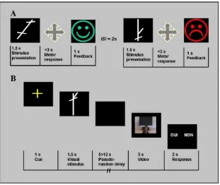
Prior to fMRI scanning, subjects learned arbitrary associa-tions between abstract visual stimuli (linear segments com-bined to form white shapes on a black background—Fig. 1) and motor responses (joystick movements). Each subject had to learn one set of three visuomotor associations by trial and error and another set by observing an expert actor performing the visuomotor associations in front of him or her. The order of the two learning sessions was randomized between subjects.
During the trial and error learning session, visual stimuli were presented on a computer screen in front of the subjects. Motor responses were recorded with a joystick. After presentation of the visual stimulus (1.5 s), the subject had to choose one of four possible joystick movements (up, down, right or left). The visual feedback (a green/happy or red/sad smiley-like face) then indicated whether the movement was correct or incorrect. The correct stimulus–response associa-tions were kept constant throughout the experiment. To ensure that participants had learned the associations, the session ended when the subject had given 4 consecutive correct responses to each of the 3 visual stimuli. A pilot study was conducted on 5 subjects in order to validate the learning criterion. A mean of 46.36 trials was necessary to reach the criterion.
During learning by observation, subjects had to learn through the observation of an expert individual demonstrating the correct visuomotor associations. Subjects were seated beside the demonstrator and close enough to observe t he visual stimuli
on the computer screen and the joystick movement (distance = approximately 50 cm). A pilot study was conducted on 5 subjects, in order to estimate the approximate number of trials repetition needed to learn the associations. To ensure that the participants had learned the visuomotor associations, the session ended when the subjects told the experimenter they knew the associations. The behavioral performances obtained during fMRI scanning were used to control that learning by observation had occurred prior to scanning (cf.Supplementary Table 1). A mean of 15.27 trials was necessary to learn the associations.
Scanning session
Stimuli and experimental design. Subjects were scanned approximately 30 min after the learning sessions using an event-related fMRI paradigm with three trial types (condi-tions). Each trial started with afixation cross (1 s), immedi-ately followed by the presentation of the visual stimulus (1.5 s). In the trial and error (TE) condition, the correct action associated to the presented visual stimulus had been learned by trial and error previous to scanning. In the learning by observation (LeO) condition, the stimuli had been previously learned by observation. A third trial type served as control condition (CONT), in which arrows pointing in one of four directions were used as visual stimuli (Fig. 1). The stimulus properties in the control condition were very similar to those in the TE and LeO condition. Subjects were not informed that the arrow indicated the correct direction of movement. For all conditions, the stimulus presentation was followed by a video sequence of 3 s showing the hand of an actor executing a joystick movement. A variable delay between 5 and 12 s was introduced between the stimulus and the video presentation in order to dissociate the BOLD responses produced by the two events. After each movie, the subjects had to judge whether the joystick movement executed by the actor was correct or not. Subjects responses were collected using a two-button computer mouse; the button associated to the “correct”
response was randomly assigned across trials. The actor's responses were correct in 50% of the trials. Subjects performed a total of 40 trials of each of the three conditions, presented pseudo-randomly in four blocks. Each block consisted of 30 trials (10 of each condition), and the presentation order was randomized between subjects. Behavioral data (accuracy and reaction time) were collected during the scanning sessions. The stimuli were projected onto a screen positioned at the back of the scanner. Subjects could see the video reflected in a mirror (15 × 9 cm) suspended 10 cm in front of their face and subtending visual angles of 42° horizontally and 32° vertically.
Image acquisition. Images were acquired using a 3-T whole-body imager. For each participant, wefirst acquired a high-resolution structural T1-weighted anatomical image (inver-sion–recovery sequence, 1 × 0.75 × 1.22 mm) parallel to the AC–PC plane, covering the whole brain. For functional imaging, we used a T2*-weighted echo-planar sequence at 36 interleaved 3.5-mm-thick axial slices with 1 mm gap (TR = 2400 ms, TE = 35 ms, flip angle = 80°, FOV = 19.2 × 19.2 cm, 64 × 64 matrix of 3 × 3 mm voxels).
Data processing and statistical analysis
Image processing and analysis of fMRI data were conducted with SPM2 software (http://www.fil.ion.ucl.ac.uk/spm/software
/spm2/). Thefirstfive volumes of each participant's data were discarded to allow for longitudinal relaxation time equilibration. Functional images for each subject were slice-time corrected to a slice acquired halfway through image acquisition in order to correct for temporal differences (up to 2.4 s) between slices acquired early, and those acquired late in the image volume. All volumes were realigned to thefirst volume to correct for head movement between scans. A mean image was created using the realigned volumes. The mean image was spatially normal-ized to the standard EPI template given in the SPM software. All images were then spatially normalized using the norma-lization parameters determined during the normanorma-lization of mean image to EPI template. Data were then smoothed using an 8 mm full-width-at-half-maximum isotropic Gaussian kernel to accommodate inter-subject differences in anatomy. Finally, the data were normalized and high-passfiltered (128 s).
The statistical analysis of the pre-processed BOLD signals was performed using a generalized linear model (GLM) approach. To dissociate two events per trial (the stimulus and video presentation), two regressors were constructed per trial type by convolving the canonical haemodynamic response function (HRF) with delta functions aligned either on the time of stimulus or video presentation. The design matrix contained a total of six regressors (two regressors for each of the three conditions). Since we hypothesized that association retrieval occurs at stimulus presentation, we aligned our events of interest to the onset of the stimulus.
The regression coefficients (the beta values) were esti-mated for each subject, and were then taken to the random-effects level. All the fMRI statistics andp values arise from group random-effects analyses. We considered as activated brain regions those clusters of more than 10 contiguous voxels with pb0.001 at the voxel level (uncorrected for multiple comparisons).
Results
Eleven subjects participated in the fMRI study. Prior to the scanning session, subjects learned two sets of three arbitrary visuomotor associations. One set was learned by trial and error and one set by observation of an expert actor performing the task. During the scanning session, the subjects were tested in three different conditions: i) retrieval of associations learned by trial and error; ii) retrieval of rules learned by observation; and iii) a control condition using arrows as stimuli (see me-thods,Fig. 1).
The mean percentage of correct responses for each exper-imental condition acquired during fMRI scanning confirmed that subjects could perfectly retrieve the visuomotor asso-ciation for all stimuli (Supplementary Table 1). There was no significant difference across conditions (TE, LeO and CONT) in reaction times (F(2,768) = 0.238, pN0.05), suggesting that task difficulty was comparable in all experimental conditions (Supplementary Table 2).
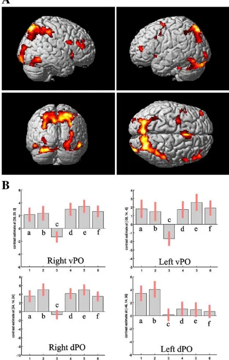
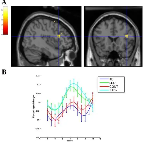
subtracted the brain activity in the control condition (CONT) from the TE and LeO conditions. We then performed a conjunction analysis between the contrasts‘TE’–‘CONT’and ‘LeO’–‘CONT’ to explore the brain areas commonly activated both in the LeO and TE conditions. This analysis revealed BOLD signal increases bilaterally in three distinct foci of the lateral prefrontal cortex (BA46; ventral pars opercularis BA44; dorsal pars opercularis BA44/45). This network spread from the inferior to the superior posterior parietal cortex bilaterally, in the dorsal posterior cingulate gyrus extending dorsally to the pre-SMA, in the posterior ventro-temporal visual areas, and in the thalamus (Fig. 2;Table 1A). This network includes areas that activate during the retrieval of associations recently learned either by trial and error or by observation. To assess which brain areas are specifically involved in rule retrieval after observational learning, we performed a direct compar-ison between conditions LeO and TE. Significant activations were found in the right prefrontal cortex (pars triangularis, BA45), in the inferior parietal lobule (BA40) and in the occipital primary visual areas (BA17/18;Fig. 3andTable 1B).
Discussion
Although various aspects of rule retrieval and their neural correlates have been explored in recent studies (Bunge et al., 2003; Crone et al., 2006; Donohue et al., 2005), none have addressed whether these cerebral areas can be influenced by the type of preceding learning. In this study, we manipulated how visuomotor associations were learned prior to scanning and investigated their brain correlates during retrieval.
We compared the brain activations triggered by the stimulus presentation, that is, when subjects retrieve from memory the correct action associated to the stimulus. We used abstract visual stimuli to minimize verbalization (inter-nal speech) effects that could account for differences in brain activations between the TE/LeO conditions and the CONT condition. In addition, the training sessions prior to scanning were matched to ensure that subjects performed the task during scanning without errors. Indeed, the analysis of behavioral data confirmed that the level of performance and reaction times were statistically identical in all conditions. In order to control for effects not deriving from the type of learning, we used a control condition (CONT) where the visual stimuli were arrows pointing in the 4 joystick directions. This control condition was subtracted from the TE and LeO con-ditions. Therefore, we argue that differences in brain activa-tions between condiactiva-tions TE and LeO are exclusively related to the type of learning.
Common networks involved in retrieval of associations learned by trial and error and by observation
The results from the conjunction analysis confirmed the involvement of a brain network composed of the right ventrolateral and anterior prefrontal cortices, pre-SMA, and parietal cortex when retrieving newly acquired rules (Bunge et al., 2003; Bunge, 2004; Crone et al., 2006; Donohue et al., 2005). This is consistent with a role of the inferior frontal junction in the retrieval of actions associated with symbolic cues (Donohue et al., 2005). It also confirms that the posterior medial temporal gyrus plays a general role in storing action knowledge, more specifically in representing arbitrary asso-ciations between symbols and associated rules for how to act
(Donohue et al., 2005). Our results show that the same network is recruited also during the retrieval of appropriate action representations that have been learned via the observation of others' behaviors. A straightforward interpre-tation is that during both types of learning, the brain builds an internal model linking an executed or observed action with a visual stimulus and a feedback. The generation of such inter-nal models necessitates the activation of brain areas coding for action execution and observation. Except for the most anterior frontal area (BA46), the activations in the dorsal and ventral part of pars opercularis and in the parietal cortex have been consistently reported in studies where subjects execute, observe, imitate or understand actions, intentions or emo-tions, pretend to use tools, or generate verbs (Buccino et al., 2001; Gazzola et al., 2006, 2007; Grèzes et al., 2003; Iacoboni et al., 2005; Jeannerod, 2001; Rizzolatti et al., 1996; 2001; Wicker et al., 2003a). These brain areas are thought to be core components of the mirror neuron system (MNS) in humans and may mediate action and intention understanding mecha-nisms (Rizzolatti and Craighero, 2004). In the present study,
Fig. 2B illustrates that the prefrontal and parietal regions activate during stimulus perception and association retrieval, and also during observation of movies (i.e., observation of
joystick movements). This demonstrates their role in action observation. Although we did not have an experimental condition to map the brain areas involved in action execution, our results suggest that a subset of the mirror system mediates the retrieval of visuomotor associations recently learned by trial and error or by observation. Subject's own actions and observed actor's actions are stored in the same
‘mirror’areas, suggesting the presence of a mirror-like system that stores the representations of recently learned visuomotor associations. This interpretation of ourfindings implies that, in addition to the classically described mirror neurons thatfire during the execution and observation of the same or broadly identical motor act (e.g. a joystick movement), a subset of these neurons might be visually triggered by the presentation of an abstract stimulus that has been previously associated to a specific action. The ability to store visuomotor associations learned by observation might rely on a kind ofmirror activity
of prefrontal areas, more precisely in the ventral and dorsal part of the pars opercularis. Similarly to the understanding of sensations (Keysers et al. 2004), emotions (Wicker et al., 2003b) and pain (Singer et al., 2004), observational learning might be supported by a reenactment of the experience of the model in the observer. Albeit theoretically (Gallese and Goldman, 1998), literature in the field of rule retrieval and learning has not been linked to literature on mirror neuron system. Here we provide suggestive evidence of a possible role of the mirror system in observational learning of visuomotor associations.
A specific network for the retrieval of associations learned by observation
The observation of abstract stimuli and their association to specific movements produced activations in a network of brain areas comprising the right pars triangularis (BA 45), the right inferior parietal lobule and the posterior visual areas. To better understand the role of the right pars triangularis (BA45), we performed a Student'st-test on the average percent change in BOLD signal in the right pars triangularis during retrieval and movie observation. The analysis revealed similar BOLD responses when subjects retrieved the motor action associated with a visual stimulus in the LeO condition and when subjects watched a video sequence of a hand performing a joystick movement (Fig. 3). This suggests that the pars triangularis is engaged both during observation of action and during the retrieval of the correct movement previously associated with a stimulus by observation of another's action. This is consistent with recent data showing that the right pars triangularis is involved in action observation, but not in imitation, hence not in execution (Molnar-Szakacs et al., 2005). Because of these properties, authors have suggested that this region should not be considered as part of the mirror system, but rather as related to the suppression of movement execution during action observation and motor imagery (Deiber et al., 1998; Molnar-Szakacs et al., 2006). Our results thus suggest that the mere perception of an abstract stimulus previously associated with a specific movement is sufficient to activate this area and hence that this region plays an important role in storing information about other's actions performed in a given context. One could further propose that this area monitors theflow of information within the fronto-parietal network, to store the information as
Table 1
Anatomical regions BA MNI coordinates
x y z Peak
Zscore A
R superior parietal lobe 7 24 −72 52 5.42
L superior parietal lobe 7 −16 −78 52 5.13
R inferior parietal lobe 40 48 −34 50 5.00
L inferior parietal lobe 40 −34 −50 50 3.56
R anterior cingulate gyrus (also pre-SMA)
24 4 16 44 5.15
R posterior cingulate gyrus 31 10 −46 18 3.51
R inferior temporal gyrus 19 52 −60 −12 4.95
L fusiform gyrus 37 −28 −74 −24 4.81
L fusiform gyrus 37 −30 −36 −32 4.13
R pulvinar 10 −26 12 4.74
L middle frontal gyrus 46 −42 48 12 4.70
R middle frontal gyrus 46 48 42 14 4.57
L middle frontal gyrus 6 −22 0 50 4.40
R inferior frontal gyrus 44/45 54 14 24 4.06 L inferior frontal gyrus 44/45 −46 14 36 3.56
R inferior frontal gyrus 44 38 20 0 4.13
L inferior frontal gyrus 44 −36 14 −6 3.87
L inferior frontal gyrus 44 −50 4 42 3.58
R middle occipital gyrus 19 28 −98 −8 4.54
L middle occipital gyrus 19 −26 96 −6 4.05
Lingual gyrus 18 0 −72 −16 3.48
R hippocampal gyrus 20 8 −18 3.90
R calcarine sulcus 2 −64 0 3.75
B
R cuneus 17/18 10 −104 18 5.18
R inferior frontal gyrus (GFi) 45 42 20 18 3.91 R inferior parietal lobule 40 60 −24 30 3.88
R caudate nucleus 12 18 4 3.58
(A) Coordinates and peakZvalue of functional activations common to LeO and TE conditions (FDR correctedpb0.01). (B) Location and peak Zvalue of functional activations found in LeO vs TE (pnot corrected, cluster sizeN10 functional voxels). R, right hemisphere; L, left hemisphere.
an internal representation about the actions of the demonstra-tor in relation with the visual stimulus and the feedback. Interestingly, the rostral part of the IPS has been recently claimed to play a key role in observational learning of complex action sequences by forming representations of the temporal ordering of actions needed to guide subsequent performances (Frey and Gerry, 2006). We extend thisfinding by showing that this region is also engaged when the level of representation necessary for retrieval of the correct answer is abstract.
What could explain the specific activation of early visual areas depending on the way the visuomotor association was learned? Since conditions were identical in terms of complexity of the visual stimuli, we suggest that the activation of the visual areas results from top–down modulations. Indeed, results of several neuroimaging studies have suggested that a frontal–parietal network controls attention by sending “top–down” signals modulating the activity of the visual cortex during the execution of given tasks (Hopfinger et al., 2000; for review, seeCorbetta and Shulman, 2002). In the present study, attention to the hand is an intrinsic characteristic of learning by observation. The retrieval process might necessitate the reactivation of the observed move-ment during learning and the top–down modulation might affect distinct visual areas depending on whether the rule was learned in LeO or in a TE context (Vidyasagar and Pigarev, 2007; Supèr et al., 2001). In a recent study using magnetoencephalography, Nueuwenhuis et al. showed an increased gamma activity in the visual areas (BA17 and BA18) when labile compared to stabilized memories are recalled (Nieuwenhuis et al., 2008). This result further increases the evidence that the visual system is engaged in tasks beyond visual perception. These tasks include directed attention, working memory maintenance and long-term mem-ory encoding and recall (Jensen et al., 2007).
To conclude, we suggest that the mere perception of an abstract visual stimulus that has been previously associated with a motor response by trial and error or by observation of an actor is sufficient to trigger the activation of a set of brain areas typically involved in action observation or execution. This suggests the existence of a common neural system responsible for the storage of stimulus–response mappings learned either by trial and error or by observation of another's actions (Table 1). Retrieving an arbitrary visuomotor association learned by observation engages the pars triangularis of the right prefrontal cortex, reflecting the potential value of other's actions in such context and the need for its privileged processing.
The results of the present study try to bridge the gap between two widely explored cognitive functions: individual learning and action observation and understanding. Visuo-motor individual learning is thought to be mediated by the fronto-striatal system (Wise and Murray, 2000; Hadj-Bouziane and Boussaoud, 2003), whereas action observation engages the fronto-parietal mirror system. The fact that we can learn both through trial and error and from observation of other's behavior suggests that these two systems interact to allow the transfer of other's experience from the fronto-parietal system to the fronto-striatal system. If so, the fronto-striatal and the fronto-parietal systems cooperate to ensure the learning of behavioral patterns via observation of other's actions.
Acknowledgments
The authors wish to thank B. Nazarian, M. Roth and J.L. Anton for assistance with scanning, and Dr. G. Prabhu for correcting the English. This work was funded by Action Concertée
Incitative NIC0050. EM is supported by funding from EC-contract number 27654 and AB is supported by a two-year fellowship from the“Fondation pour la Recherche Médicale”.
Conflicts of interest
The authors have declared that no conflicts of interest exist.
Appendix A. Supplementary data
Supplementary data associated with this article can be found, in the online version, atdoi:10.1016/j.neuroimage.2008. 05.043.
References
Bandura, A., 1977. Self-efficacy: toward a unifying theory of behavioral change. Psychol. Rev. 84, 191–215.
Boyd, R., Richerson, P.J., 1985. Culture and the Evolutionary Process. University of Chicago Press, Chicago.
Boyd, R., Richerson, P.J., 1988. An evolutionary model of social learning: the effects of spatial and temporal variation. In: Zentall, T.R., Galef, B.G. (Eds.), Social Learning: A Psychological and Biological Approaches. Erlbaum, Hillsdale NJ, pp. 29–48. Brovelli A., Laksiri N., Nazarian B., Meunier M., Boussaoud D., in press. Understanding
the Neural Computations of Arbitrary Visuomotor Learning Through fMRI and Associative Learning Theory. Cereb. Cortex. PMID: 18033767.
Buccino, G., Binkofski, F., Fink, G.R., Fadiga, L., Fogassi, L., Gallese, V., Seitz, R.J., Zilles, K., Rizzolatti, G., Freund, H.J., 2001. Action observation activates premotor and parietal areas in a somatotopic manner: an fMRI study. Eur. J. Neurosci. 13, 400–404. Bunge, S.A., 2004. How we use rules to select actions: a review of evidence from
cognitive neuroscience. Cogn. Affect. Behav. Neurosci. 4, 564–579.
Bunge, S.A., Kahn, I., Wallis, J.D., Miller, E.K., Wagner, A.D., 2003. Neural circuits subserving the retrieval and maintenance of abstract rules. J. Neurophysiol. 90, 3419–3428.
Castro, L., Toro, M., 2004. The evolution of culture: from primate social learning to human culture. Proc. Natl. Acad. Sci. 101, 10235–10240.
Cavalli-Sforza, L.L., Feldman, M.W., 1981. Cultural Transmission and Evolution: A Quantitative Approach. Princeton Univ Press, Princeton.
Corbetta, M., Shulman, G.L., 2002. Control of goal-directed and stimulus-driven attention in the brain. Nat. Rev. Neurosci. 3, 201–215.
Crone, E.A., Donohue, S.E., Honomichl, R., Wendelken, C., Bunge, S.A., 2006. Brain regions mediatingflexible rule use during development. J. Neurosci. 26, 11239–11247. Deiber, M.P., Wise, S.P., Honda, M., Catalan, M.J., Grafman, J., Hallett, M., 1997. Frontal and
parietal networks for conditional motor learning: a positron emission tomography study. J. Neurophysiol. 78, 977–991.
Deiber, M.P., Ibanez, V., Honda, M., Sadato, N., Raman, R., Hallett, M., 1998. Cerebral processes related to visuomotor imagery and generation of simplefinger move-ments studied with positron emission tomography. NeuroImage 7, 73–85. Donohue, S.E., Wendelken, C., Crone, E.A., Bunge, S.A., 2005. Retrieving rules for
behavior from long-term memory. Neuroimage 26, 1140–1149.
Eliassen, J.C., Souza, T., Sanes, J.N., 2003. Experience-dependent activation patterns in human brain during visual–motor associative learning. J. Neurosci. 23, 10540–10547. Frey, S.H., Gerry, V.E., 2006. Modulation of neural activity during observational learning
of actions and their sequential orders. J. Neurosci. 26, 13194–13201.
Gazzola, V., Aziz-Zadeh, L., Keysers, C., 2006. Empathy and the somatotopic auditory mirror system in humans. Curr. Biol. 16, 1824–1829.
Gazzola, V., Rizzolatti, G., Wicker, B., Keysers, C., 2007. The anthropomorphic brain: the mirror neuron system responds to human and robotic actions. NeuroImage 35, 1674–1684.
Gallese, V., Goldman, A., 1998. Mirror neurons and the simulation theory of mind-reading. Trends Cogn. Sci. 2, 493–501.
Grèzes, J., Armony, J.L., Rowe, J., Passingham, R.E., 2003. Activations related to“mirror”and “canonical”neurons in the human brain: an fMRI study. NeuroImage 18, 928–937. Hadj-Bouziane, F., Boussaoud, D., 2003. Neuronal activity in the monkey striatum
during conditional visuomotor learning. Exp. Brain Res. 153, 190–196.
Hopfinger, J.B., Buonocore, M.H., Mangun, G.R., 2000. The neural mechanisms of top– down attentional control. Nat. Neurosci. 3, 284–291.
Iacoboni, M., Molnar-Szakacs, I., Gallese, V., Buccino, G., Mazziotta, J.C., Rizzolatti, G., 2005. Grasping the intentions of others with one's own mirror neuron system. PLoS Biol. 3, 529–535.
Jeannerod, M., 2001. Neural simulation of action: a unifying mechanism for motor cognition. NeuroImage 14, 103–109.
Jensen, O., Kaiser, J., Lachaux, J.P., 2007. Human gamma-frequency oscillations associated with attention and memory. Trends Neurosci. 30, 317–324.
Keysers, C., Wicker, B., Gazzola, V., Anton, J.L., Fogassi, L., Gallese, V., 2004. A touching sight: SII/PV activation during the observation and experience of touch. Neuron 42, 335–346.
Molnar-Szakacs, I., Iacoboni, M., Koski, L., Mazziotta, J.C., 2005. Functional segregation within pars opercularis of the inferior frontal gyrus: evidence from fMRI studies of imitation and action observation. Cereb. Cortex 7, 986–994.
Molnar-Szakacs, I., Kaplan, J., Greenfield, P.M., Iacoboni, M., 2006. Observing complex action sequences: the role of the fronto-parietal mirror neuron system. NeuroImage 33, 923–935.
Nieuwenhuis, I.L., Takashima, A., Oostenveld, R., Fernández, G., Jensen, O., 2008. Visual areas become less engaged in associative recall following memory stabilization. Neuroimage 40, 1319–1327.
Rizzolatti, G., Craighero, L., 2004. The mirror-neuron system. Annu. Rev. Neurosci. 27, 169–192.
Rizzolatti, G., Fadiga, L., Gallese, V., Fogassi, L., 1996. Premotor cortex and the recognition of motor actions. Cogn. Brain Res. 3, 131–141.
Rizzolatti, G., Fogassi, L., Gallese, V., 2001. Neurophysiological mechanisms underlying the understanding and imitation of action. Nat. Rev. Neurosci. 2, 661–670. Singer, T., Seymour, B., O'Doherty, J., Kaube, H., Dolan, R.J., Frith, C.D., 2004. Empathy for
pain involves the affective but not sensory components of pain. Science 303, 1157–1162.
Supèr, H., Spekreijse, H., Lamme, V.A., 2001. A neural correlate of working memory in the monkey primary visual cortex. Science 293, 120–124.
Toni, I., Passingham, R.E., 1999. Prefrontal–Basal ganglia pathways are involved in the learning of arbitrary visuomotor associations: a PET study. Exp. Brain. Res. 127, 19–32. Toni, I., Ramnani, N., Josephs, O., Ashburner, J., Passingham, R.E., 2001. Learning arbitrary visuomotor associations: temporal dynamic of brain activity. Neuroimage 14, 1048–1057.
Vidyasagar, T.R., Pigarev, I.N., 2007. Modulation of neuronal responses in macaque primary visual cortex in a memory task. Eur. J. Neurosci. 25, 2547–2557. Wicker, B., Perrett, D.I., Baron-Cohen, S., Decety, J., 2003a. Being the target of another's
emotion: a PET study. Neuropsychologia 41, 139–146.
Wicker, B., Keysers, C., Plailly, J., Royet, J.P., Gallese, V., Rizzolatti, G., 2003b. Both of us disgusted in My insula: the common neural basis of seeing and feeling disgust. Neuron 40, 655–664.
Wise, S.P., Murray, E.A., 2000. Arbitrary associations between antecedents and actions. Trends Neurosci. 23, 271–276.