April 2017 7 *Corresponding author:
E-mail: [email protected]
Strengthening No: 36b/E/KPT/2016 Available online at http://medpet.journal.ipb.ac.id/
Insulin-Like Growth Factor-I Concentration in the Follicular Fluid of Bali Cattle
and Its Role in the Oocyte Nuclear Maturation and Fertilization Rate
H. Hasbia,b, S. Gustinab,c, N. W. K. Karjab, I. Supriatnab, & M. A. Setiadib,* aDepartment of Animal Production, Faculty of Animal Science, Hasanuddin University
Jalan Perintis Kemerdekaan Km. 10, Makassar 90245, Indonesia
b Division of Reproduction and Obstetric, Department of Veterinary Clinic, Reproduction and Pathology, Faculty of Veterinary Medicine, Bogor Agricultural University
Jalan Agatis, Kampus IPB Darmaga Bogor 16680, Indonesia
cDivision of Animal Science, Faculty of Animal Science and Fisheries, Sulawesi Barat University
Majene, Sulawesi Barat, Indonesia
(Received 24-01-2017; Reviewed 15-03-2017; Accepted 10-04-2017)
ABSTRACT
The objective of this study was to determine the concentration of IGF-I in the follicular fluid (FF) of Bali cattle and its role in the nuclear maturation and fertilization rate. The follicular fluid was col -lected by the aspiration technique, then it was centrifuged at 1500 g for 30 min at 24oC. The superna-tant was collected and stored at -20oC until being used in the experiment for analysis of IGF-1. A total of 1105 oocytes were used in this study. The oocytes were matured in M199 without supplementation of bovine serum albumin, with supplementation of BSA, and with supplementations of 10% FF (v/v)
from the follicle with diameter Ø<4 mm, 4≤Ø<6 mm, 6≤Ø<8 mm, and Ø≥8 mm at the luteal phase
and then fertilized. The results showed that the concentrations of IGF-I in the FF obtained during
the luteal phase was significantly higher (P<0.05) compared to those obtained during follicular phase. The IGF-I concentrations in the follicular fluid of follicle with diameter smaller than 6 mm were significantly higher (P<0.05) compared to those with diameters larger than 6 mm. The percentage of
nuclear maturation rate of oocytes cultured with FF obtained from follicle with diameter <4 mm was
significantly higher (P<0.05) compared to those obtained from the other groups of follicle diameters.
The supplementation of maturation media with BSA and FF were able to improve fertilization rate
significantly (P<0.05) compared to maturation media without BSA. In conclusion, the concentration of IGF-I in the follicular fluid obtained during the luteal phase was higher compared to those obtained during the follicular phase. The IGF-I concentrations in the follicular fluid of smaller follicles (diam
-eter <6 mm) were higher compared to those in the large follicles (diam-eter ≥6 mm). The supplementa -tion of FF can improve the nuclear matura-tion and fertiliza-tion rate.
Keywords: IGF-I, luteal, follicular, Bali cattle, oocytes
ABSTRAK
Penelitian ini bertujuan untuk menentukan konsentrasi IGF-I cairan folikel sapi Bali dan pengaruhnya pada angka maturasi inti dan fertilisasi. Cairan folikel dikumpulkan dengan teknik as-pirasi, selanjutnya disentrifugasi pada kecepatan 1500 g selama 30 menit pada suhu 24oC. Supernatan dikoleksi selanjutnya disimpan pada suhu -20oC hingga digunakan untuk penelitian dan untuk analisis IGF-I. Total oosit yang digunakan pada penelitian ini adalah 1105. Oosit dimaturasi dalam M 199 tanpa penambahan BSA, dengan penambahan BSA, dengan penambahan 10% cairan folikel
yang berasal dari fase luteal dengan diameter folikel Ø<4 mm, 4≤Ø<6 mm, 6≤Ø<8 mm, dan Ø≥8 mm
yang selanjutnya difertilisasi. Hasil penelitian menunjukkan bahwa konsentrasi IGF-I cairan folikel yang berasal dari fase luteal nyata lebih tinggi (P<0.05) dibandingkan dengan cairan folikel yang
ber-asal dari fase folikuler. Konsentrasi IGF-I dalam cairan folikel berdiameter <6 mm nyata lebih tinggi (P<0.05) dibandingkan dengan cairan folikel berdiameter >6 mm. Tingkat maturasi inti oosit yang
ber-HASBI ET AL. / Media Peternakan 40(1):7-13
INTRODUCTION
Follicular fluid (FF) is a natural media for the
nuclear and cytoplasmic maturation and ovulation
of mammalian oocytes in vivo (Coleman et al., 2007;
Revelli et al., 2009), which is the result of a complex fluid
secretion by the follicle (Nandi et al., 2008). Coleman et al. (2007) described that during the growth of oocyte,
metabolic processes were occurred in the oocyte and
follicular cells. Follicular cells (granulosa and theca cells) produce a number of steroids and certain types
of proteins, including growth factors. FF consists of a variety nutrients, growth factors, hormones, electrolytes, metabolites, enzymes, and regulatory molecules that play critical roles in the development and maturation
of oocytes (Nandi et al., 2008; Sinclair et al., 2008; Bender
et al., 2010; Petro et al., 2012). FF protects oocyte from factors leading to premature resumption of meiosis,
prevents proteolytic process, facilitates the extrusion of oocyte during ovulation, and enhances sperm attraction, motility, and acrosome reaction (Avery et al., 2003).
Insulin-like growth factor-I (IGF-I) is one of complex components of IGF superfamily which plays
an important role in mammalian reproduction and is produced in the hypothalamus, ovaries, oviducts, and
uterus (Velazquez et al., 2008). The IGF-I in the follicu
-lar fluid is mainly produced by the follicle and corpus luteum (Woad et al., 2000; Velazquez et al., 2008), and its
concentration increases progressively during follicular growth until the dominant follicle is ovulated. It was reported that the average concentration of IGF-I in
fol-licular fluid obtained from the small follicle was lower
than that obtained from the large follicle of porcine
(Oberlender et al., 2013). Furthermore, Silva et al. (2009) explain that the IGF-I plays an important role in the
development of primordial follicles to pre-ovulatory follicles. Insulin-like growth factor-I acts to initiate the growth of primordial follicles to primary follicles and the growth of primary follicles to secondary follicles. At the stage of follicle development from secondary follicle to antral follicle, IGF-I controls the follicle growth and
survival, proliferation and differentiation of granulosa cells, the production of estradiol and induces the expres -sion of FSH-R on the granulosa cells, the cells survival, and the formation of cortical granules. During the stage of development from the antral to pre-ovulatory fol-licles, IGF-I controls the proliferation of the granulosa cells, the production of estradiol and progesterone by the granulosa cells, increases the sensitivity of the fol-licle to gonadotropins, viability of oocytes, increases the LH-R on granulosa and theca cells, the dominant follicle and multiple ovulation, oocyte maturation, and secretion of inhibin A, activin A, and folistatin by the granulosa cells.
The use of follicular fluid for in vitro embryos
production is rather contradictive. Follicular fluid as a maturation media had an inhibitory effect on germinal
vesicle breakdown, meiotic progression, and nuclear
maturation (Ducolomb et al., 2013). In contrary, numer -ous studies reports that the supplementation of
matura-tion media with follicular fluid have beneficial effects on the oocyte development competence (Ito et al., 2008;
Agung et al., 2010), promotes sperm penetration during
in vitro fertilization (Somfai et al., 2012) and embryo quality (Valckx et al., 2015). However, the use of com
-mercial IGF-I in maturation medium requires the high
cost for in vitro embryo production.
In vivo, IGF-I in follicular fluid induces mitotic divi
-sion in granulosa cells (Spanos et al., 2000) and resume
meiosis to stimulate the formation of LH receptors on the granulosa cells that cause the follicle more
respon-sive to LH (Hurk & Zhao, 2005). Therefore, the aim of
the present study was to determine the concentration of
IGF-I in follicular fluid obtained from different diam -eters of follicles and its role in oocyte nuclear
matura-tion and fertilizamatura-tion rate. In this experiment we used Bali cattle as a source of Indonesian original germplasm. Bali cattle is characterized with the high reproductive performance, good carcass quality, and adaptive to the tropical environmental conditions (Supriyantono et al.,
2008).
MATERIALS AND METHODS
Collection, Preparation, and Analysis of IGF-I Concentration in FF
The ovaries of Bali cattle were collected from local abattoir. The collected ovaries were transported to the laboratory in 0.9% NaCl solution (Merck, Darmstadt Germany) supplemented with 100 IU/mL penicillin G (Pharmaceutical Industries, Meiji Indonesia) and 100 µg/ mL streptomycin sulphate (Pharmaceutical Industries, Meiji Indonesia). The follicular fluid was aspirated using an 18 G needle connected to a 5 mL disposable syringe.
The follicles as sources of IGF-I were grouped into 4
categories: follicle with diameter (Ø) of smaller than 4 mm, 4≤Ø<6 mm, 6≤Ø<8 mm, and Ø≥8 mm based on reproductive cycle (luteal and follicular phases). The
pool of FF obtained in each category was centrifuged at
1500 g for 30 min at 24 oC (Park et al., 2009). The super
-natant was filtered through a 0.20 µm filter (Sartorius Stedim, Minisart Biotech), and then aliquoted into 2 mL
microtubes and stored at -20 oC until being used and
analyzed. The concentration of IGF-I in FF was analyzed
using KIT IGF-I 600 ELISA, EIA-4140 (DRG Instruments GmbH, Germany).
asal dari fase folikuler. Konsentrasi IGF-I dalam cairan folikel berdimater kecil (diameter <6 mm) lebih tinggi dibandingkan dengan pada folikel berdiameter besar (diameter ≥8 mm). Penambahan
cairan folikel dapat meningkatkan angka maturasi inti dan fertilisasi.
April 2017 9
Oocyte Collection and in Vitro Maturation
Cumulus oocyte complexes (COCs) were recovered by slicing technique using phosphate buffered saline (Gibco by life technologies, USA) supplemented with 0.2% bovine serum albumin (BSA) (Sigma-Aldrich, USA). Oocytes selection was performed with a ste
-reomicroscope (Olympus SZ51, Japan). Only oocytes
with a homogeneous cytoplasm and surrounded by more than three layers of cumulus cells were used. The
selected COCs were cultured in maturation media M199 (Gibco, USA) supplemented with 10 IU/mL of pregnant mare serum gonadotrophin (PMSG, Folligon, Intervet International B.V. Boxmeer, The Netherlands), 10 IU/ mL of human chorionic gonadotrophin (hCG, Chorulon, Intervet International B.V. The European Union), 50 µg/ mL of gentamycin (Sigma-Aldrich, USA), without BSA supplementation (-BSA), supplemented with 0.3% BSA (+BSA), and 10% FF (v/v) from the follicle with diameter Ø<4 mm, 4≤Ø<6 mm, 6≤Ø<8 mm, and Ø≥8 mm during
the luteal phase. Oocytes maturation was done in 100
µL droplets (approximately 10–15 COCs) in petridishes with diameter of 35 mm (Nunclon, Denmark) under mineral oil (Sigma-Aldrich, USA) and incubated for 24 h at 38.5 oC in 5% CO
2 in air (Modification of Pereira et
al., 2013).
Evaluation of Nuclear Maturation Rate
A total of 553 oocytes were processed to determine
the nuclear maturation rate with the supplementation of FF in maturation media. After 22-24 h maturation period, cumulus cells were denuded from the oocyte by
repeated pipetting in PBS containing 0.25% hyaluroni
-dase (Sigma-Aldrich, USA). Denuded oocytes were then
mounted on a slide and overlaid with a cover slip sup-ported by vaselin stripes. The preparation was put into
a fixative solution containing acetic acid and ethanol (1:3 v/v) for 3 d. The preparation was stained with 2% aceto-orcein and rinsed with 25% acetic acid. The stained oocytes were examined under the epi-fluorescence microscope (Zeiss Axio Imager A2 with a Zeiss Axiocam HRc, Germany). Evaluation of the nuclear maturation was classified by chronological meiosis changes such as germinal vesicle (GV), germinal vesicle breakdown (GVBD), metaphase I (MI), anaphase I (AI), telophase I (TI), or metaphase II (MII) (Shirazi & Sadeghi, 2007).
The nuclear maturation rate was calculated based on the percentage of oocytes reached the stage of metaphase II
(MII) (Bijttebier et al., 2008).
In Vitro Fertilization
A total of 552 oocytes were used to determine the effects of FF supplementation in maturation media on
fertilization rate. Frozen semen collected from one Bali
cattle bull was used for in vitro fertilization. Frozen
se-men was thawed at 37 oC for 20 s and then centrifuged
at 700 g for 5 min in fertilization media (Suzuki et al.,
2000). After centrifugation, the sperm pellet was diluted in fertilization media to obtain final concentration of 1.5×10⁶ mL-1. Oocytes fertilization was done in 100 µL
drop fertilization media under mineral oil (Sigma-Aldrich, USA) and incubated for 14-16 h at 38.5 oC in
5% CO2. After incubation period, evaluation of the fer-tilization rate was done in accordance with the staining procedure for the nuclear maturation. The fertilization rate was calculated based on the number and pronuclear
(PN) formation.
Statistical Analysis
The experiments of IGF-I concentration and fer -tilization rate were replicated 8 times, meanwhile the
experiment of nuclear maturation rate were replicated
seven times. All data are presented as mean ± standard
error of the mean (SEM) and analyzed by ANOVA. When there was a significant effect by ANOVA, the dif -ferences among means were analyzed by Duncan’s test.
A significance level of 5% was considered to indicate a statistically meaningful difference. All statistical analy -ses were performed using the statistical package SPSS for windows version 21.
RESULTS
IGF-I Concentration in FF Bali Cattle
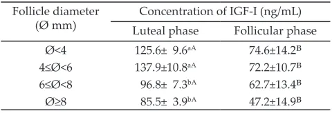
The concentrations of IGF-I in the follicular fluid of Bali cattle with diameter Ø<4 mm, 4≤Ø<6 mm, 6≤Ø<8 mm, and Ø ≥8 mm during the luteal phase were signifi
-cantly higher (P<0.05) than those during the follicular phase (Table 1). Results of the experiment indicated
that IGF-I concentrations in the follicles with diameters
smaller than 6 mm had significantly higher (P<0.05)
compared to those in the follicle diameters larger than 6 mm. Meanwhile, the concentrations of IGF-I in the
follicular fluid obtained from the ovaries in the fol
-licular phase did not show a significant effect of follicle
diameter.
Nuclear Maturation Rate
Evaluation of the nuclear maturation was classified
by chronological meiosis changes such as the stage of
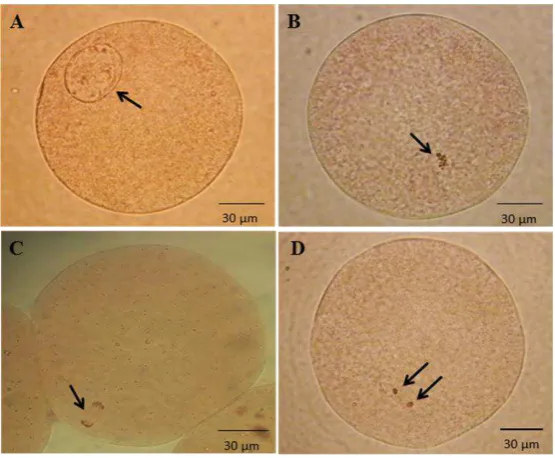
germinal vesicle (GV) to metaphase II (MII) after 22-24 h of maturation period (Figure 1). The percentages of
oocytes reached MII in the media supplemented with
Table 1. Concentrations of IGF-I in the follicular fluid of Bali cattle with different follicle diameters and reproduc -tive cycles*
Follicle diameter
(Ø mm) Luteal phaseConcentration of IGF-I (ng/mL)Follicular phase Ø<4 125.6± 9.6aA 74.6±14.2B
4≤Ø<6 137.9±10.8aA 72.2±10.7B
6≤Ø<8 96.8± 7.3bA 62.7±13.4B
Ø≥8 85.5± 3.9bA 47.2±14.9B Note: *Concentrations are expressed as mean + SEM. Means in the same
10 April 2017
HASBI ET AL. / Media Peternakan 40(1):7-13
Note: *Percentages are expressed as mean + SEM. Means in the same columns with different superscripts differ significantly (P<0.05). GV= germinal vesicle; GVBD= germinalvesicle breakdown; MI= metaphase I; AI/TI= anaphase I/telophase I; MII= metaphase II; - BSA= without supplementa -tion of BSA; + BSA= with supplementa-tion of BSA; FF Ø<4= follicular fluid obtained from follicle with diameter <4 mm; FF4≤Ø<6= follicular fluid obtained from follicle with diameter range of 4≤Ø<6 mm; FF6≤Ø<8= follicular fluid obtained from follicle with diameter range of 6≤Ø<8; FFØ ≥8= follicular fluid obtained from follicle with diameter range of Ø≥8 mm.
Treatments No. of
Oocytes
Nuclear maturation rate (%)
GV GVBD MI AI/TI MII Degenerated
- BSA 107 0(0.0±0.0) 2(1.9±1.4) 29(27.1±1.6)a 1(0.9±0.9) 68(63.6±1.9)ᵃ 7(6.5±2.1)
+ BSA 95 2(2.1±1.6) 0(0.0±0.0) 19(20.0±3.6)ab 0(0.0±0.0) 71(74.7±2.8)ᵇ 3(3.2±2.7)
FF Ø<4 85 0(0.0±0.0) 0(0.0±0.0) 4(4.7±2.0)c 0(0.0±0.0) 78(91.8±2.0)c 3(3.5±2.1) FF 4≤Ø<6 92 0(0.0±0.0) 0(0.0±0.0) 9(9.8±2.9)bc 0(0.0±0.0) 76(82.6±4.1)bc 7(7.6±4.7)
FF 6≤Ø<8 93 0(0.0±0.0) 0(0.0±0.0) 13(14.0±5.6)bc 0(0.0±0.0) 70(75.3±3.4)ᵇ 10(10.8±4.0)
FF Ø≥8 81 0(0.0±0.0) 0(0.0±0.0) 9(11.1±3.9)c 0(0.0±0.0) 63(77.8±5.7)ᵇ 9(11.1±5.0)
Table 2. Nuclear maturation rate of bali cattle oocytes in maturation media supplemented with follicular fluid obtained from different
diameters of follicle*
BSA and FF were significantly higher (P<0.05) compared
to those cultured in the media without BSA. However, when the oocyte were cultured or matured in the media supplemented with BSA and FF, the percentage of
mat-uration was similar among the groups, except in those
cultured in the media added with FF from follicles with
diameters <4 mm. The percentage of oocytes reaching
MII in oocytes cultured with FF from follicle with
diam-eter <4 mm was significantly higher (P<0.05) compared to the other groups (Table 2).
Fertilization Rate
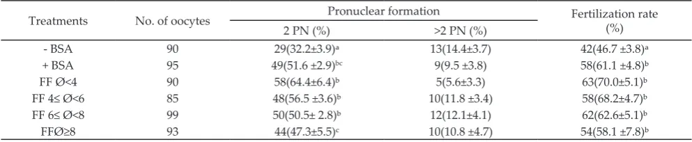
Fertilization rate was calculated based on the
number and pronuclear (PN) formation after 14-16 h of fertilization period (Figure 2). The supplementations
of maturation media with BSA and FF were able to
improve fertilization rate significantly (P<0.05) com -pared to media without BSA. The formation of 2 PN
were higher (P<0.05) in oocytes cultured in the media
supplemented with BSA and FF compared to those in the media without BSA. However, the supplementation of the media culture with FF derived from follicles with
diameters <8 mm resulted in higher 2 PN formation
compared to those derived from follicles with diameters
≥8 mm (Table 3).
DISCUSSION
IGF-I Concentration in FF Bali Cattle
Follicular fluid contains a variety of nutrients
including growth factors secreted by the follicles cells
(Nandi et al., 2008). Insulin-like growth factor-I, one of
Figure 1. Bovine oocytes after 22-24 hours in vitro maturation period. A= germinal vesicle, B= metaphase I, C= anaphase/telophase,
D= metaphase II, (arrow). Pictures of oocytes stained by 2% aceto-orcein and examined under a microscope epi-fluorescence (Zeiss Axio Imager A2 with a Zeiss Axiocam HRc, Germany).
464 465 466 467 468 469
April 2017 11 HASBI ET AL. / Media Peternakan 40(1):7-13
Note: *Percentages are expressed as mean + SEM. Means in the same columns with different superscripts differ significantly (P<0.05). PN= pronuclei; - BSA= without supplementation of BSA; + BSA= with supplementation of BSA; FF Ø<4= follicular fluid obtained from follicle with diameter <4 mm; FF4≤Ø<6= follicular fluid obtained from follicle with diameter range of 4≤Ø<6 mm; FF6≤Ø<8= follicular fluid obtained from follicle with diameter range of 6≤Ø<8; FFØ ≥8= follicular fluid obtained from follicle with diameter range of Ø≥8 mm.
Treatments No. of oocytes Pronuclear formation Fertilization rate (%)
2 PN (%) >2 PN (%)
- BSA 90 29(32.2±3.9)ᵃ 13(14.4±3.7) 42(46.7 ±3.8)ᵃ
+ BSA 95 49(51.6 ±2.9)bc 9(9.5 ±3.8) 58(61.1 ±4.8)ᵇ
FF Ø<4 90 58(64.4±6.4)ᵇ 5(5.6±3.3) 63(70.0±5.1)ᵇ
FF 4≤ Ø<6 85 48(56.5 ±3.6)ᵇ 10(11.8 ±3.4) 58(68.2±4.7)ᵇ FF 6≤ Ø<8 99 50(50.5± 2.8)ᵇ 12(12.1±4.1) 62(62.6±5.1)ᵇ
FFØ≥8 93 44(47.3±5.5)c 10(10.8 ±4.7) 54(58.1 ±7.8)ᵇ
Table 3. Fertilization rate of bali cattle oocytes in maturation media supplemented with follicular fluid obtained from different
dia-meters of follicle*
Figure 2. Bovine oocytes after 14-16 hours fertilization period. A= 2 PN and B= >2 PN. Pronuclei= PN (arrow). Pictures of oocytes stained by 2% aceto-orcein and examined under a microscope epi-fluorescence (Zeiss Axio Imager A2 with a Zeiss Axiocam HRc, Germany).
465 466 467 468 469
470 471 472 473 474
A B
complex components of IGF superfamily, plays an im
-portant role in mammalian reproduction (Velazquez et al., 2008). Coleman et al. (2007) reported that Insulin-like growth factor-I in the follicular fluid was produced dur -ing follicular growth as indicated by the IGF-I receptor
expressions in the cumulus cells, granulosa cells, and theca cells. Whereas others studies reported that IGF-I in the follicular fluid was mainly produced by the follicle and corpus luteum (Woad et al., 2000; Velazquez et al.,
2008), and its concentration increased progressively dur -ing the follicular growth until the ovulation of dominant follicle.
In our study, we found that the concentrations
of IGF-I in follicular fluid collected from follicles with
diameters of less than 4 mm to more than 8 mm
dur-ing follicular phase were similar (P>0.05). On the other
hand, at the luteal phase, the IGF-I concentrations in the
follicular fluid collected from follicle with diameter ≥6 mm was significantly lower (P<0.05) than those collected from follicle with diameter <6 mm. These results were
probably due to the increased size of the follicle dur-ing follicular growth in accordance with the decreased size and function of the corpus luteum, therefore the secretion of IGF-I in corpus luteum also decreased. This
result is different from those reported previously by
Oberlender et al. (2013) that the concentrations of IGF-I
in large follicles were greater than in small follicles.
By comparing different phases of the ovarian
reproductive status, we found that IGF-I concentrations in all follicles diameters during the luteal phase was
higher than during the follicular phase (P<0.05). These
results might be due to the contributions of the corpus luteum and the follicles to secrete IGF-I during the luteal phase of reproductive cycle. This assumption is
consistent with previous report by Woad et al. (2000) that the bovine corpus luteum showed the expression of
IGF-I receptor mRNA and a site of IGF-I reception and production. Insulin-like growth factor-I produced in the corpus luteum may enter the FF through the blood vessels of the follicle having increased vascularization and permeability during follicular development. Hurk
& Zhao (2005) described that high IGF-I concentration
made the follicle more sensitive to LH that result in the formation of angiogenic factors like vascular
endothe-lial growth factor (VEGF). Vascular endotheendothe-lial growth
factor increases vascularization and permeability of the blood vessels. On the other hand, IGF-I plays roles to stimulate progesterone production by the corpus luteum so that during the luteal phase the higher
HASBI ET AL. / Media Peternakan 40(1):7-13
Nuclear Maturation Rate
Data on the maturation rate in this study showed
that only FF derived from follicle with diameter <4
mm that was able to improve the percentage of oocytes
reaching the MII stage. These different results may be due to the differences in the concentrations of IGF-I in FF obtained from follicles with different sizes. In this study, we used follicular fluid from luteal phase, in which the concentration of IGF-I in follicular fluid ob
-tained from follicle with diameter <4 mm (125.6±9.6 ng/ mL) was higher than the other diameters. Oberlender et al. (2013) reported that the optimum concentrations of IGF-I to have positive effects on maturation rate were 129 ng/mL. Hurk & Zhao (2005) reported that the role of
IGF-I is to resume meiosis by stimulating the formation of LH receptors on the granulosa cells that causes the
follicle more responsive to LH. It is further explained
that the process of oocyte maturation is a response to the LH surge that causes some changes in the regulatory
pathway in the oocyte (Gordo et al., 2001). The mecha -nism for meiotic resumption begins with the activation of G protein that further activates phospholipase C
that hydrolases phosphatidylinositol 4,5-bisphosphate (PIP2) to form inositol triphosphate (IP3) causing the
mobilization of intracellular Ca2+ followed by an influx
of extracellular Ca2+ (Ajduk et al., 2008). The influx of extracellular Ca2+ in addition to inhibit adenylyl
cyclase which causes lower of cAMP/PKA, also enable
calmodulin-dependent protein kinase (CaM kinase II) to modify/activate the maturation promoting factor (MPF) (Hurk & Zhao, 2005;Oh et al., 2010; Conti et al., 2012).
Fertilization Rate
The success of fertilization could be assessed by the formation of male and female pro-nucleus. In this study, we found that the fertilization rate of oocytes
only about 70.0% even though the maturation rate
of oocyte matured in the media supplemented with
follicular fluid was high (75.3%-91.8%). These results
revealed that the supplementation of the maturation
media with follicular fluid did not influence fertilization
rate of oocytes. It was suspected that the IGF-I in the
fol-licular fluid only acted on promoting oocyte maturation (Oberlender et al., 2013). The ability of oocyte to be fertil -ized is not only determined by the nuclear maturation of oocytes but also by the cytoplasmic maturation that are
required for the progress of the maturation and to pre
-vent polyspermy (Hyttel et al., 1997). The cytoplasmic maturation is influenced by several factors including
the successive transformations of mitochondria, cortical granules, and smooth and rough endoplasmic reticulum
(Hyttel et al., 1997). Furthermore, the distributions of
organelles in the cytoplasm are closely correlated with
the maturation and competence of oocyte (Brevini et al., 2007). Therefore, incomplete cytoplasmic matura -tion of oocytes at the MII stage may be one of reasons for the low rate of in vitro embryo production (Blanco
et al., 2011). This is in contrary to previous reports that the follicular fluid promotes cytoplasmic maturation of oocyte during IVM (Hong & Lee, 2007; Papanikolaou et
al., 2008; Grupen & Armstrong, 2010). Agung et al. (2013) reported that the use of porcine follicular fluid as a sole
maturation media resulted in the formation of male pro-nuclear after in vitro fertilization in the matured porcine oocytes. Furthermore, Cruz et al. (2014) reported that
the addition of follicular fluid at lower concentration in
maturation media enhanced the total cell numbers in
bovine embryos produced in vitro.
CONCLUSION
The concentration of IGF-I in follicular fluid ob -tained from the ovary in the luteal phase was higher than that in the follicular phase. The IGF-I
concentra-tion in a smaller follicle (diameter <6 mm) was higher compared to a large follicle (diameter ≥6 mm). The supplementation of follicular fluid improves the nuclear maturation and fertilization rate of oocytes of Bali cattle.
ACKNOWLEDGEMENT
This study was supported by Doctoral Research
Grant No. 019/SP2H/LT/DPRM/II/2016 of the Ministry
of Research, Technology, and Higher Education of the Republic of Indonesia.
REFERENCES
Agung, B., Y. Piao, D. Fuchimoto, S. Senbon, A. Onishi, T. Otoi, & T. Nagai. 2010. Effects of oxygen tension and follicle cells on maturation and fertilization of porcine oocytes during in vitro culture in follicular fluid. Theriogenology 73:893–
899. https://doi.org/10.1016/j.theriogenology.2009.11.013
Agung, B., T. Otoi, D. Fuchimoto, S. Senbon, A. Onishi, & T. Nagai. 2013. In vitro Fertilization and development of
porcine oocytes matured in follicular fluid. J. Reprod. Dev. 59:103-106. https://doi.org/10.1262/jrd.2011-050
Ajduk, A., A. Małagocki, & M. Maleszewski. 2008. Cytoplasmic maturation of mammalian oocytes:
develop-ment of a mechanism responsible for sperm-induced Ca2+ oscillations. Reprod. Biol. 8:3-22. https://doi.org/10.1016/ s1642-431x(12)60001-1
Avery, B., L. Strobech, T. Jacobsen, I. B. Bogh, & T. Greve. 2003.
In vitro maturation of bovine cumulus oocyte complexes in undiluted FF: effect on nuclear maturation, pronucle -us formation and embryo development. Theriogenology
59:987–999. https://doi.org/10.1016/S0093-691X(02)01139-1
Bender, K., S. Walsh, A. Evans, T. Fair, & L. Brennan. 2010.
Metabolite concentrations in follicular fluid may explain differences in fertility between heifers and lactating cows. Reproduction 139:1047-1055. https://doi.org/10.1530/
REP-10-0068
Bijttebier, J., A. Van Soom, E. Meyer, B. Mateusen, & D. Maes.
2008. Preovulatory follicular fluid during in vitro matura-tion decreases polyspermic fertilizamatura-tion of cumulus-intact porcine oocytes in vitro maturation of porcine oocytes.
Theriogenology 70:715–724. https://doi.org/10.1016/j.
theriogenology.2008.04.046
Blanco, M. R., S. Demyda, M. Moreno Millán, & E. Genero. 2011. Developmental competence of in vivo and in vitro matured oocytes. A review. Biotechnol. Mol. Biol. Rev.
6:155-165.
Brevini, T. L., F. Cillo, A. Antonini, & F. Gandolfi. 2007.
Cytoplasmic remodeling and the acquisition of develop -mental competence in pig oocytes. Anim. Reprod. Sci.
April 2017 13 Coleman, N. V., G. A. Shagiakhmetova, I. Y. Lebedeva, T. I.
Kuzmina, & A. K. Golubev. 2007. In vitro maturation and early developmental capacity of bovine oocytes
cul-tured in pure follicular fluid and supplementation with follicular wall. Theriogenology 67:1053–1059. https://doi. org/10.1016/j.theriogenology.2006.10.019
Conti, M., M. Hsieh, A. M. Zamah, & J. S. Oh. 2012. Novel sig-naling mechanisms in the ovary during oocyte maturation
and ovulation. Mol. Cell. Endocrinol. 356:65-73. https:// doi.org/10.1016/j.mce.2011.11.002
Cruz, M. H. C., N. Z. Saraiva, J. F. da Cruz, C. S. Oliveira, M. D. Collado, H. Fernandes, F. C. de Castro, & J. M. Garcia.
2014. Effect of follicular fluid supplementation during in
vitro maturation on total cell number in bovine blastocysts produced in vitro. R. Bras. Zootec. 43:120-126. https://doi.
org/10.1590/S1516-35982014000300003
Ducolomb, Y., H. Gonzalez-Marquez, R. Fierro, R. Fierro, I. Jimenez, E. Casas, D. Flores, E. Bonilla, Z. Salazar, & M. Bitancourt. 2013. Effect of porcine follicular fluid proteins
and peptides on oocyte maturation and their subsequent effect on in vitro fertilization. Theriogenology 79:896-904.
https://doi.org/10.1016/j.theriogenology.2013.01.024
Gordo, A. C., C. L. He, S. Smith, & R. A. Fissore. 2001. Mitogen
activated protein kinase plays a significant role in meta -phase II arrest, spindle morphology, and maintenance of maturation promoting factor activity in bovine oocytes.
Mol. Reprod. Dev. 59:106–114. https://doi.org/10.1002/
mrd.1012
Grupen, C. & D. Armstrong. 2010. Relationship between cumu-lus cell apoptosis, progesterone production and porcine
oocyte developmental competence: temporal effects of fol
-licular fluid during IVM. Reprod. Fertil. Dev. 22:1100-1109. https://doi.org/10.1071/RD09307
Hong, J. & E. Lee. 2007. Intrafollicular amino acid
concentra-tion and the effect of amino acids in a defined maturaconcentra-tion
medium on porcine oocyte maturation, fertilization, and preimplantation development. Theriogenology
68:728-335. https://doi.org/10.1016/j.theriogenology.2007.06.002
Hurk, R. V. N. & J. Zhao. 2005. Formation of mammalian oo
-cytes and their growth, differentiation and maturation within ovarian follicles. Theriogenology 63:1717–1751. https://doi.org/10.1016/j.theriogenology.2004.08.005
Hyttel, P., T. Fair, H. Callensen, & T. Greve. 1997. Oocyte growth capacitation and final maturation in cattle. Theriogenology 47:23-32. https://doi.org/10.1016/S0093-691X(96)00336-6
Ito, M., H. Iwata, M. Kitagawa, Y. Kon, T. Kuwayama, & Y. Monji. 2008. Effect of follicular fluid collected from vari -ous diameter follicles on the progression of nuclear mat-uration and developmental competence of pig oocytes.
Anim. Reprod. Sci. 106:421-430. https://doi.org/10.1016/j.
anireprosci.2007.06.003
Nandi, S., V. G. Kumar, B. M. Manjunatha, H. S. Ramesh,
& P. S. P. Gupta. 2008. Follicular fluid concentrations of
glucose, lactate and pyruvate in buffalo and sheep, and their effects on cultured oocytes, granulosa and cumulus cells. Theriogenology 69:186–196. https://doi.org/10.1016/j.
theriogenology.2007.08.036
Oberlender, G., L. D. S. Murgas, M. G. Zangeronimo, A. C.
da Silva, T. A. Menezes, T. P. Pontelo, & L. A. Vieira. 2013. Role of insulin-like growth factor-I and
follicu-lar fluid from ovarian follicles with different diameters
on porcine oocyte maturation and fertilization in vitro.
Theriogenology 80:319–327. https://doi.org/10.1016/j.
theriogenology.2013.04.018
Oh, J. S., S. J. Han, & M. Conti. 2010. Wee1B, Myt1, and Cdc25 function in distinct compartments of the mouse oocyte
to control meiotic resumption. J. Cell. Biol. 188:199-207. https://doi.org/10.1083/jcb.200907161
Papanikolaou, T., G. Amiridis, T. Dimitriadis, E. Vainas, & C.
Rekkas. 2008. Effect of plasmin, plasminogen activators
and a plasmin inhibitor on bovine in vitro embryo
pro-duction. Reprod. Fertil. Dev. 20:320-327. https://doi.
org/10.1071/RD07108
Park, C. H., S. G. Lee, D. H. Choi, & C. K. Lee. 2009. A modified swim-up method reduces polyspermy during in vitro
fer-tilization of porcine oocytes. Anim. Reprod. Sci. 115:169– 181. https://doi.org/10.1016/j.anireprosci.2008.12.004
Pereira, G. R., P. L. Lorenzo, G. F. Carneiro, B. A. Ball, L. M. C. Pegoraro, C. A. Pimentel, & I. K. M. Liu. 2013. Influence of
equine growth hormone, insulin-like growth factor-I and
its interaction with gonadotropins on in vitro maturation
and cytoskeleton morphology in equine oocytes. Animal 7:1493–1499. https://doi.org/10.1017/S175173111300116X
Petro, E. M., J. L. Leroy, A. Covaci, E. Fransen, D. De Neubourg, A. C. Dirtu, I. De Pauw, & P. E. Bols. 2012.
Endocrine-disrupting chemicals in human follicular fluid impair in
vitro oocyte developmental competence. Hum. Reprod.
27:1025-1033. https://doi.org/10.1093/humrep/der448
Revelli, A., L. Delle-Piane, S. Casano, E. Molinari, M. Massobrio, & P. Rinaudo. 2009. Folicular fluid content
and oocyte quality: from single biochemical markers to metabolic. Reprod. Biol. Endocrinol. 7: 1-13. https://doi.
org/10.1186/1477-7827-7-40
Shirazi, A. & N. Sadeghi. 2007. The effect of ovine oocyte di
-ameter on nuclear maturation. Small Rum. Res. 69:103-107. https://doi.org/10.1016/j.smallrumres.2005.12.022
Silva, J. R. V., J. R. Figueiredo, &R.van den Hurk. 2009. Involvement of growth hormone (GH) and insulin-like growth factor (IGF) system in ovarian folliculogenesis. Theriogenology 71:1193–1208. https://doi.org/10.1016/j. theriogenology.2008.12.015
Sinclair, K., L. Lunn, W. Kwong, K. Wonnacot, R. Linforth, & J. Craigon. 2008. Amino acid and fatty acid composition
of follicular fluid as predictors of in vitro embryo
devel-opment. Reprod. BioMed. Online. 16:859-868. https://doi. org/10.1016/S1472-6483(10)60153-8
Somfai, T., Y. Inaba, S Watanabe, M Geshi, & T Nagai. 2012.
Follicular fluid supplementation during in vitro matura-tion promotes sperm penetramatura-tion in bovine oocytes by
en-hancing cumulus expansion and increasing mitochondrial activity in oocytes. Reprod. Fertil. Dev. 24:743-752. https:// doi.org/10.1071/RD11251
Spanos, S., D. L. Becker, R. M. L. Winston, & K. Hardy. 2000. Anti-apoptotic action of insulin-like growth fac-tor-I during human preimplantation embryo
develop-ment. Biol. Reprod. 63:1413–1420. https://doi.org/10.1095/ biolreprod63.5.1413
Supriyantono, A., L. Hakim, Suyadi, & Ismudiono. 2008.
Performance of bali cattle in three areas in the provinces of bali. Berk. Penel. Hayati 13:147–152.
Suzuki, K., B. Eriksson, H. Shimizu, T. Nagai, & H. Rodrigues-Martines. 2000. Effect of hyaluronan on monospermic penetration of porcine oocytes fertilized in vitro. Int. J.
Androl. 23:13-21. https://doi.org/10.1046/j.1365-2605.2000. t01-1-00198.x
Valckx, S. D. M., J. De Bie, E. D. Michiels, I. G. Goovaerts, U.
Punjabi, P. Ramos-Ibeas, A. Gutierrez-Adan, P. E. Bols, & J. L. Leroy. 2015. The effect of human follicular fluid on bovine oocyte developmental competence and embryo
quality. Reprod. BioMed. Online. 30:203–207. https://doi. org/10.1016/j.rbmo.2014.10.008
Velazquez, M. A., L. J. Spicer, & D. C. Wathes. 2008. The role
of endocrine insulin-like growth factor-I (IGF-I) in female bovine reproduction. Domestic Anim. Endocrinol. 35:325– 342. https://doi.org/10.1016/j.domaniend.2008.07.002
Woad, K. J., G. Baxter, C. O. Hogg, T. A. Bramley, R. Webb, & D. G. Armstrong. 2000. Expression of mRNA encoding insulin-like growth factors I and II and the type 1 IGF