www.elsevier.com / locate / livprodsci
Post-weaning multisystemic wasting syndrome (PMWS) in pigs
in France: clinical observations from follow-up studies on
affected farms
a ,
*
a a a a aF. Madec
, E. Eveno , P. Morvan , L. Hamon , P. Blanchard , R. Cariolet ,
b b a a a a
´
N. Amenna , H. Morvan , C. Truong , D. Mahe , E. Albina , A. Jestin
a
´ ´ ˆ
AFSSA Agence Franc¸aise de Securite Sanitaire des Aliments, Zoopole, BP 53 22440, Ploufragan, France
b
´ ˆ
LDA22 Laboratoire de Developement et d’Analyses, Zoopole, BP 54 22440, Ploufragan, France Received 6 May 1999; received in revised form 12 July 1999; accepted 30 July 1999
Abstract
A disease affecting weaned pigs and known as Postweaning Multisystemic Wasting Syndrome (PMWS) is described on French farms. Follow-up studies were designed on cohorts of pigs on a group of 12 severely affected farrow-to-finish operations. Three of them were free of Porcine Reproductive and Respiratory Syndrome Virus (PRRSV) infection. Three thousand and seventy eight pigs were included in the study. Mortality, from weaning to slaughter, was 11% and weeks 11–13 were the most critical. The first clinical sign reported was unthriftiness, then pallor and often fever with associated respiratory or digestive disorders. Wasting could follow rapidly and when clearly established in an individual the prognosis was grave especially when the sick pigs were kept with their penmates. Antibiotics were administered without real efficacy. The lesions affected several viscera including lungs, spleen, kidneys and lymph nodes with a severe lymphoid depletion. The disease did not show a collective impact. From preliminary epidemiological investigations, a strong litter effect on disease susceptibility was suspected. PRRS virus was excluded as a major agent. On the other hand, a porcine circovirus (PCV2) was found associated to the lesions. The environment was suspected as an important determining factor for the effect of PMWS in the herds.
´ ´ Resume
´ ´ ´ ´
Les manifestations pathologiques associees au syndrome du deperissement du porcelet (PMWS) aussi appele Maladie de
´ ´ ´
l’Amaigrissement du Porcelet (MAP) sont exposees. Pour cela des suivis de cohortes de porcs ont ete mis en place dans un
´ ´ ´ ` ´
groupe de 12 elevages. Il s’agissait d’elevages naisseurs-engraisseurs severement affectes par la maladie. Trois d’entre eux
´ ´ ´
etaient indemnes de SDRP (Syndrome Dysgenesique et Respiratoire Porcin). Trois mille soixante dix huit porcs sont inclus ˆ
´ ´ ` ` ´ ´
dans l’etude. Le taux de mortalite du sevrage a l’abattage a atteint 11 % et les semaines 11 a 13 se sont averees etre les plus ˆ
` ´ `
critiques a cet egard. Le premier signe perceptible est la perte de vigueur puis la paleur et souvent la fievre. Les
´ ´ ´ `
manifestations respiratoires (toux, dyspnee) et digestives (diarrhee) sont frequentes. Il s’en suit un amaigrissement tres rapide ´
(2–3 jours). Lorsque l’amaigrissement est clairement exprime le pronostic est sombre notamment lorsque les animaux
*Corresponding author. Tel.: 133-2-9601-6222; fax: 133-2-9601-6253. E-mail address: [email protected] (F. Madec)
´ ´ ` ´ ´
atteints ne sont pas retires du groupe. Les medicaments s’averent inefficaces. Les lesions affectent differents organes: les
` ´ ` ´ ´ ¨
poumons, la rate, les reins et surtout les ganglions lymphatiques ou on observe une severe depletion lymphoıde. La maladie
` ` ´ ´ ´ ´
ne possede pas d’expression collective. Les premieres donnees epidemiologiques mettent en relief une susceptibilite accrue
´ ´ ´
de certaines portees. Le virus du SDRP est exclu comme agent causal au benefice d’un circovirus porcin (PCV2). Les
´ ´ ´ ´ ´
conditions d’environnement offertes aux animaux sont suspectees decisives dans la severite de l’impact de la maladie dans ´
les elevages. 2000 Elsevier Science B.V. All rights reserved.
Keywords: Wasting syndrome; Pig; Postweaning; Epidemiology; PMWS
1. Introduction 2. Material and methods
Late in Spring 1996, two farmers complained 2.1. The farms
about unexpected mortality occurring in growing
pigs in Brittany (France). The pigs were two to three Farms were selected by field veterinarians. The
months old and no obvious other health disturbance latter, in turn, had been called to these farms because
could be detected in the farms. In a preliminary of high and persistent mortality in weaned pigs
study, piglets showing the first clinical signs of despite medication. The authors visited the farms and
wasting were transferred to our experimental their selection was based on the willingness of the
facilities and SPF pigs of the same age were placed farmer to co-operate and also on the availability of
in contact. Surprisingly, the wasting piglets pro- information. Twelve farms in the western part of
gressively recovered without any medication, whilst France working with six different farm organisations
the contact SPF pigs became severely ill. They (co-operatives) were included in the survey. They
developed typical Glasser’s disease. At that time the were all farrow-to-finish operations like the majority
situation was rather confusing. Porcine Respiratory of farms in the area. On 11 farms the replacement
and Reproductive Syndrome (PRRS) was detected gilts came from a multiplier. On the other the
on those farms affected by the piglet wasting prob- replacement stock was home-produced. Artificial
lem as well as several other enzootic agents includ- insemination from bought-in specialised AI centres
ing M. hyopneumoniae, P. multocida, H. parasuis, S. was widely used. The average herd size was 343
suis and enterotoxigenic E. coli. During the second sows (range 105–600). Eleven farms had intensive
half of 1996 we tried to determine the presence or confined production (slatted floor, fan ventilated
absence of the specific new syndrome. Field case rooms) and on one (Farm 2, Table 1) the sows were
observations, often associated with experimental kept outdoors. The productivity of the herds was
trials, were designed. By the end of 1996 we came to good (24.4 piglets weaned / sow / year). Age at
wean-the conclusion that since wean-the condition could be ing was either 26–28 days (n57 herds) or 21 days
transmitted to naive pigs in our facilities (Le Cann et on average (n55 herds). They were free of
Pseu-al., 1998), it was infectious and transmissible. Final- dorabies but 9 of them were PRRS positive. Herds
ly, the syndrome (clinical signs, macroscopic lesions, No. 2, 8 and 10 were PRRS free.
histopathology) was recognised as being similar to
the descriptions made in North America and called 2.2. The protocol on the farms
‘‘Postweaning Multisystemic Wasting Syndrome’’
(Harding, 1996, Clark, 1996). Since then, an exten- The protocol applied across the 12 farms was
sive research programme has been performed. The designed to achieve two objectives. Firstly to
char-present paper reports a series of field surveys and acterise PMWS which was new to France. No
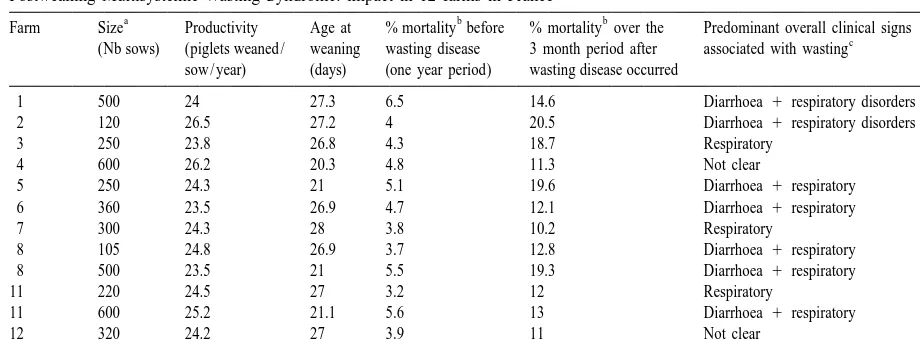
Table 1
Postweaning Multisystemic Wasting Syndrome: impact in 12 farms in France
a b b
Farm Size Productivity Age at % mortality before % mortality over the Predominant overall clinical signs
c
(Nb sows) (piglets weaned / weaning wasting disease 3 month period after associated with wasting sow / year) (days) (one year period) wasting disease occurred
1 500 24 27.3 6.5 14.6 Diarrhoea 1respiratory disorders
2 120 26.5 27.2 4 20.5 Diarrhoea 1respiratory disorders
3 250 23.8 26.8 4.3 18.7 Respiratory
4 600 26.2 20.3 4.8 11.3 Not clear
5 250 24.3 21 5.1 19.6 Diarrhoea 1respiratory
6 360 23.5 26.9 4.7 12.1 Diarrhoea 1respiratory
7 300 24.3 28 3.8 10.2 Respiratory
8 105 24.8 26.9 3.7 12.8 Diarrhoea 1respiratory
8 500 23.5 21 5.5 19.3 Diarrhoea 1respiratory
11 220 24.5 27 3.2 12 Respiratory
11 600 25.2 21.1 5.6 13 Diarrhoea 1respiratory
12 320 24.2 27 3.9 11 Not clear
TOTAL 343 24.4 24.5 4.6 14.6
a
Farrow-to-finish farms.
b
Mortality (for any reason) from weaning to slaughter.
c
Individual signs: unthriftiness, pallor, etc . . . were present but not indicated here
laboratory examinations and clinical observations. On most of the farms, there were numerous
Retrospectively, the 12 farms were diagnosed as differences in husbandry. On four farms (3, 4, 7 and
having PMWS on histopathology and by in situ 9) the hygiene policy, space per pig, ventilation,
hybridisation using a specific circovirus type II segregation of subsequent batches and mixing of pigs
probe. The second objective was to investigate the varied and changes occurred at the different stages
epidemiology of PMWS and to try to build up (suckling, weaning, fattening). The objective was not
hygiene proposals for the affected farms (cohort to test the relevance of each of these changes but to
studies were carried out on these farms). The piglets look at the overall consequences of a global
come-were identified at birth. They come-were weighed at back to standard recommendations (ITP, 1993;
weaning and then, at different times depending on Madec et al., 1999).
the farms. On farm 9, early weaning was carried out. Within the whole group of 12 farms, the
prod-The cohorts (one or two per farm) were visited by us uctivity and mortality data were recorded. All the
every 14 days. The farmer was asked to take special herds were managed according to a batch system
care with recording (signs of illness, medications, (groups of sows weaned the same day every 3
death etc). If illness occurred numerous ‘‘extra weeks; each week for the largest units, ITP, 1993)
visits’’ were scheduled. A proportion of the pigs and therefore we were able to know retrospectively
dying were necropsied at the laboratory. At the end the evolution of mortality in the subsequent groups
of the critical period the pigs were weighed and bled. of contemporary pigs.
On three farms (5, 6 and 7) a more detailed protocol
was applied since the pigs were visited every 2 days 2.3. The protocol at the laboratory
on average during the critical phase. Rectal
tempera-ture was recorded in a group of 40 pigs randomly At the laboratory, the pigs were euthanased and
selected. The pigs were weighed at weaning, at 8 subjected to a standard necropsy. Bacteriological
weeks of age and at 12 weeks. On each occasion examination was performed on the tonsils and on
they were blood sampled. An additional blood certain viscera showing gross lesions: particularly the
Histopathology was undertaken with special em- 3.2. The overall results of the cohort studies phasis on lymphoid tissues associated with lung and
intestine. Viral inclusions were looked for following The total number of pigs considered was 3078.
the protocol of Ellis et al. (1998). Three hundred and thirty eight (11%) died or were
Lastly, an in situ hybridisation specific for cir- sacrificed principally for animal welfare reasons. Fig.
covirus Type II, was performed in cases where there 1 shows the mortality according to their age. The
was microscopic damage. twelfth week was the most critical and 68% of the
losses occurred during weeks 11–13. Signs of illness were noticed in all the piglets before death. When
3. Results recorded, rectal temperature was found to be 40.58C
or above, on at least one day in all these fatal cases.
3.1. Impact of PMWS in the herds The first sign reported to us by the farmers was
unthriftiness, then most of the pigs became pale and
The first warning signs usually came post-weaning their flanks looked empty since their feed intake had
when the pigs were around two months old. They been reduced. This was confirmed later by opening
were weaned at four to five weeks but in most cases, the stomach at necropsy. Wasting was established
the critical phase for disease occurrence ended at rapidly and the back bone soon became apparent.
around three months of age (13 weeks). Clear signs Cough and diarrhoea were frequent but not always
were still observed after the third month especially found. Dermatitic skin lesions especially on the rear
when the disorders occurred in week twelve. In a of the body were noticed in the cohorts on six farms
given batch of pigs only some individuals clearly (n515 pigs) and transient cyanosis (especially on
showed clinical signs (i.e. low morbidity). A wide the ears, and in the perineal area) on four farms
range of drugs was used without any significant (n516 pigs). Icterus was detected in only six pigs.
positive effect. There was no detrimental effect on When the wasting process was clearly visible,
reproductive function. Prolificacy and farrowing despite a wide range of treatments, the prognosis was
rates were maintained at the previous level. Abor- grave. When these pigs were removed from the pens
tions, stillbirth or mummifications were not noticed. and placed in hospital accommodation, about half of
Muscle wasting in the piglets was in every case the them survived, but only half later showed a correct
major and most typical sign, but was associated with weight gain. One hundred and eight pigs from 12
other manifestations (Table 1). The mortality rate farms were sent to the laboratory. They were 81 days
was rather high, both post-weaning and in the old on average (612 days). Table 2 gives the results
finishing stage and depended on the age at which the of necropsy. Most of the pigs were in poor body
pigs were moved to the finishing house. On these condition when submitted to the laboratory. It is
severely affected farms, mortality level reached remarkable that a high percentage of the pigs were
14.6% on average for the three months preceding our affected by pneumonia (67.5%). The lesions were
intervention. The corresponding figures before often of severe bronchopneumonia and extended to
PMWS onset are also shown in Table 1. In every all lung lobes. A high proportion (53%) of pigs
case, when PMWS signs appeared, a sudden increase showed a disturbance of the large bowel (oedema,
in mortality was noticed by the farmers and con- dilatation). The lymph nodes were often enlarged but
firmed by the recordings. In a given herd, consider- those affected differed from pig to pig. Lymphoid
able differences could be seen in mortality impact depletion in the lymph nodes and Peyer’s Patches
between the subsequent batches. However, a baseline was also a common finding but was not the general
was observed. This was around 10–15% (from rule. Eleven pigs with dermatitic skin lesions type
weaning until slaughter) for the majority of the farms were sent to the laboratory and kidney lesions were
(8 farms), but it was closer to 20% for four farms. mainly found in these pigs. Bacteriology was not
On three of the farms the problem remained acute for systematically performed since the largest number of
Fig. 1. Age of the pigs at death in the cohort study carried out on severely PMWS affected farms (n5338 pigs, dead or euthanized).
necropsy. A variety of infectious agents were iso- 3.3.1. Farm 5 cohort
lated from lesions in the viscera and from the tonsils. Two hundred and ten pigs (19 litters) were
Haemophilus parasuis, Pasteurella multocida and studied. Some pigs could be heard coughing after
Streptococcus suis were commonly found (50, 41 weaning (on week 6 and 7) without any apparent
and 10% of the pigs respectively). detrimental consequences. At the end of the 10th
week of age and during the 11th, several pigs
3.3. The results of the detailed on-farm clinical became progressively hyperthermic, and 46% of the
observation ( farms 5, 6 and 7) pigs reached $40.58C at least once, most of them without any sign of depression. A proportion of the
On these three farms, there were no obvious signs pigs looked pale (12%) and diarrhoea was seen in
of disease during lactation. Average weaning weight 13% of the pigs. Respiratory signs (cough and
was 6.2 kg, 7.5 kg and 8.2 kg for herds 5, 6 and 7 sneezing) were also detected. Different treatments
respectively. The pigs were weaned at 21 days on were administered especially antibiotics (tiamuline
farm 5; and at 27 and 28 days on average respective- and / or colistine and neomycine depending on the
ly in herds 6 and 7. Average litter size at weaning individuals). Anti-inflammatory compounds
(Dexa-was 11, 10.2 and 9.5 for the three cohorts. During methasone) were also injected to affected pigs
with-the first month post-weaning, things went on normal- out real success. Twenty three piglets (11%) died
ly and only some mild diarrhoea was noticed during between weeks 10 and 13 (2, 7, 10 and 4
respective-the second week post-weaning. Growth rate re- ly for the 4 weeks). Average weight at the end of
mained moderate. Average liveweight at 8 weeks of week 12 was 35 kg (67 kg). One week later, at the
age was 18.8 kg (63.7), 19 kg (63.9) and 19.7 kg end of week 13 the group of pigs looked unequal
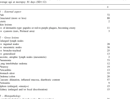
Table 2
External aspect and lesions observed in a sample of piglets taken on farms severely affected by PMWS n5108 piglets from 12 farms, average age at necropsy: 81 days (SD612)
n %
1 – External aspect
Pale 72 66
Emaciated (more or less) 88 81
icteric 2 1.8
skin lesions
⇒of dermatitis type: papules or red-to-purple plaques, becoming crusty 11 10.1
⇒ cyanosis (ears, Perineal area) 3 2.7
2 – Gross lesions Enlarged lymph nodes
⇒ inguinal nodes 28 25.9
⇒ mesenteric nodes 36 33.3
⇒ broncho-tracheal 25 23.1
⇒ generalised 13 12
necrotic, atrophic lymph nodes (mesenteric) 2 1.8
Pneumonia 73 67.5
Lung interlobular oedema 12 11.1
Pleuresy 19 17.5
Pericarditis 13 12
Stomach ulcer 33 30.5
Oedema / colon 20 18.5
Caecum: dilatation, inflamed mucosa, diarrhoeic content 57 53.2
Peritonitis 9 8.3
Spleen (enlarged, «meaty») 15 13.8
Kidney (enlarged and / or focal discoloration) 15 13.8
3 – Histopathology ¨
Lymphoıd depletion (lymph nodes, Peyer patches) 76 70.3
Intertitial pneumonia 83 76.9
Multinucleated giant cells 65 60
Hystiocitic infiltration 60 55.5
Intracytoplasmic viral inclusion bodies 62 57.4
be removed from the room and by adding the cull numerous (up to 418C was recorded). The pigs
piglets to the wasting ones total losses reached exhibited diarrhoea (12%) at clinical examination in
15.7%. the middle of week 11 and treatment was rapidly
It became obvious that some of the litters were prescribed (Colistine 1 Neomycine, oral route).
more affected than others. (For example seven pigs A recrudescence of coughing was also observed
that died were from the same litter of 11 weaned and patent signs of wasting became visible soon in
piglets). the first sick pigs. During week 12, dermatitic skin
lesions were present in three pigs and cyanosis in
3.3.2. Farm 6 cohort two other individuals. Dermatitis was first seen as
Three hundred and two pigs (30 litters) were papules, then as erythematous and crusty lesions (1
studied. Until the 10th week of age there was no to 3 cm diameter) located first in the perineal area
obvious sign of disease. By the end of the 10th week, and later covering the whole rear part of the body
one pig showed fever and unthriftiness and during including the legs. Cyanosis, when it occurred, lasted
Table 3
also on the ears. When cyanosis occurred the pigs
Weight changes in the pigs of Fig. 2 (kg)
had already started to decline, coughed and had been
No. pigs Week 4 Week 8 Week 12
pyrexic ($ 418C) for two days. One pig became
a b
icteric in week 12 after starting to lose weight. Fig. 2 A 4.6 13.2 15
shows the clinical pattern of four selected pigs raised B 9.5 23.6 28.2
C 7.8 19.4 25.4
in the same room. At the laboratory, typical lesions
D 7.7 22 37
of PMWS were observed in the pigs. Table 3 gives
their liveweight at weaning, at 8 and at 12 weeks. Group average
Pig D was still apparently healthy at the end of week (n5302 pigs) 7.5 19.7 34.7
12. At that time, average live weight of the cohort a
Week 3 at weaning for this pig (A) and the corresponding
was 34.7 kg (67 kg). Wasting stopped after week 13. litter; weight later on at week 7 and 11.
b
Within the cohort 21 pigs had died by week 14 and 7 At death.
pigs were in a bad physical state (total: 9.3%).
3.3.3. Farm 7 cohort in the room but they were littermates of four sows
Two hundred and fifty nine pigs (24 litters) were (out of the 24 sows studied). Respiratory signs were
studied. The first clinical sign was pyrexia associated also common. Three pigs died, 2 with signs of
with depression in 5 pigs during week 10, and the wasting. They became pale, dyspnoeic and died
proportion increased (15%) with time. The same within 3 days. In the remaining pigs, external signs
week one pig showed typical lesions of skin der- of wasting were obvious in 12 pigs by the end of
matitis and a rectal temperature of 418C. The pig week 12. Amongst these, 10 died or were euthanased
died 3 days later. The pigs that showed the first high later (total loss of 13 pigs, 5%). Average liveweight
rectal temperatures were spread over different pens at 12 weeks was 37 kg (66.5 kg).
4. Preliminary epidemiology had seroconverted showing clear evidence of PRRS virus infection on the farms. However, only some of
4.1. The role of the sow (or «litter-effect») the sows which gave birth to the piglets were PRRS
seropositive at weaning time (36 sows out of 75,
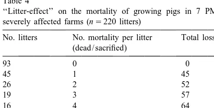
As had already been mentioned, some litters 48%). The litters of PRRS seronegative sows tended
seemed to react more severely. On the farms where to be more predisposed to exhibiting signs especially
cross-fostering was limited during the suckling phase fever than those of PRRS seropositive sows. On the
and where it could be properly recorded, the litter other hand no difference could be seen regarding
effect was investigated (Table 4). Whilst 42% of the wasting and mortality.
litters were not at all affected, 16% accounted for The PRRS virus was found in only one pig (Pig B,
54% of the losses. The figures obtained were sig- farm 6) but the results strongly suggested circulating
nificantly different (P,0.001) from the theoretical PRRS virus in the growing pigs during the survey on
distributions especially the Poisson distribution. On the 3 farms.
the other hand, no clear relationship could be found In conclusion, from our observations on all 12
with the parity of the sow. farms, the real impact of PRRS infection on PMWS
could not be established but was estimated to be low. 4.2. The role of PRRS
4.3. The role of environment Three of the 12 farms (No. 2, 8 and 10) were
seronegative for PRRS and had never experienced In the cohorts which were followed, mortality was
infection (no clinical signs were observed and regu- reduced (11%) compared to the previous situation of
lar serology remained negative over the years). On the farms (14.6%). Part of the explanation was found
these farms the clinical picture of PMWS was not in the changes in the environment offered to the pigs
different and the level of mortality was also very especially on four farms (No. 3, 4, 7 and 9). The
similar to that of the seropositive farms (Table 1). technical conditions of production were investigated
In the detailed on-farm investigations carried out and in agreement with the farmers, husbandry was
in farms 5, 6 and 7 all the piglets except six in farm modified. The changes were directed at reduction of
7 were seronegative to PRRS at 12 weeks of age. infection pressure in the herds through better hygiene
Fever, cough and cyanosis were observed starting in and management. Over-crowding and mixing were
week 10, the illness culminating during weeks 11 reduced and the cleaning strategy was improved. An
and 12 after transfer to the finishing pens. The time all-in / all-out hygiene policy system (one batch of
lapse between the PRRS-like troubles and blood contemporary pigs per room) was strictly applied and
sampling for serology was probably too short to other measures including emptying the pits below the
demonstrate seroconversion. By week 16 all the pigs slatted floors introduced on those farms. Whereas
wasting was still observed, mortality from weaning to slaughter was considerably reduced (from 12% or
Table 4 more to 6% or less for farms 3, 4, 7 and 9).
‘‘Litter-effect’’ on the mortality of growing pigs in 7 PMWS
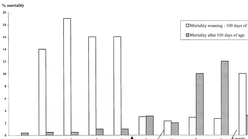
In herd 9, three batches of pigs, each born one
severely affected farms (n5220 litters)
week apart, were observed (total5350 pigs, 123
No. litters No. mortality per litter Total losses litters). In each batch, half of the randomly selected (dead / sacrified)
pigs were weaned at 12 days of age and housed in
93 0 0 appropriate accommodation off the farm. The
re-45 1 45
maining litters were weaned at 21 days with strict
26 2 52
hygiene and husbandry. From weaning to 100 days
19 3 57
of age, mortality was limited to less than 1% for all
16 4 64
10 5 50 pigs. After the trial the farmer tried to continue the
8 6 48 strict husbandry. Mortality from wasting was
3 7 21
Un-Fig. 3. Mortality of pigs (subsequent batches of contemporary animals, one week between each batch). Farm 9: farrow-to-finish unit;h, mortality weaning – 100 days of age;j, mortality after 100 days of age.
fortunately after remission in 4 subsequent batches published on-farm observations remain rather
gener-the troubles re-occurred during gener-the finishing phase, at al.
15–17 weeks of age. Mortality (wasting mainly but The present work which focused on follow-up
also dermatitis-nephropathy) was 9% on average and studies on severely affected farms (some for several
persisted for 5 months (from 8 to 12% mortality, Fig. months), describes detailed clinical observations. Our
3). results show a real critical phase for disease
occur-After 5 months, the problem recurred at an earlier rence. On our French farms, it starts at around eight
age (9–10th week). A failure to adhere to the weeks of age and ends at about 13 weeks. Obviously,
previous hygiene recommendations was cleary ob- signs of the disorders are still clear after this period
served. especially the weight heterogeneity. Under Canadian
conditions, Harding and Clark (1997) observed an earlier occurrence (5–6 weeks). The average
mortali-5. Discussion ty prevalence registered on the 12 farms from
weaning to slaughter was 14.6% for the 3 months
Post-Weaning Multisystemic Wasting Syndrome preceding our first visit. This figure, although it
(PMWS), a new condition affecting the pig was includes all the losses for any reason, is high. It is
reported first in Canada (Clark, 1996; Harding, higher than the levels already published (Harding
1996). Since then it has been recognised in several and Clark, 1997). These authors only described the
other countries (Daft et al., 1996; Segales et al., post-weaning phase and there were limited losses
1997; Le Cann et al., 1997; Kennedy et al., 1998; associated with PMWS. On our farms, by comparing
Kiupel et al., 1998). The descriptions highlight the overall mortality levels after and before PMWS
fact that the post-weaning / growing period is the occurrence, the losses attributable to the disease were
to the data published. Another source of variation From follow-up studies of individual pigs, we
may have come from the farm status with respect to learned that wasting could occur in heavy pigs at
potential pathogens. The reports from Canada dealt weaning as well as in light pigs for a given age. On
with farms known to be of high health level. the other hand, a strong ‘‘litter effect’’ was observed,
The clinical signs we have seen are similar to some litters being decimated whilst others did not
those reported earlier in the different countries show adverse effects. No obvious parameter has
(Harding and Clark, 1997, Segales et al., 1997). been found to have a predictive value for the
However icterus was uncommon on our affected phenomenon. However it can be suggested that the
farms. Our laboratory findings are also in general sow plays a key role, which may be the infection
accordance with the literature. Lung consolidation load transferred to the piglets since the sow is
was common and histopathology showed interstitial supposed to be the reservoir for circovirus and the
pneumonia in a high percentage of the pigs. In the other pathogens found on conventional commercial
abdominal cavity, the caecum and colon were the farms. On the other hand the protection level present
most often affected. The caecum was often dilated (natural constitution, acquired protection from
suck-and had a diarrhoeic content. The lymph nodes were ling etc. . . . ) may be important. The interaction of
enlarged and a severe lymphoid depletion was these antagonistic forces might influence the
vul-observed. In the lymph nodes and Peyer’s patches, nerability of the littermates in PMWS expression.
viral inclusion bodies were present. On the other However, this reasoning is limited by the strong
hand, as suggested by Clark (1997) and Segales et individual (nearly binary or bimodal) expression of
al. (1998) the question of whether skin lesions and the disease. Certain pigs (the large majority) did not
´
especially those of dermatitis type (Helie et al., exhibit any wasting illness whereas others were
1995) are part of the PMWS or not, cannot be severely affected. Since the exposure level to the
answered. Such lesions are not regularly observed. infectious agents is ‘‘a priori’’ identical for
litter-The PMWS-associated circovirus might enhance mates, only individual resistance capability may be
different infections. The PRRS virus was suggested involved. Several years ago it was reported that there
to be involved (Thibault et al., 1998) but a priori, our was in the pig an important inter-individual
vari-findings can hardly support this hypothesis, since ability of response to antigenic challenge (Buschman
dermatitic skin lesions were also observed, though at et al., 1974).
a low prevalence, on PRRS-free farms. It was From our study, a causative role of PRRS virus in
surprising to notice that the disease expression was PMWS expression can be excluded. At the best,
centred around the period eight to thirteen weeks of under particular circumstances, it can be a
complicat-age. Even at the beginning of the PMWS outbreak, ing factor. Confusion comes from the frequent
no impact was detected on reproductive parameters. concomitant PRRS virus activity in eight to thirteen
Sow productivity was and remained good-to-excel- week-old pigs. Severe PMWS sequelae were
re-lent (according to the French ITP recording scheme corded on three of the 12 farms which were PRRS
covering about 5000 farms, ITP, 1997). Up to seronegative and that had never experienced the
weaning no health disturbances could be seen in the disese (PMWS persisted 2 years in Farm 2).
piglets. It is uncommon to have such a disease, In conclusion, our observations favour an
im-strongly believed to be infectious (Ellis et al., 1998; portant influence of environment on the overall
Kiupel et al., 1998; Allan et al., 1998), and capable severity of PMWS on the farms. Our proposals were
of killing 10% of growing pigs, without any detri- based on 2 main points. The first, from our
ex-mental consequences in other categories of pigs. perimental studies, suggests that a certain infection
The literature on PMWS tends to describe an pressure is necessary for the disease to be fully
intermittent or epidemic picture to PMWS (Kiupel et expressed and therefore for wasting to occur.
Sec-al., 1998; Harding and Clark, 1998). On our group of ondly, the immune system was also involved.
Pro-12 farms, three experienced problems at a severe posals were made to reduce the microbial load
level for about 2 years, showing the persistence of through technical means by better hygiene,
over-ing on plasma cortisol concentration and lymphocytes
blasto-crowding. In addition, an effort was directed at
genesis of peripheral blood mononuclear cells induced by
reduction in mixing. This is because the higher risk
mitogens in piglets. J. Vet. Med. Sci. 60, 149–153.
of pathogen transfer. On the other hand, mixing of Ellis, J., Hassard, L., Clark, E., Harding, J., Allan, G., Willson, P.,
unacquainted litters may lead to intensive fighting. Strokappe, J., Martin, K., Mc Neilly, F., Meehan, B., Todd, D.,
This might interfere with lymphocyte blastogenesis Haines, D., 1998. Isolation of circovirus from lesions of pigs
with Postweaning Multisystemic Wasting Syndrome. Can. Vet.
(Deguchi and Akuzawa, 1998).
J. 39, 44–51.
The preliminary response in four farms has looked
Harding, J.C., 1996. Postweaning Multisystemic Syndrome:
pre-promising despite wasting signs remaining visible liminary epidemiology and clinical findings. Proc. Western
(Madec et al., 1999). These investigations are to be Can. Assoc. Swine Pract. 21.
continued. In this task, veterinary practitioners and Harding, J.C., Clark, E.G., 1998. Postweaning Multisystemic
Wasting Syndrome (PMWS) Preliminary epidemiology and
other professionals will be greatly helped when new
clinical presentation. Proceedings IPVS congress, P213.
relevant diagnostic tools and, in particular serology
Harding, J.C., Clark, E.G., 1997. Recognizing and diagnosing
become available. Postweaning Multisystemic Wasting Syndrome (PMWS).
Swine Health and Production 5, 201–203. ´
Helie, P., Drolet, R., Germain, M.C., Bourgault, A., 1995. Systemic necrotizing vasculitis and glomerulonephritis in
Acknowledgements
´
grower pigs in southwestern Quebec. Can. Vet. J. 36, 150–154.
´ ´
ITP, 1993. Memento de l’eleveur de Porc. 381 pages, 5th edition.
The authors thank the different farmers and Vet- ITP ed., Paris.
erinarians involved in the present work. They also ITP, 1997. Gestion Technico-economique, Gestion Technique des´
´ ´ ´
thank «la Region Bretagne» and «le Comite Regional Troupeaux de Truies. Techni-Porc 20, 13–25.
Kennedy, S., Allan, G., Mc Neilly, F., Adair, B.M., Hughes, A.,
Porcin» for their financial contribution. The authors
Spillane, P., 1998. Porcine circovirus infection in Northern
are finally indebt to Dr Stan DONE from the
Ireland. The Vet. Rec. 142, 495–496.
Veterinary Laboratories Agencies (Weybridge, UK) Kiupel, M., Stevenson, G.W., Mittal, S.K., Clark, E.G., Haines,
for his precious help in the preparation of the D.M., 1998. Circovirus like viral associated disease in weaned
manuscript. pigs in Indiana. Vet. Pathol. 35, 303–307.
Le Cann, P., Blanchard, P., Arnauld, C., Albina, E., Hutet, E., Madec, F., Morvan, P., Eveno, E., Cariolet, R., Jestin, A., 1998. Identification of a porcine circovirus associated with piglet
References wasting disease. Proceedings IPVS Congress, Birmingham,
July 1998, p. 402.
Allan, G., Meehan, B., Todd, D., Kennedy, S., McNeilly, F., Ellis, Le Cann, P., Albina, E., Madec, F., Cariolet, R., Jestin, A., 1997. J., Clark, E.G., Harding, J.C., Espuna, E., Bofner, A., Char- Piglet wasting disease. The Vet. Rec. 141, 600.
reyre, C., 1998. Novel porcine circoviruses from pigs with Madec, F., Eveno, E., Morvan, P., Hamon, L., Morvan, H., Albina, wasting disease syndromes. The Vet. Rec. 142, 467–468. E., Truong, C., Hutet, E., Cariolet, R., Arnauld, C., Jestin, A., Buschman, H., Radzikowski, A., Krausslich, H., Schmid, D.O., 1999. La Maladie de l’Amaigrissement du Porcelet (MAP) en
¨ ´
Cwik, S., 1974. Untersuchungen uber Immunantwort France. 1 – Aspects descriptifs, impact en elevage. J. Rech. gegenuber DNP-Hapten in Mehreren Schweinerassen Zentrabl. Porcine en France 31, 347–354.
Vet. Med. 22B, 155–161. Segales, J., Sitjar, M., Domingo, M., Dee, S., Del Pozzo, M., Clark, E.G., 1996. Postweaning Multisystemic Wasting Syndrome: Noval, R., Sacristan, C., De Las Heras, A., Ferro, A., Latimer, preliminary epidemiology and clinical findings. Proc. Western K.S., 1997. First report of Postweaning Multisystemic Wasting Can. Assoc. Swine Pract. 22–25 Syndrome in pigs in Spain. The Vet. Rec. 141, 600–601. Clark, E.G., 1997. Skin lesions, a diagnostic dilemma. Proc. Segales, J., Domingo, M., Latimer, K.S., 1998. Porcine circovirus
Swine Disease Conf. For Swine Pract. 5, 15–18. is present in cases of porcine Dermatitis and Nephropathy Daft, B., Nordhausen, R.W., Latimer, K.S., Niagro, F.D., 1996. Syndrome (PDNS), Proceedings IPVS Congress, Birmingham,
Interstitial pneumonia and lymphadenopathy associated with July 1998, p. 215.