www.elsevier.com/locate/jinsphys
Host plant urease in the hemolymph of the silkworm, Bombyx
mori
Chikara Hirayama
*, Masahiro Sugimura, Hitoshi Saito, Masatoshi Nakamura
National Institute of Sericultural and Entomological Science, Ohwashi, Tsukuba, Ibaraki 305-8634, Japan
Received 11 January 2000; accepted 13 March 2000
Abstract
Urease activity was detected in the hemolymph of the silkworm, Bombyx mori from the beginning of spinning to the pharate adult stage if the larvae were reared on mulberry leaves throughout the 5th-instar (the last larval instar). In contrast, no urease activity was detected in the hemolymph of insects fed artificial diets, resulting in accumulation of urea during the spinning stage. To identify the hemolymph urease, the enzyme was highly purified from the hemolymph of the spinning larvae that had been reared on mulberry leaves and the properties of the purified enzyme were compared with those of the mulberry leaf urease. Four out of six monoclonal antibodies raised against jack bean seed urease cross-reacted equally with the silkworm hemolymph urease and the mulberry leaf urease. Under reducing conditions, the hemolymph urease and the mulberry leaf urease migrated at 90.5 kDa on SDS–PAGE gels. The first 20 N–terminal sequence of the hemolymph urease revealed complete identity with that of the leaf urease. The optimum pH for activity and Km value for urea were almost the same for the two enzymes. In conclusion, these two ureases are very likely identical, strongly suggesting that the mulberry leaf urease passes through the larval gut wall into the hemolymph without being digested. In addition, oral administration of mulberry leaf urease just before spinning induced considerable urease activity in the hemolymph of the larvae, but the same treatment did not induce enzyme activity in the hemolymph of the larvae three days before the onset of spinning. These results suggest that the silkworm larvae acquire the host plant urease specifically at the end of the feeding stage in order to degrade urea accumulated in the hemolymph.2000 Elsevier Science Ltd. All rights reserved.
Keywords: Urease; Urea; Ammonia; Silkworm (Bombyx mori); Enzyme purification
1. Introduction
Urea has been detected in the hemolymph and in the excreta of many insects (Burrsell, 1967; Cochran, 1985), although it is generally considered as a minor end pro-duct of nitrogen metabolism in insects. In the silkworm,
Bombyx mori, a considerable amount of urea was
detected in the hemolymph (Yamada et al., 1983; Sum-ida et al., 1990). It has been reported that urea concen-tration in the hemolymph of the larvae reared on artificial diets steeply increases from the beginning of spinning, while it decreases rapidly in this stage when larvae are reared on fresh mulberry leaves (Yamada et al., 1983). Interestingly, urease activity was found in the hemo-lymph of the spinning larvae when they had been reared
* Corresponding author. Tel.:+81-298-38-6087; fax:+ 81-298-38-6028.
E-mail address: [email protected] (C. Hirayama).
0022-1910/00/$ - see front matter2000 Elsevier Science Ltd. All rights reserved. PII: S 0 0 2 2 - 1 9 1 0 ( 0 0 ) 0 0 0 6 3 - 9
on mulberry leaves throughout the 5th instar, while no urease was detected in larvae reared on artificial diets (Yamada et al., 1984). Since the synthetic process of urea was the same both in larvae fed the artificial diet and in those fed mulberry leaves (Yamada and Inokuchi, 1985), the change of urea concentration in the hemo-lymph following the onset of spinning is considered to be associated with the presence of urease activity in the hemolymph.
The origin of urease in the silkworm hemolymph, however, is not clear due to the lack of biochemical and structural information about the enzyme. Yamada et al. (1984) speculated that the urease activity found in the larvae fed the mulberry leaves originates from the mul-berry leaves; the host plant urease itself could pass across the gut wall by an unknown mechanism. How-ever, we can not rule out the possibility that urease could be produced in the tissues of the silkworm reared on the mulberry leaves.
In the present study, we purified and characterized the urease from the hemolymph of larvae reared on mulberry leaves. Our results show that mulberry leaf urease passes across the larval gut wall into the hemolymph without digestion. In addition, quantitative analysis on the efficiency of the enzyme transport from the midgut into the insect hemolymph was made by feeding larvae with artificial diets containing defined amounts of the mul-berry leaf urease.
2. Materials and methods
2.1. Chemicals
Q–Sepharose FF, Superdex 200 pg, and Mono Q 5/5 were purchased from Amersham Pharmacia Biotech (Uppsala. Sweden). Jack bean seed urease (type C–3) was obtained from Sigma (St Louis, USA). Mulberry leaf urease was purified using the purification procedure as described (Hirayama et al., 2000). All other chemicals used were of the highest purity commercially available.
2.2. Insects and collection of hemolymph
Silkworm larvae, Bombyx mori, N601·2×C602·3 race,
were reared on commercial artificial diet (Silk mate, Nihon Nosan Kogyo Co, Japan) from hatching through
the 4th larval instar at 25°C. Newly ecdysed 5th instar
larvae were reared on an artificial diet (Hirayama et al., 1997) or fresh mulberry leaves (Morus alba L. cv. Shin-ichinose). In the present experiments, almost all larvae began to spin six days after the 4th ecdysis. These mature larvae were transferred into cages for spinning.
Hemolymph was collected by cutting the abdominal legs. To prevent melanogenesis, 1/3 volume of 0.5% asc-orbic acid solution was added to the collected hemo-lymph.
2.3. Urease assay
Urease was assayed as previously described
(Hirayama et al., 2000). Briefly, the standard reaction mixture containing 0.1 M–Tris HCl buffer (pH 9.0), 30
mM urea and the enzyme in a total volume of 100 µl
was incubated at 30°C. The reaction was terminated by
adding 10 µl 1 N H2SO4 to the mixture and ammonia
produced was determined using the ammonia assay reagent (171–C, Sigma). One unit of the enzyme was
defined as the amount of enzyme that hydrolyses 1µmol
of urea per min under the assay condition.
2.4. Urease purification from the hemolymph
All the purification procedures were carried out at 0–
4°C, unless stated otherwise. Hemolymph was collected
from spinning larvae reared on fresh mulberry leaves seven days after the 4th ecdysis. Twenty ml of ice cooled 1 M Tris–HCl buffer (pH 7.5) containing 50 mM EDTA and 50 mM 2–mercaptoethanol was added to 200 ml of the collected hemolymph. The floating debris in the sol-ution was discarded after centrifugation at 22,000 g for 20 min. The resultant supernatant was considered to be a crude urease preparation and was loaded on a Q–
Sepharose FF column (5.0×10 cm), which had been
equilibrated with buffer A [100 mM Tris–HCl, 5 mM EDTA, 5 mM 2–mercaptoethanol, pH 7.5]. After wash-ing with this buffer, the column was extensively washed with buffer A containing 0.2 M KCl at a flow rate 5 ml/min. The urease activity was released when the KCl concentration was increased to 0.35 M and the fractions containing urease were pooled. Solid ammonium sulfate was added to the pooled fraction to 60% saturation, then centrifuged at 22,000 g for 20 min. The supernatant was discarded and the precipitated proteins were resuspended in a minimum volume of buffer B [20 mM Tris–HCl, 1 mM EDTA, 1 mM 2–mercaptoethanol, pH 7.5] contain-ing 0.15M KCl. Insoluble material was removed by cen-trifugation. The solution obtained in the above step was further purified by gel filtration using a Superdex 200
pg column (1.6×60 cm) previously equilibrated with
buffer B containing 0.15 M KCl. Fractions of 2.0 ml were collected at a flow rate of 1.0 ml/min and assayed for urease activity. The urease active fractions were combined and concentrated by ultrafiltration (Ultrafree 15, Millipore, Bedford, MA, USA). The concentrated solution was applied to a Mono Q 5/5 column connected to HPLC (Model Bio–LC System, TOSOH, Tokyo, Japan), which had been equilibrated with buffer B con-taining 0.15 M KCl at room temperature. The protein was eluted with a linear gradient of KCl (0.15 to 0.5 M) in buffer B at a flow rate of 0.5 ml/min. The urease
active fractions were pooled and stored at 4°C.
2.5. Protein assay
Protein concentration was determined by using a
pro-tein assay kit (CoomassiePlus Protein Assay Reagent,
2.6. SDS–PAGE analysis
Sodium dodecyl sulfate (SDS) polyacrylamide gel electrophoresis (PAGE) was carried out on a 7.5% poly-acrylamide gel containing 0.1% SDS according to Laemmli (1970). Proteins separated by SDS–PAGE were transferred to PVDF membrane in 25 mM Tris– 197 mM glycine (pH 8.3) according to the method of Towbin et al. (1979). A constant 100 volts were applied
for 2hr at 4°C. Proteins on the membranes were stained
with Coomassie Brilliant Blue R–250. Molecular mass of ureases under the reducing condition was determined by comparison to a standard curve using a high molecu-lar weight calibration kit purchased from Amersham
Pharmacia Biotech (myosin, 212 kDa; α
2–macroglobu-lin, 170 kDa; β–galactosidase, 116 kDa; transferrin, 76
kDa; glutamate dehydrogenase, 53 kDa).
2.7. N–terminal amino acid sequence analysis
Purified urease from the hemolymph was concentrated using an Ultrafree 15 apparatus and subjected to SDS– PAGE, electroblotted onto PVDF membrane. The pro-teins on the membrane were visualized by Coomassie Brilliant Blue staining. The 90.5 kDa protein band was cut out and subjected to N–terminal sequence analysis using a gas phase protein sequencer (Model LF–3400 DT, Beckman, USA).
2.8. Preparation of monoclonal antibodies (Mabs)
A BALB/c mouse was immunized with 50µg of jack
bean urease in Freund’s complete adjuvant followed by three additional injections at intervals of two weeks. Spleen cells were removed three days after the last boosting and fused with P3U1 mouse myeloma cells using polyethylene glycol 1500 as described by Ko¨hler and Milstein (1975). Cells were fused at a ratio of five spleen cells to one myeloma and plated into 96-well plates in RPMI1640 medium (GIBCO BRL, USA) con-taining 15% (V/V) fetal calf serum and HAT sup-plements. Hybridomas producing jack bean
anti-body were selected using a Mini–PROTEAN II
multiscreen apparatus (Bio–Rad, USA) on Western blots. Positive hybrid cells were cloned by the limiting dilution method and six stable hybridomas were cloned. Ascitic fluids were obtained by introducing hybridoma cells into pristane primed BALB/c mice. Ascitic fluids were applied on an ion-exchange column and sub-sequently a protein A column in order to purify anti-bodies. The antibodies obtained were designated as MabU3, MabU10, MabU13, MabU21, MabU23, and MabU27, respectively.
2.9. Western blot analysis
Proteins separated by SDS–PAGE were transferred to PVDF membrane as described above. The membrane was then blocked for 1 hr at room temperature with 1% (w/v) BSA in 50 mM Tris–HCl (pH 7.5) containing 150 mM NaCl (Tris–buffered saline; TBS). After washing three times with TBS, the membrane was hybridized with mouse monoclonal IgGs raised against jack bean urease in 1% BSA in 50 mM Tris–HCl (pH 7.5) contain-ing 0.05% Tween 20 and 150mM NaCl (Tween–Tris– buffered saline; TTBS) for 1 hr at room temperature. After washing with TTBS, the membrane was incubated with goat anti-mouse IgG conjugated with alkaline phos-phatase (Bio–Rad) at 1:3000 dilution in 1% BSA–TTBS. Color development was carried out with 5–bromo, 4– chloro, 3–indolyl phosphate (BCIP) as the substrate.
2.10. Oral administration of mulberry leaf urease to the silkworm larvae
Mulberry leaf urease was partially purified by combi-nation of heat treatment, ammonium sulfate fraction-ation, acetone fractionation and ion-exchange chromato-graphy from frozen leaves as described (Hirayama et al., 2000). The urease solution was diluted with 0.2 M phos-phate, 5 mM EDTA, 0.1% 2–mercaptoethanol to make solutions of different concentrations (1, 3, and 9 U/ml)
and 50µl of the enzyme solution was applied on a small
piece of the artificial diet. Three-day or five-day-old 5th instar male larvae that had been reared on the normal artificial diet were used in the experiment. Each larva was given four pieces of the diet treated with the enzyme solutions at 6 hr intervals. We confirmed that the larvae used here ate all the diets given. Hemolymph was col-lected from the larvae 12 hr after receiving the last diet piece and urease activity in the hemolymph was measur-ed.
3. Results
3.1. Purification of urease from the silkworm hemolymph
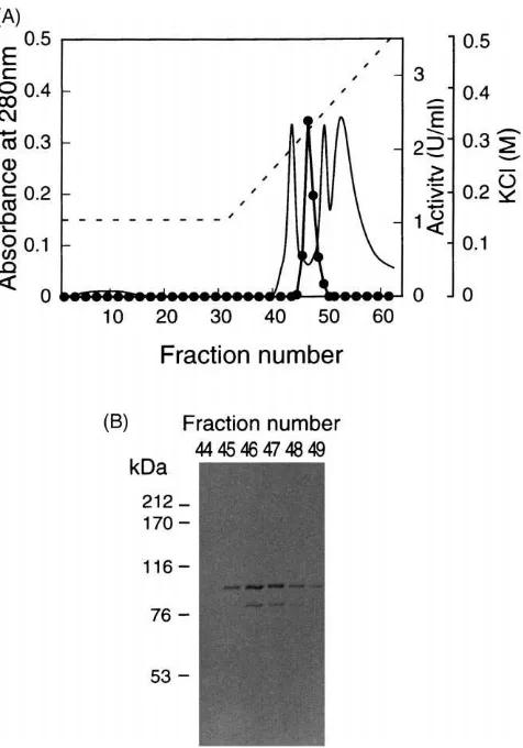
Fig. 1. Elution profile of urease from the silkworm hemolymph on a Mono Q column (A) and distribution of immuno reactive polypeptide (B). The protein obtained from Superdex 200 pg column chromato-graphy was loaded onto a Mono Q column and eluted in 0.15–0.5 M KCl gradient (- - -). Fractions were monitored at 280 nm for protein (———) and assayed for enzyme activity (I). For Western blot, 5µl
of each fraction was subjected to SDS–PAGE and blotted onto PVDF membrane. The membrane was immunostained with a monoclonal antibody (MabU23) raised against jack bean seed urease.
Table 1
Purification of urease from the hemolymph of the spinning larvae
Specific Activity
Purification Step Total Protein (mg) Total Activitya(units) Yield (%) Purification (fold) (units/mg protein)
Hemolymph 8,189 12.65 0.00154 100 1
Q–Sepharose FF 36.5 4.57 0.125 36.1 81.2
Superdex 200 pg 4.82 2.16 0.448 17.1 291
Mono Q 0.22 1.89 8.59 14.9 5,578
aA unit of enzyme is defined as the amount of the enzyme which hydrolyze 1µmol urea per min at 30°C.
Urease active fractions were checked by immuno blot probed with a mouse monoclonal antibody MabU 23 raised against jack bean urease (Fig. 1B). The intensity of the major immunostained band correlated well with the strength of urease activity and the molecular mass of this band was estimated to be 90.5 kDa. These results suggest that the 90.5 kDa polypeptide was the subunit
of the urease from the silkworm hemolymph. A minor stained band of 79 kDa was also present, which might be a degraded product from the 90.5 kDa polypeptide (Fig. 1B).
3.2. Properties of urease from the silkworm hemolymph
At first, we studied the antigenicity of the urease from the silkworm hemolymph using the panel of monoclonal antibodies produced against jack bean urease. All of these clones recognized the jack bean urease on immu-noblots, while four clones (MabU3, MabU21, MabU23, and MabU27) cross-reacted with the hemolymph urease as well as the urease purified from mulberry leaf (Fig. 2). We also confirmed that the hemolymph urease and the mulberry leaf urease migrated at the same position on the blots, whereas the jack bean urease migrated slightly slower than these enzymes. In this experiment,
Fig. 2. Reactivity of a panel of monoclonal antibodies in Western blot analysis with different urease sources. Used monoclonal anti-bodies were MabU3 (A), MabU10 (B), MabU13 (C), MabU21 (D), MabU23 (E), and MabU27 (F). Lane 1; 0.1µg jack bean seed urease, lane 2; 0.1µg mulberry leaf urease, lane 3; 0.3 µg partially purified urease from the silkworm hemolymph.
silk-Fig. 3. Sequence alignment of N–terminal amino acid sequence of urease from the silkworm hemolymph compared with those of mul-berry leaf urease and jack bean seed urease. Shown are the N–terminal 20 amino acid residues of urease from the silkworm hemolymph (the present study); mulberry leaf urease (Hirayama et al., 1999); jack bean seed urease (Takishima et al., 1988). Asterisks represent identical amino acid residues to those of the silkworm urease.
worm hemolymph and the urease from the mulberry leaf had similar antigenic properties.
The 90.5 kDa polypeptide present in the fraction after Mono Q column chromatography was subjected to N– terminal sequencing. The first 20 amino acids of the hemolymph urease were determined and the results are shown with the relevant amino acid sequences of ureases from mulberry leaf and jack bean seed (Fig. 3). Com-plete homology was found with the mulberry leaf urease within the 20 amino acids, with 16 out of the 20 amino acids (80%) matching the sequence of the jack bean ure-ase. In addition, the N–terminal sequence of the hemo-lymph urease showed much lower similarities with the relevant sequences, if compared with microbial ureases (40–70%) (data not shown).
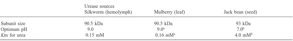
Subsequently, we investigated the kinetic properties of the hemolymph urease (Table 2). For the substrate urea, the Km value was very near to that obtained from the mulberry leaf urease. These two ureases show much higher affinity for urea than jack bean urease. The opti-mum pH of the both enzymes was 9.0, whereas most ureases, including jack bean seed urease, have a neutral pH optimum (Fishbein, 1969; Mobley and Hausinger, 1989).
3.3. Urease activity in the larval hemolymph after feeding of mulberry leaf urease
Three-day or five-day-old male fifth instar larvae that had been reared on an artificial diet were fed the diet coated with several concentrations of mulberry leaf ure-ase, and urease activity in the hemolymph of the larvae
Table 2
Comparison of properties of ureases from different organisms
Urease sources
Silkworm (hemolymph) Mulberry (leaf) Jack bean (seed)
Subunit size 90.5 kDa 90.5 kDa 93 kDa
Optimum pH 9.0 9.0a 7.0b
Km for urea 0.15 mM 0.16 mMa 4.0 mMb
aData from Hirayama et al. (2000). b Data from Fishbein (1969).
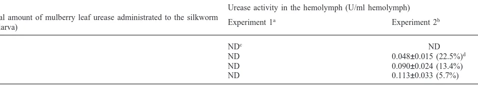
was measured (Table 3). Oral administration to the three-day-old larvae induced no urease activity in the hemolymph, even when the total amount of urease given was 1.8 U/larva (The dose was slightly larger than the total amount that a single larva ingests during the 5th instar from the mulberry leaves). In contrast, significant urease activity appeared in the hemolymph of the larvae by the same treatment of five-day-old larvae. When a larva ingested 0.2 U urease, 0.048 U urease was detected in 1 ml of the larval hemolymph. By the administration of 0.6 U and 1.8 U urease per larva, activity of 0.090 U/ml hemolymph and 0.113 U/ml hemolymph was detected, respectively. In these cases, urease activities in the hemolymph were very similar to that in the hemo-lymph of larvae normally fed fresh mulberry leaves (data not shown).
We estimated the hemolymph volume by comparing weights of the larvae before and after collection of hem-olymph, according to Nittono (1960), and the efficiency of incorporation of the enzyme was calculated based on the enzyme activity using the equation: (Total urease
activity in the hemolymph)/(Total urease activity
ingested by the insect)×100%. Although the efficiency
of incorporation would depend on the amount of urease administrated to the larvae, at least 5.7% of ingested ure-ase could pass through the gut wall into the hemolymph in this experiment (Table 3).
4. Discussion
Urease activity in several insects has been reported (Robinson and Baker, 1939; Rosenthal et al., 1977; Ble-iler and Rosenthal, 1988). However, to our knowledge, there have been no descriptions concerning molecular properties of insect ureases. Thus, the origins of urease activity detected in insects have not been clarified. In the present study, we first identified urease enzyme from an insect at the molecular level. We demonstrated that ure-ase from hemolymph of spinning larvae of the silkworm,
Bombyx mori, was identical with the mulberry leaf
Table 3
Urease activity in the hemolymph of the silkworm fed with mulberry leaf urease
Urease activity in the hemolymph (U/ml hemolymph) Total amount of mulberry leaf urease administrated to the silkworm
Experiment 1a Experiment 2b
(U/larva)
0 NDc ND
0.2 ND 0.048±0.015 (22.5%)d
0.6 ND 0.090±0.024 (13.4%)
1.8 ND 0.113±0.033 (5.7%)
aThree-day-old 5th instar male larvae were used. b Five-day-old 5th instar male larvae were used. cNon-detectable.
d Efficiency of incorporation of mulberry leaf urease is expressed in parenthesis. Values are expressed as mean±SD of five replications.
and enzymatic characteristics. The obtained results strongly suggest that the mulberry leaf urease can pass through the larval gut wall into the hemolymph without being digested. Similarly, ureases in some other insects might originate from their host plants as shown in this study.
It has been demonstrated that host serum components are incorporated into the hemolymph across the digestive tract without digestion in an intact form in many blood sucking insects. For example, incorporated IgG retained the antibody activity for several days after blood-feeding (Hatfield, 1988; Lackie and Gavin, 1989; Vaughan et al., 1990; Allingham et al., 1992). Passage of the host blood components across the gut appears to be a normal physiological process in these species. But it is unclear whether blood meal proteins which enter the hemolymph have significant functions and also how such large mol-ecules pass through the gut wall. Since the gut junctions of these haematophagous insects are quite leaky, rapid incorporation of blood components into the hemolymph of these insects may be by passive transfer involving these junctions (Billingsley and Lehane, 1996). In these heamatophagous insects, the concentration of IgG in the
hemolymph was reported to be 1/5000|1/200 of the host
serum (Vaughan et al., 1990; Allingham et al., 1992), suggesting that only a small part of ingested proteins is incorporated into the hemolymph non-selectively. In contrast, urease incorporation across the silkworm midgut seems to be much more efficient. We observed
that 5|23% of orally administrated urease passed
through the gut wall into the hemolymph (Table. 3). In the present study, we observed that the transport of mulberry leaf urease across the insect gut could be apparently regulated in a stage-specific manner. This result was consistent with the previous report that urease activity was detected in the hemolymph after the onset of spinning when larvae were reared on fresh mulberry leaves through the 5th instar (Yamada et al., 1984). It has been reported that starvation for 72 hr after the 4th ecdysis caused an accumulation of urea in the hemo-lymph of the spinning larvae due to the lack of ureae
activity, even if they had been reared on fresh mulberry leaves before the starvation (Sumida et al., 1990), sup-porting the conclusion that the silkworm takes up the host plant urease just before the beginning of spinning. Previously, we found urease activity in the digestive tract of larvae fed mulberry leaves at the feeding stage and reported that urea secreted from the intestinal epi-thelium was decomposed into ammonia by this enzyme activity. In addition, ammonia produced from urea was
reabsorbed and used for silk–protein synthesis
(Hirayama et al., 1999). Since the silkworm constantly ingests mulberry leaf urease during the feeding stage, the enzyme activity is always present in the digestive tract to work this urea recycling system. However, midgut contents should be removed by “gut purge” at the beginning of the spinning stage. Therefore, the silkworm needs to absorb the enzyme through the midgut wall to utilize urea as a nitrogen source even after “gut purge”. The stage specific transport of the host plant urease across the gut seems to be reasonable, and the midgut epithelium may be developmentally specialized to trans-fer the host plant urease into the hemolymph, however the actual mechanism involving this phenomenon is unclear. Passage of intact antibodies is known to occur across the guts of certain new born mammals by a recep-tor-mediated mechanism. During the suckling period, the intestinal epithelium of neonatal rat expresses a receptor specific for the Fc portion of IgG that mediates the uptake of maternal IgG from milk (Rodewald and Kraehenbuhl, 1984). In the same manner mulberry leaf urease might be specifically and actively absorbed across the silkworm gut wall, although it remains to be studied whether the other leaf components could also pass across the gut.
Acknowledgements
We thank Ms. Mayumi Hazeyama for her research assistance. The manuscript was critically reviewed Dr. Kotaro Konno and his comments were greatly appreci-ated. This research was supported by Enhancement of Center of Excellence, Special Coordination Funds for Promoting Science and Technology, Science and Tech-nology Agency, Japan.
References
Allingham, P.G., Kerlin, R.L., Tellam, R.L., Briscoe, S.J., Standfast, H.A., 1992. Passage of host immunoglobulin across the mid-gut epithelium into the haemolymph of blood-fed buffalo flies
Haema-tobia iriitans exigua. Journal of Insect Physiology 38, 9–17.
Billingsley, P.F., Lehane, M.J., 1996. Structure and ultrastracture of the insect midgut. In: Lehane, M.J., Billingsley, P.F. (Eds.), Biology of the Insect Midgut. Chapman and Hall, London, pp. 3–30. Bleiler, J.A., Rosenthal, G.A., 1988. Biochemical ecology of
canavan-ine-eating seed predators. Ecology 69, 427–433.
Burrsell, E., 1967. The excretion of nitrogen in insects. Advance of Insect Physiology 4, 33–67.
Cochran, D.G., 1985. Nitrogen excretion. In: Kerulkut, G.A., Gilbert, L.I. (Eds.). Comparative Insect Physiology Biochemistry and Phar-macology, vol. 4. Pergamon Press, Oxford, pp. 467–506. Fishbein, W.N., 1969. Urease catalysis. III. Stoichiometry, kinetics,
and inhibitory properties of a third substrate dihydroxyurea. Journal of Biological Chemistry 244, 1188–1193.
Hatfield, P.R., 1988. Detection and localization of antibody ingested with a mosquito bloodmeal. Medical and Veterinary Entomology 2, 339–345.
Hirayama, C., Konno, K., Shinbo, H., 1997. The pathway of ammonia assimilation in the silkworm Bombyx mori. Journal of Insect Physi-ology 43, 959–964.
Hirayama, C., Saito, H., Konno, K., Shinbo, H., 1998. Purification and characterization of NADH-dependent glutamate synthase from the silkworm fat body (Bombyx mori). Insect Biochemistry and Mol-ecular Biology 28, 473–482.
Hirayama, C., Sugimura, M., Shinbo, H., 1999. Recycling of urea asso-ciated with the host plant urease in the silkworm larvae, Bombyx
mori. Journal of Insect Physiology 45, 15–20.
Hirayama, C., Sugimura, M., Saito, H., Nakamura, M., 2000. Purifi-cation and properties of urease from the leaf of mulberry, Morus
aluba. Phytochemistry 53, 325–330.
Ko¨hler, G., Milstein, C., 1975. Continuous cultures of fused cells secreting antibody of predefined specificity. Nature 256, 495–497. Lackie, A.M., Gavin, S., 1989. Uptake and persistence of ingested anti-body in the mosquito Anopheles stephensi. Medical and Veterinary Entomology 3, 225–230.
Laemmli, U.K., 1970. Cleavage of structural proteins during the assembly of the head of bacteriophage T4. Nature 227, 680–685. Mobley, H.L.T., Hausinger, R.P., 1989. Microbial ureases:
signifi-cance, regulation, and molecular characterization. Microbiological Reviews 53, 85–108.
Nittono, Y., 1960. Studies on the blood cells in the silkworm, Bombyx
mori L. (in Japanese with English Summary). Bulletin of
Sericurtu-ral Experimental Station 16, 171–266.
Robinson, W., Baker, F.C., 1939. The enzyme urease and the occur-rence of ammonia in maggot-infected wounds. Journal of Parasit-ology 25, 149–155.
Rodewald, R., Kraehenbuhl, J.P., 1984. Receptor-mediated transport of IgG. Journal of Cell Biology 99, 159–164.
Rosenthal, G.A., Janzen, D.H., Dahlman, D.L., 1977. Degradation and detoxification of canavanine by a specialized seed predator. Science 196, 658–660.
Sumida, M., Yamada, Y., Tanaka, Y., Shimabukuro, J., Ohnishi, M., Mori, H., Matsubara, F., 1990. Changes in urea in the haemolymph of the silkworm, Bombyx mori in the fourth and the fifth larval instars and effect of starvation in the fifth instar on the level of urea in the pharate adults. Comparative Biochemistry and Physi-ology 97A, 373–379.
Takishima, K., Suga, T., Mamiya, G., 1988. The structure of jack bean urease. European Journal of Biochemistry 175, 151–165. Towbin, H., Staehelin, T., Gordon, J., 1979. Electeophoretic transfer
of proteins from polyacrylamide gels to nitrocellulose sheets: pro-cedure and some applications. Proceeding of National Academy of Science, USA 76, 4350–4354.
Vaughan, J.A., Wirtz, R.A., Do Rosario, V.E., Azad, A.F., 1990. Quan-titation of antisporozoite immunoglobulins in the hemolymph of
Anopheles stephensi after blood feeding. American Journal of
Trop-ical Medicine and Hygiene 42, 10–16.
Yamada, M., Inokuchi, T., 1985. Dietary effects on conversion of argi-nine to urea in the silkworm, Bombyx mori (Lepidptera: Bombycidae) (in Japanese with English Summary). Japanese Jour-nal of Applied Entomology and Zoology 29, 31–35.
Yamada, M., Nakamura, K., Inokuchi, T., 1983. Effects of diets on urea contents in the hemolymph and silk gland of the last instar larvae of the silkworm, Bombyx mori (Lepidptera: Bombycidae) (in Japanese with English Summary). Japanese Journal of Applied Entomology and Zoology 27, 92–98.