159
CALCULATION OF BNCT DOSIMETRY FOR BRAIN CANCER BASED ON KARTINI RESEARCH REACTOR USING THE PHITS CODE
Suhendra Gunawan Ntoy1, Yohannes Sardjono2 1Department of Physics, Faculty of Science and Mathematics,
Satya Wacana Christian University, Salatiga, 50711
2Particle Physics Division, Centre for Science and Accelerator Technology-National Nuclear Energy Agency,
Jl. Babarsari Kotak Pos Ykbb 6101, Sleman - Yogyakarta, 55281 Diterima editor: 2 September 2017
Diperbaiki: 16 Oktober 2017 Disetujui untuk publikasi: 17 Oktober 2017
ABSTRACT
CALCULATION OF BNCT DOSIMETRY FOR BRAIN CANCER BASED ON KARTINI RESEARCH REACTOR USING PHITS CODE. Cancer is a dangerous disease caused by the growth of a mass of cells that are unnatural and uncontrollable. Glioblastoma, also called as glioblastoma multiforme (GBM), is one of dangerous brain cancer.The dismal prognosis associated with glioblastoma is attributable not only to its aggressive and infiltrative behavior, but also to its location typically deep in the parenchyma of the brain. In resolving this chalenge, the BNCT method can be a solution. This study aims to calculate BNCT dosimetry in different of cancer positions and irradiation geometries using PHITS code. The results show that the deeper the cancers target at brain the slower the total absorbed dose rate of cancer target. It takes a longer treatment time. Based on the treatment time and total absorbed dose rate of cancer target, the TOP irradiation geometry is an appropriate choice in treating the cancer target in this case. To achieve the histopathological cure of GBM at the primary site, the absorbed dose of brain was calculated to be 1.07 Gy and 1.64 Gy for the LLAT and PA irradiation geometry, respectively. While, for cancer position of 3 cm, 5 cm, 7.15 cm, 9 cm, and 11 cm, the absorbed dose of brain is 0.25 Gy, 0.48 Gy, 0.85 Gy, 1.33 Gy, and 2.01 Gy, respectively. In addition to the stochastic effect, it was found also deterministic effects that may be produced such as cataracts.
Keywords: BNCT dosimetry; GBM; brain cancer cases; PHITS; MIRD phantom
ABSTRAK
PERHITUNGAN DOSIMETRI BNCT PADA KANKER OTAK BERBASIS REAKTOR RISET KARTINI MENGGUNAKAN PROGRAM PHITS. Kanker merupakansalahsatu penyakit berbahaya
yang diakibatkan oleh tumbuhnya sekumpulan massa sel-sel yang tidak wajar dan tidak terkendali. Salah satu penyakit kanker otak yang berbahaya adalah Glioblastoma atau yang biasa disebut Glioblastoma Multiforme (GBM). Prognosis suram terkait dengan GBM tidak hanya untuk perilaku agresif dan infiltrasi, tetapi juga terhadaplokasi yang jauh di dalam parenkim otak. Untuk menjawab hal tersebut, Boron Neutron Capture Therapy (BNCT) dapat menjadi solusi. Penilitian ini bertujuan untuk menghitung dosimetri BNCT dalam berbagai posisikan kerdan geometri penyinaran dengan menggunakan program PHITS. Hasil perhitungan menunjukkan bahwa semakin dalam target kanker di otak maka semakin kecil total laju dosis serap dari target kanker. Semakin dalam target kanker di otak dibutuhkan waktu pengobatan yang semakin lama. Berdasarkan waktu pengobatan dan laju dosis serap dari target kanker, bidang penyinaran TOP merupakan pilihan yang tepat dalam mengobati target kanker dalam kasus ini. Untuk mencapai penyembuhan GBM secara histopatologis di lokasi utama, dosis serap dari otak dihitung berturut-turut sebesar 1,07 Gy dan 1,64 Gy untuk bidang penyinaran LLAT dan PA. Sedangkan, untuk posisi kanker 3 cm, 5 cm, 7,15 cm, 9 cm, dan 11 cm, berturut-turut dosis serap dari otak adalah 0,25 Gy, 0,48 Gy, 0,85 Gy, 1,33 Gy, and 2,01 Gy. Selain adanya efek stokastik, ditemukan juga efek deterministik yang mungkin dihasilkan seperti katarak.
Kata kunci: Dosimetri BNCT, GBM, kasuskankerotak, geometripenyinaran, posisikanker, ORNLMIRD phantom.
_______________________________
160
INTRODUCTION
Cancer is a dangerous disease caused by the growth of a mass of cells that is unnatural and uncontrollable. According to the World Health Organization (WHO), approximately 8.2 million people die annually from cancer or an estimated 13% of all deaths worldwide [1]. In Indonesia, the GLOBOCAN estimated in 2012, for all ages and both sexes, the numbers of incidence and mortality of brain cancer reached 3402 and 4902 [2], respectively. According to that, Indonesia occupied the first rank in the Southeastern Asia. Glioblastoma, also called as glioblastoma multiforme (GBM), is one of dangerous brain cancer. GBM is a group of gliomas grade IV according to the WHO classification based on the increasing degree of anaplasia and aggressiveness [3]. Most studies reported that the survival of patients with GBM is quite short as compared with anaplastic astrocytoma and low-grade gliomas [4]. The dismal prognosis associated with glioblastoma is attributable not only to its aggressive and infiltrative behavior, but also to its location typically deep in the parenchyma of the brain [5]. Location GBM generally located in supratentorial areas such as frontal, temporal, parietal and occipital lobes [6,7]. The median overall survival of GBM patients with a deep location was 10 months and 24 months for GBM patients with superficial location [8]. It has been treated with various methods such as radiotherapy, chemotherapy, and surgery. However, the prognosis of GBM patients remain poor, median overall survival (OS) reached 11 months, and the 1-and 2 years OS rates were 45.9% and 7.7%, respectively. Therefore, the need for more effective novel treatments for this disease is urgently needed [9]. In resolving this challenge, Boron Neutron Capture Therapy (BNCT) can be a solution. Several countries have conducted research on BNCT as one of the GBM therapy types such as Japan, Finland, Taiwan, Germany, United States, Sweden, and Czech Republic [10]. It was found in the experience of BNCT clinical trials in Japan that for newly diagnosed GBM cases, median overall survival (OS) reached 25.7 months, and the 1- and 2-year OS rates were 85.7 % and 45.5 %, respectively [11]. Therefore, treatment for GBM patients with BNCT showed promising survival benefits. BNCT is a cancer treatment method designed to be selective in destroying cancer cells and saving healthy tissue cells [12]. This method is based on the irradiation of neutrons to cancer cells containing 10B compound [13]. It causes fission reaction of 7Li nuclei and alpha particles, where the linear energy transfers (LETs) value are 163 keV/µm and 210 keV/µm, respectively [14]. Both of these particles have a high-LET as well as a short range of about one cell diameter resulting in a lethal dose only on cancer cells or tumors [15]. It is known as a boron dose, which is the main dose delivered to tumor cells or cancer. In addition to the boron dose, other major dose components such as nitrogen doses, gamma doses, and neutron doses are considered in the calculation of BNCT dosimetry [16].
161
MATERIAL AND METHODS
Beam source
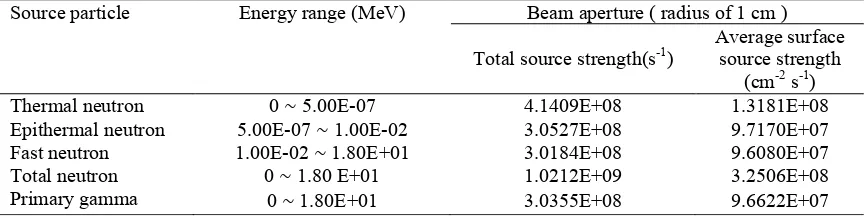
The beam source was obtained from the design of collimator in the radial piercing beam port of Kartini Reactor for BNCT [26]. It was operated at 100 kW. The beam source is made in the form of a circle with a radius of 1 cm. It consists of four source components, namely thermal neutron, epithermal neutron, fast neutron, and primary gamma-ray. The distance between the beam source and the phantom surface was set as 10 cm. Table 1 lists the source strength of each of the four components of the beam source for Kartini Reactor operated at 100 kW.
Table 1. Source strengths of the different components of the Kartini Reactor beam source.
Patient model
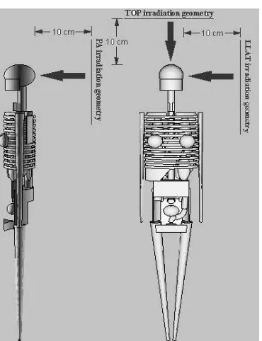
In this study, GBM patient was defined as ORNL MIRD phantom [27] that was simulated in the PHITS code. The ORNL MIRD phantom has been used for BNCT-related research [28,29]. It was created by researchers at Hanyang University in Korea. The ORNL MIRD phantom is ready for use for BNCT dosimetry calculations and it is available at the Los Alamos National Laboratory database [30] and the Computational Medical Physics Working Group [31]. Three types of material compositions, namely, soft tissue, lung, and bone were used in the phantom. Fig. 1 shows the outline of the phantom without skin made with the PHITS code in three dimensions. Cancer target was assumed as the clinical target volume (CTV). It was defined as a sphere with radius of 1.5 cm.
According to the data of 22 recurrent glioma patients were treated with BNCT in Finland, the 10B
concentration in blood in lies the range from 11 to 22 ppm, from 19 to 46 ppm in brain, and from 44 to 93 ppm in tumor under BPA-f infusion of 290-450 mg/kg during 2 hours [32]. Therefore, the 10
B concentration in the cancer target was reasonably taken as 60 and 20 ppm for the normal organs
assuming that the 10B concentrations in normal tissue and in blood are the same.
Source particle Energy range (MeV) Beam aperture ( radius of 1 cm )
Total source strength(s-1)
Average surface source strength
(cm-2 s-1)
Thermal neutron 0 ~ 5.00E-07 4.1409E+08 1.3181E+08
Epithermal neutron 5.00E-07 ~ 1.00E-02 3.0527E+08 9.7170E+07 Fast neutron 1.00E-02 ~ 1.80E+01 3.0184E+08 9.6080E+07
Total neutron 0 ~ 1.80 E+01 1.0212E+09 3.2506E+08
162
Figure 1.Outline and irradiation geometries of the ORNL MIRD phantom.
Calculation of BNCT dosimetry
The Monte Carlo code PHITS version 2.81[33] was applied to calculate BNCT dosimetry for clinical cases. The clinical cases that were selected to cover the study of brain cancer (GBM) are presented. This set of cases included superficial and deep cancer. In addition, the selection of appropriate irradiation geometry for the treatment of brain cancer was also involved. Therefore, the simulation in this study was conducted in two stages. In the first stage, it was performed to investigate the effect of different cancer positions toward the total absorbed dose rates of cancer target and some representative organs. Note that in the first stage, the TOP irradiation geometry was made constant. In the second stage, the simulation was performed to investigate the effect of different irradiation geometries toward the total absorbed dose rates of cancer target and some representative organs. Note that in the second stage, the cancer position of 7.15 cm was made constant. The cancer positions and irradiation geometry is shown in Table 2.
Table 2.Different of cancer positions and irradiation geometries.
Cancer positiona
(cm)
Irradiation geometry
3 TOPb
5 AP
7.15c LLAT
8 -
11 -
a
The cancer position is the depth of cancer in the brain. It is defined as the distance between the skin and the center of mass of the tumor.
b
The irradiation geometry is made constant in the first stage. c
The cancer position is made constant in the second stage. It defines the center of mass of the tumor.
The absorbed dose rate due to the neutron, gamma, 14N (n, p)14C reaction and 10B (n, α)7Li reaction
were determined separately. The absorbed dose due to 14N (n, p)14C and 10B (n, α)7Li reaction were
[T-163
TRACK] tally and [MULTIPLIER] section considering kerma coefficients in the Dosimetry System 2002 (DS02) [34]. Note that the gamma dose is the sum of the secondary gamma dose
which is generated of the 1H (n, γ)2H reaction and the primary gamma dose which is generated
from primary gamma-ray sources.
Table 3.Relative- and compound biological effectiveness of different dose components for use in the calculation of BNCT dosimetry.
In term of BNCT, the weighted dose or commonly known as the total biologically weighted dose, is defined as [35]:
D! =CBEx D! + RBE x D!"+ RBE x D!+ RBE xD! (1)
Where Dnf, Dγ, DB dan DN are the absorbed dose due to neutron, gamma,
10
B(n, α)7Li
reaction, and 14N(n, p)14C reaction, respectively. The values of Relative Biological Effectiveness
(RBE) and Compound Biological Effect (CBE) are shown in Table 3[36,37,18]. RBE is comparison of absorbed doses of radiation reference sources with test radiation that produce the same biological effect, whereas CBE is a factor of the biological effect used on boron carrier compound [38,39]. To illustrate the difference of the absorbed and weighted doses, the letter W in parenthesis is added to the symbol Gy writing one space between the symbol and the additional specification for the weighted dose (Gy (w) [40].
RESULTS AND DISCUSSION
The results of BNCT dosimetry calculations obtained in the first and second stages are shown in Table 4. In the first stage, Table 4 lists the total absorbed dose rates of cancer target and some representative organs for different cancer positions. Note that in the first stage, the TOP irradiation geometry is made constant. In the second stage, Table 4 also lists the total absorbed dose rates of cancer target and some representative organs for different irradiation geometry. Note that in the second stage, the cancer position of 7.15 cm is constant. In addition, the total weighted dose rates of cancer targets and some representative organs are included in Table 4. As shown in Table 4, the total weighted dose rate is greater than the total absorbed rate by a factor of 1.42-3.
Table 4.The total absorbed and weighted dose of cancer target and some representative organ
Cancer Positiona (cm) 3 5 7.15b 9
Cranium 3.29E-05 3.30E-05 3.31E-05 (3.33E-05) (3.28E-05) 3.31E-05
Head and Neck Skin
1.41E-05 1.41E-05 1.41E-05 (1.46E-05) (1.43E-05) 1.41E-05
Thyroid 2.02E-06 2.05E-06 1.97E-06 (5.24E-06) (3.89E-06) 2.03E-06
Weighted dose rate (Gy)
Cancer 8.62E-03 4.96E-03 2.98E-03 (2.44E-03)c (1.61E-03)d 1.97E-03
Brain 1.18E-04 1.35E-04 1.45E-04 (1.45E-04) (1.55E-04) 1.50E-04
Cranium 7.99E-05 8.02E-05 8.03E-05 (8.06E-05) (8.05E-05) 8.02E-05
Head and Neck Skin
3.59E-05 3.59E-05 3.59E-05 (3.68E-05) (3.66E-05) 3.59E-05
164
a
The cancer position is the depth of cancer in the brain. It is defined as the distance between
the skin and the center of mass of the tumor, bThe center of mass of the tumor, cLLAT irradiation
geometry, dPA irradiation geometry
As shown in Table 4, for the first stage, the total absorbed dose rate of target cancer is significantly different at each cancer position. In other words, the deeper the cancer target at brain means the slower the total absorbed dose rate of cancer target. Table 4 also shows that the total absorbed dose rate of some representative organs such as cranium, head and neck skin, and thyroid are not significantly different in each cancer position. Meanwhile, the total absorbed dose rate of brain is significantly different in each cancer position. Seen in Table 4, the total absorbed dose rate of brain was increased in each cancer position. As expected, the total absorbed dose rate of target cancer is greater than the total absorbed dose rate of some representative organs for different cancer positions. For the second stage, the total absorbed dose rate of cancer target is significantly different in the irradiation geometry. The total absorbed dose rate of target cancer for the TOP irradiation geometry is greater than the total absorbed dose rate of target cancer for the LLAT and PA irradiation geometry. Regarding the average absorbed dose rate of brain and skin (skin of head and neck) as shown in Table 4, the TOP irradiation geometry is smaller than the LLAT and PA irradiation geometry. For the total absorbed dose rate of cranium, the LLAT irradiation geometry is greater than the TOP and PA irradiation geometry. For the total absorbed dose rate of thyroid, the TOP irradiation geometry is greater than the LLAT and PA irradiation geometry. However, the TOP irradiation geometry is an appropriate choice in treating the cancer target in this case. To achieve the histopathological cure of GBM at the primary site, the optimal minimal dose to the CTV was 44 Gy(w) [41]. Based on it and from the data in Table 4, the treatment time has been calculated for the first and second stages. These results are presented in Table 5.
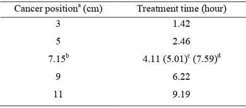
Table 5. The treatment time for different of cancer positions and irradiation geometries
Cancer positiona (cm) Treatment time (hour)
3 1.42
5 2.46
7.15b 4.11 (5.01)c (7.59)d
9 6.22
11 9.19
a
The cancer position is the depth of cancer in the brain. It is defined as the distance between
the skin and the center of mass of the tumor. bThe center of mass of the tumor cLLAT irradiation
geometry dPA irradiation geometry
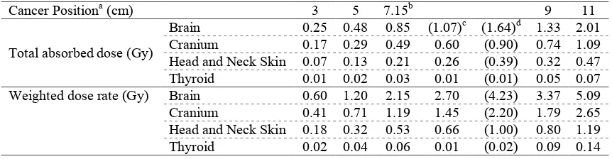
165 Table 6. The total absorbed and weighted dose of cancer target and some representative organ
Cancer Positiona (cm) 3 5 7.15b 9 11
Total absorbed dose (Gy)
Brain 0.25 0.48 0.85 (1.07)c (1.64)d 1.33 2.01 Cranium 0.17 0.29 0.49 0.60 (0.90) 0.74 1.09 Head and Neck Skin 0.07 0.13 0.21 0.26 (0.39) 0.32 0.47 Thyroid 0.01 0.02 0.03 0.01 (0.01) 0.05 0.07 Weighted dose rate (Gy) Brain 0.60 1.20 2.15 2.70 (4.23) 3.37 5.09 Cranium 0.41 0.71 1.19 1.45 (2.20) 1.79 2.65 Head and Neck Skin 0.18 0.32 0.53 0.66 (1.00) 0.80 1.19 Thyroid 0.02 0.04 0.06 0.01 (0.02) 0.09 0.14
a
The cancer position is the depth of cancer in the brain. It is defined as the distance between
the skin and the center of mass of the tumor, bThe center of mass of the tumor, cLLAT irradiation
geometry, dPA irradiation geometry
In addition to the stochastic effect related to the calculated absorbed dose, it was also found the deterministic effects that cannot be ignored in Table 6. According to the International Commission on Radiological Protection (ICRP), Assessment of the lens of the eye, skin, and extremity doses and methods of protection is necessary to improve occupational protection in interventional and nuclear medicine procedures [42]. Although the absorbed dose of the lens of the eye is not calculated on the ORNL MIRD phantom, it should be comparable to the absorbed dose of brain. Since the threshold absorbed dose of the lens of the eye is now considered to be 0.5 Gy[43], Cataracts may occur in GBM patients treated with therapy BNCT for cancer positions of 7.15 cm, 9 cm, and 11 cm.
CONCLUSION
BNCT dosimetry in brain cancer cases has been calculated using the PHITS code. The results show that the deeper the cancers target at brain means the slower the total absorbed dose rate of cancer target. It takes a longer treatment time. Based on the treatment time and total absorbed dose rate of cancer target, the TOP irradiation geometry is an appropriate choice in treating the cancer target in this case. To achieve the histopathological cure of GBM at the primary site, the absorbed dose of brain was calculated to be 1.07 Gy and 1.64 Gy for the LLAT and PA irradiation geometry, respectively. While, for cancer position of 3 cm, 5 cm, 7.15 cm, 9 cm, and 11 cm, the absorbed dose of brain is 0.25 Gy, 0.48 Gy, 0.85 Gy, 1.33 Gy, and 2.01 Gy, respectively. In addition to the stochastic effect, it was found also deterministic effects that may be produced such as cataracts.
ACKNOWLEDGMENT
166
REFERENCES
1. World Health Organization. Programmes and projects, Cancer, 2017. Available from:
http://www.who.int/cancer/en/. Last accessed 08 January 2017.
2. As Ferlay J, Soerjomataram I, Ervik M, Dikshit R, Eser S, Mathers C et al.
GLOBOCAN 2012 v1.0, Cancer Incidence and Mortality Worldwide: IARC CancerBase No. 11 [website]. Lyon, France: International Agency for Research on Cancer; 2013 (http://globocan.iarc.fr/old/bar_site.as, accessed 9 January 2017).
3. A. Omuro and L. M. DeAngelis, “Glioblastoma and Other Malignant Gliomas: A
Clinical Review,” JAMA, vol. 310, no. 17, pp. 1842–1850, Nov. 2013.
4. M. Penas-Prado, T. S. Armstrong, and M. R. Gilbert, “Chapter 32 - Glioblastoma,” in
Handbook of Clinical Neurology, 3rd series., vol. 105, W. G. and R. Soffietti, Ed. Elsevier, 2012, pp. 485–506.
5. R. D. Alkins, P. M. Brodersen, R. N. S. Sodhi, and K. Hynynen, “Enhancing drug
delivery for boron neutron capture therapy of brain tumors with focused ultrasound,”
Neuro-Oncology, vol. 15, no. 9, pp. 1225–1235, May 2013.
6. J. P. Thakkar et al., “Epidemiologic and Molecular Prognostic Review of
Glioblastoma,” Cancer Epidemiology and Prevention Biomarkers, vol. 23, no. 10, pp.
1985–1996, Oct. 2014.
7. C. C. Koet al., “Differentiation between Glioblastoma Multiforme and Primary Cerebral
Lymphoma: Additional Benefits of Quantitative Diffusion-Weighted MR Imaging,”
PLOS ONE, vol. 11, no. 9, pp. 1–15, Sep. 2016.
8. K. K. Das et al., “Pediatric glioblastoma: clinico-radiological profile and factors
affecting the outcome,” Child’s Nervous System, vol. 28, no. 12, pp. 2055–2062, Dec.
2012.
9. N. Ahmadlooet al., “Treatment outcome and prognostic factors of adult glioblastoma
multiforme,” Journal of the Egyptian National Cancer Institute, vol. 25, no. 1, pp. 21–
30, Mar. 2013.
10. R. F. Barth et al., “Current status of boron neutron capture therapy of high grade
gliomas and recurrent head and neck cancer,” Radiation Oncology, vol. 7, no. 1, pp.
146–166, Jan. 2012.
11. K. Nakai, T. Yamamoto, H. Kumada, and A. Matsumura, “Boron Neutron Capture
Therapy for Glioblastoma: A Phase-I/II Clinical Trial at JRR-4,” European Association
of Neuro Oncology Magazine, vol. 4, no. 3, pp. 116–123, Jun. 2014.
12. R. Ahangariand H. Afarideh, “Therapeutic gain prediction for evaluation and
optimization of neutron spectra in BNCT,” Annals of Nuclear Energy, vol. 49, pp. 212–
217, Nov. 2012.
13. Y. Kasesaz, H. Khalafi, and F. Rahmani, “Design of an epithermal neutron beam for
BNCT in thermal column of Tehran research reactor,” Annals of Nuclear Energy, vol.
68, pp. 234–238, Jun. 2014.
14. N. Kondo et al., “Detection of γH2AX foci in mouse normal brain and brain tumor after
boron neutron capture therapy,” Reports of Practical Oncology & Radiotherapy, vol.
21, no. 2, pp. 108–112, Mar. 2016.
15. Y. Kasesazet al., “A feasibility study of the Tehran research reactor as a neutron source
for BNCT,” Applied Radiation and Isotopes, vol. 90, pp. 132–137, Aug. 2014.
16. M. Ziegneret al., “Confirmation of a realistic reactor model for BNCT dosimetry at the
TRIGA Mainz,” Med. Phys., vol. 41, no. 11, p. 111706, Nov. 2014.
17. F. S. Rasouli and S. F. Masoudi, “Simulation of the BNCT of Brain Tumors Using
MCNP Code: Beam Designing and Dose Evaluation,” Iranian Journal of Medical
Physics, vol. 9, no. 3, pp. 183–192, Sep. 2012.
18. M. S. Herrera, S. J. González, D. M. Minsky, and A. J. Kreiner, “Evaluation of
performance of an accelerator-based BNCT facility for the treatment of different tumor
targets,” PhysicaMedica: European Journal of Medical Physics, vol. 29, no. 5, pp. 436–
167
19. M. e. Capoulat, M. s. Herrera, D. m. Minsky, S. j. González, and A. j. Kreiner, “9Be (d,
n)10B-based neutron sources for BNCT,” Applied Radiation & Isotopes, vol. 88, pp.
190–194, Jun. 2014.
20. S. F. Brandão and T. P. R. Campos, “Intracavitary moderator balloon combined with
252Cf brachytherapy and boron neutron capture therapy, improving dosimetry in brain
tumour and infiltrations,” Br. J. Radiol., vol. 88, no. 1051, pp. 1–11, Jul. 2015.
21. S. Hang et al., “Monte Carlo study of the beam shaping assembly optimization for
providing high epithermal neutron flux for BNCT based on D–T neutron generator,”
Journal of Radioanalytical and Nuclear Chemistry, vol. 310, no. 3, pp. 1289–1298, Dec. 2016.
22. M. Hsieh, Y. Liu, F. Mostafaei, J. M. Poulson, and L. H. Nie, “A feasibility study of a
deuterium–deuterium neutron generator-based boron neutron capture therapy system for
treatment of brain tumors,” Med. Phys., vol. 44, no. 2, pp. 637–643, Feb. 2017.
23. K. Takada et al., “Evaluation of the radiation dose for whole body in boron neutron
capture therapy,” Prog. Nucl. Sci. Technol., vol. 4, pp. 820– 823, 2014.
24. H. Kumada, K. Takada, K. Yamanashi, T. Sakae, A. Matsumura, and H. Sakurai,
“Verification of nuclear data for the Tsukuba plan, a newly developed treatment
planning system for boron neutron capture therapy,” Appl. Radiat. Isot., vol. 106, pp.
111–115, Dec. 2015.
25. C. H. Clement et al., “Conversion Coefficients for Radiological Protection Quantities
for External Radiation Exposures,” Ann. ICRP, vol. 40, no. 2–5, pp. 1–257, Apr. 2010.
26. M. limaMuslih, Y. Sardjono, and A. Widiharto, “PerancanganKolimator Di Beam Port
TembusReaktorKartiniUntuk Boron Neutron Capture Therapy,” Pus. Sains Dan
TenologiAkselerator - BATAN, pp. 163–178, Jun. 2014.
27. EckermanKF, Cristy M, Ryman JC. The ORNL mathematical phantom series. Oak
Ridge, TN: Oak Ridge National Laboratory. 1996 Dec.
28. D. Krstic, V. M. Markovic, Z. Jovanovic, B. Milenkovic, D. Nikezic, and J.
Atanackovic, “Monte Carlo calculations of lung dose in ORNL phantom for boron
neutron capture therapy,” Radiat. Prot. Dosimetry, vol. 161, no. 1–4, pp. 269–273, Oct.
2014.
29. J. N. Wang, C. K. Huang, W. C. Tsai, Y. H. Liu, and S. H. Jiang, “Effective dose
evaluation for BNCT treatment in the epithermal neutron beam at THOR,” Appl.
Radiat. Isot., vol. 69, no. 12, pp. 1850–1853, Dec. 2011.
30. A. D. Lazarine, Medical physics calculations with MCNP: a primer. Texas A&M
University, 2006. [Online]. Available: http://oaktrust.library.tamu.edu/handle
/1969.1/4297 [Accessed: 02-Juni2017].
31. R. Zhang and ArzuAlpan, “Computational Medical Physics Working Group.” [Online].
Available: http://cmpwg.ans.org/members.html. [Accessed: 29-May-2017].
32. H. Koivunoroet al., “Biokinetic analysis of tissue boron (10B) concentrations of glioma
patients treated with BNCT in Finland,” Applied Radiation and Isotopes, vol. 106, pp.
189–194, Dec. 2015.
33. T.Sato et al., J.Nucl.Sci.Technol, vol. 50, pp. 913-923, 2013.
34. Kerr, George D., Pace, Joseph V. III, & Egbert, Stephen D. Young, Robert W., & Kerr,
George D. (Eds.). (2005). Survivor dosimetry Part aFluence-tokerma conversion coefficients (INISJP--102). Young, Robert W., & Kerr, George D. (Eds.). Japan
35. N. Ghal–Eh, H. Goudarzi, and F. Rahmani, “FLUKA simulation studies on in–phantom
dosimetric parameters of a LINAC–based BNCT,” Radiat. Phys. Chem., vol. 141, pp. 36–40, Dec. 2017.
36. Z. A. Ganjeh and S. F. Masoudi, “Neutron beam optimization based on a 7 Li(p,n) 7 Be
reaction for treatment of deep-seated brain tumors by BNCT,” Chin. Phys. C, vol. 38, no. 10, p. 108203, 2014.
37. F. Torabi, S. FarhadMasoudi, F. Rahmani, and F. Rasouli, “BSA optimization and
dosimetric assessment for an electron linac based BNCT of deep-seated brain tumors,”
168
38. L. Evangelista, G. Jori, D. Martini, and G. Sotti, “Boron neutron capture therapy and
18F-labelled borophenylalanine positron emission tomography: A critical and clinical
overview of theliterature,” Appl. Radiat. Isot., vol. 74, pp. 91–101, Apr. 2013.
39. T. Watanabe, H. Tanaka, S. Fukutani, M. Suzuki, M. Hiraoka, and K. Ono,
“L-phenylalanine preloading reduces the 10B (n, α)7Li dose to the normal brain by
inhibiting the uptake of boronophenylalanine in boron neutron capture therapy for brain
tumours,” Cancer Lett., vol. 370, no. 1, pp. 27– 32, Jan. 2016.
40. International Atomic Energy Agency, Vienna (Austria) (2001). Current status of
neutron capture therapy (IAEA-TECDOC--1223). International Atomic Energy Agency (IAEA)
41. T. Kageji, Y. Mizobuchi, S. Nagahiro, Y. Nakagawa, and H. Kumada, “Correlation
between radiation dose and histopathological findings in patients with gliblastoma
treated with boron neutron capture therapy (BNCT),” Applied Radiation and Isotopes,
vol. 88, pp. 20–22, Jun. 2014.
42. ICRP, 2017 ICRP Statement on “Areas of Research to Support the System of
Radiological Protection”, [Online].
Available:http://www.icrp.org/docs/ICRP%20Research%20Priorities%202017.pdf[Acc essed: 8-August-2017].
43. ICRP, 2012 ICRP Statement on Tissue Reactions / Early and Late Effects of Radiation