1/24
SOFT TISSUE REPLACEMENTS
[
Adopsi dari:
Joseph D. Bronzino, “Biomedical Engineering Fundamentals”, CRC Press, third edition, 2006, Section V, CHAPTER 44,by K.B. Chandran, K.J.L. Burg, S.W. Shalaby.]
44.1 BLOOD INTERFACING IMPLANTS [K.B. Chandran]
44.1.1 Introduction
Blood comes in contact with foreign materials for a short term in extracorporeal devices such as dialysers, blood oxygenators, ventricular assist devices, and catheters. Long-term vascular implants include heart valve prostheses, vascular grafts, and cardiac pacemakers among others. In this section, we will be concerned with development of biomaterials for long-term implants, specifically for heart valve prostheses, total artificial heart (TAH), and vascular grafts. The primary requirements for biomaterials for long-term implants are biocompatibility, nontoxicity, and durability. Furthermore, the material should be nonirritating to the tissue, resistant to platelet and thrombus deposition, nondegradable in the physiological environment, and neither absorb blood constituents nor release foreign substances into the blood stream [Shim and Lenker, 1988]. In addition, design considerations include that the implant should mimic the function of the organ that it replaces without interfering with the surrounding anatomical structures and must be of suitable size and weight. The biomaterials chosen must be easily available, inexpensive, easily machinable, sterilizable, and have a long storage life. The selection of material will also be dictated by the strength requirement for the implant being made. As an example, an artíficial heart valve prosthesis is required to open and close on an average once every second. The biomaterial chosen must be such that the valve is durable and will not fail under fatigue stress after implantation in a patient. As sophisticated measurement techniques and detailed computational analyses become available with the advent of super computers, our knowledge on the complex dynamics of the functioning of the implants is increasing. Improvements in design based on such knowledge and improvements in selection and manufacture of biomaterials will minimize problems associated with blood interfacing implants and significantly improve the quality of life for patients with implants. We will discuss the development of biomaterials for the blood interfacing implants, problems associated with the same, and future directions in the development of such implants.
2/24
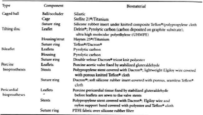
44.1.2 Heart Valve ProsthesesAttempts at replacing diseased natural human valves with prostheses began about four decades ago. The details of the development of heart valve prostheses, design considerations, in vitro functional testing, and durability testing of valve prototypes can be found in several monographs [Shim and Lenker, 1988; Chandran, 1992]. The heart valve prostheses can be broadly classified into mechanical prostheses (made of non-biological material) and bioprostheses (made of non-biological tissue). Currently available mechanical and tissue heart valve prostheses in the United States are listed in Table 44.1.
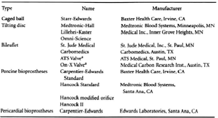
44.1.2.1. Mechanical Heart Valves. Lefrak and Starr [1970] describe the early history of mechanical valve development. The initial designs of mechanical valves were of centrally occluding caged ball or caged disc type. The Starr—Edwards caged ball prostheses, commercially available at the present time, was successfully implanted in the mitral position in 1961. The caged ball prostheses is made of a polished Co—Cr alloy (Stellite 21•) cage and a silicone rubber ball (Silastic) which contains 2% by weight barium sulfate for radiopacity (Figure 44.1). The valve sewing rings use a silicone rubber insert under a knitted composite polytetrafluorethylene (PTFE Teflone) and polypropylene cloth. Even though these valves have proven to be durable, the centrally occluding design of the valve results in a larger pressure drop in flow across the valve and higher turbulent stresses distal to the valve compared to other designs of mechanical valve prostheses [Yoganathan et al., 1979a,b; 1986; Chandran et al, l983]. The relatively large profile design of caged ball or disc construction also increases the possibility of interference with anatomical structures after implantation. The tilting disc valves, with improved hemodvnarnic characteristics, were introduced in the late l960s. The initial design consisted of a polyacetal (Delrin) disc with a Teflon sewing ring. Delrin acetal resins are thermoplastic polymers manufactured by the polymerization of formaldehyde [Shim and Lenker. 1988].
FIGURE 44.1 A caged-ball heart valve prosthesis. (Courtesy of Baxter Health Care. Irvine. CA.)
3/24
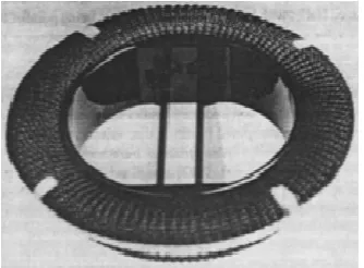
pure titanium and its alloy (Ti6A14V) exhibit excellent mechanical properties as well as resistance to corrosion and thrombus deposition. A typical commercially available tilting disc valve with a pyrolytic carbon disk is shown in Figure 44.2a. A tilting disc valve with the leaflet made of ultra high molecular weight polyethylene (Chitra valve — Figure 44.2b) is currently marketed in India. The advantages of leaflets with relatively more flexibility compared to pyrolytic carbon leaflets are discussed in Chandran et al. [1994a]. Another new concept in a tilting disc valve design introduced by Reul et al. [1995] has an S-shaped leaflet with leading and trailing edges being parallel to the direction of blood flow. The housing for the valve is nozzle-shaped to minimize flow separation at the inlet and energy loss in flow across the valve. Results from in vitro evaluation and animal implantation have been encouraging.
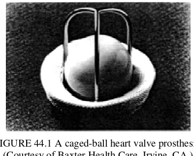
In the late 1970s, a bileaflet design was introduced for mechanical valve prostheses and several different bileaflet models are being introduced into the market today. The leaflets as well as the housing of the bileaflet valves are made of pyrolytic carbon and the bileaflet valves show improved hemodynamic characieristics especially in smaller sizes compared to tilting disc valves. A typical bileaflet valve is shown in Figure 44.3. Design features to improve the hydrodynamic characteristics of the mechanical valves indude the opening angle of the leaflets [Baldwin et al., 1997] as well as having an open-pivot design in which the pivot area protrudes into the orifice and is exposed to the washing action of flowing blood [Drogue and Villafana, 1997]. Other design modifications to improve the mechanical valve function include: the use of double polyester (Dacrons) velour material for the suture ring to encourage rapid and controlled tissue ingrowth, and mounting the cuff on a rotation ring which surrounds the orifice ring to protect the cuff mounting mechanism from deeply placed annulus sutures. A PTFE (Teflon) insert in the cuff provides pliability without excessive drag on the sutures. Tungsten (20% by weight) is incorporated into the leaflet substrate in order to visualize the leaflet motion in vivo.
a) (b)
FIGURE 44.2 (a) Photograph of a typical tilting disc valve prosthesis. (Courtesy of Medtronic Heart Valves, Minneapolis. MN.) (b) Chitra tilting disc valve prosthesis with the ocduder made of ultra high molecular weight polyethylene. (Courtesy of Sree Chitra
Tirunal Institute for Medical Sciences and Technology. India.)
4/24
(a) (b)
FIGURE 44.4 A tri-leaflett heart valve prosthesis under development. (Courtesy of Triflo Medical, Inc.,Costa Mesa, CA.)
Another attempt to design a mechanical valve which mimics the geometry and function of the tri-leaflet aortic valve is that of Lapeyre et al. (1994) (Figure 44.4a, b). The geometry of the valve affords true central flow characteristics with reduced backflow. Accelerated fatigue tests have also shown good wear characteristics for this design and the valve is undergoing further evaluation including animal studies.
Other improvements in the mechanical valves which augment performance include: machining of the valve housing to fit a disk so as to produce optimal washing and minimal regurgitation [McKenna, 1997]; a supra-annular design so that a larger-sized valve can be inserted in the aortic position in the case of patients with small aortic annulus [Bell, 1997]; and coating of a titanium alloy ring with a thin, uniform, and strongly adherent film of high-density turbostratic carbon (Carbofilm) [Bona et al., 1997] in order to integrate the structural stability of the metal alloy to the non-thrombogenecity of pyrolytic carbon. Details of contemporary design efforts in mechanical valve design and potential future biomaterials such as Boralyn• (boron carbide) are discussed in Wieting [1997].
In spite of the desirable characteristics of the biomaterials used in the heart valve prostheses, problems with thrombo-embolic complications are significant with implanted valves and patients with mechanical valves are under long-term anticoagulant therapy. The mechanical stresses induced by the flow of blood across the valve prostheses have been linked to the lysis and activation of formed elements of blood (red blood cells, white blood cells, and platelets) resulting in the deposition of thrombi in regions with relative stasis in the vicinity of the prostheses. Numerous in vitro studies with mechanical valves in pulse duplicators simulating physiological flow have been reported in the literature and have been reviewed by Chandran [1988] and Dellsperger and Chandran [1991]. Such studies have included measurement of velocity profiles and turbulent stresses distal to the valve due to flow across the valve. The aim of these studies has been the correlation of regions prone to thrombus deposition and tissue overgrowth with explanted valves and the experimentally measured bulk turbulent shear stresses as well as regions of relative stasis. In spite of improvements in design of the prostheses to afford a centralized flow with minimal flow disturbances and fluid mechanical stresses, the problems with thrombus deposition remain significant.
5/24
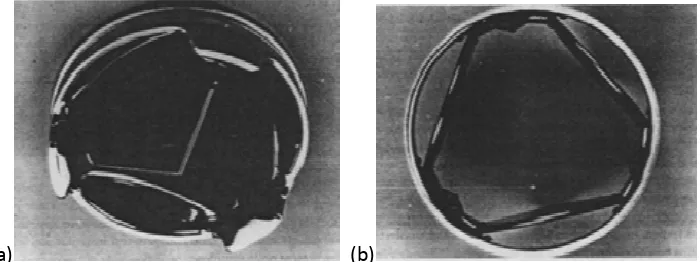
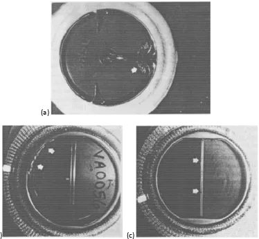
respectively, at the instant when the leaflet impacts against the seat stop or the guiding strut [Leuer, 1986; Chandran et al, 1994a]. The negative pressure transients have been shown to reach magnitudes below the liquid vapor pressure and have been demonstrated to be a function of the loading rate on the leaflet inducing the valve closure. As the magnitudes of negative pressure transients go below the liquid vapor pressure, cavitation bubbles are initiated and the subsequent collapse of the cavitation bubbles may also be a factor in the lysis of red blood cells, platelets, and valvular structures [Chandran et al., 1994a; Lee et al., 199]. Typical cavitation bubbles visualized in an in vitro study with tilting disc and bileaflet valves are shown in Figure 44.5. A correlation is also observed between the region where cavitation bubbles are present, even though for a period of time less than a millisecond after valve dosure, and sites of pitting and erosion reported in the pyrolytic carbon material in the valve housing and on the leaflets with explanted valves [Kafesiian, 1994] as well as those used in total artificial hearts [Leuer, 1987]. An electron micrograph of pitting and erosion observed in the pyrolytic carbon valve housing of an explanted bileaflet mechanical valve is shown in Figure 44.6. The pressure transients at valve dosure are substantially smaller in mechanical valves with a flexible occluder and leaflets made of ultra high molecular weight polyethylene (Figure 44.2b) may prove to be advantageous based on the dosing dynamic analysis [Chandran et al., 1994a]. A correlation between the average velocity of the leaflet edge and the negative pressure transients in the same region at the instant of valve closure, as well as the presence of cavitation bubbles has been reported recently [Chandran et al., 1997]. This study demonstrated that for the valves of the same geometry (e.g., tilting disk) and size, the leaflet edge velocity as well as the negative pressure transients were similar. However, the presence of cavitation bubbles depended on the local interaction between the leaflet and the seat stop. Hence, it was pointed out that magnitudes of leaflet velocity or presence of pressure transients below the liquid vapor pressure might not necessarily indicate cavitation inception with mechanical valve closure. Chandran et al. [1998] have also demonstrated the presence of negative pressure transients in the atrial chamber with implanted mechanical valves in the mitral position in animals, demonstrating that potential for cavitation exists with implanted mechanical valves. Similar to the in vitro results, the transients were of smaller magnitudes with the Chitra valve made of flexible leaflets, and no pressure transients were observed with tissue valve implanted in the mitral position in vivo. The demonstration of the negative pressure transients with mechanical valve closure also shows that this phenomenon is localized and the flow chamber or valve holder rigidity with the in vitro experiments will not affect the valve closing dynamics.
6/24
(a)
(b) (c)
FIGURE 44.5 Cavitation bubbles visualized on the inflow side of the valves in vitro
[Chandran et al., 1994a: a) Medtronic-Hall tilting disc valve; (b) Edwards-Duromedis bileaflet valve; (c) CarboMedics bileaflet valve.
FIGURE 44.6 Photographs showing pitting on pyrolytic carbon surface of a mechanical heart valve. (Courtesy of Baxter Health Care, Irvine, CA.)
7/24
durability and due to limited availability except in a few centers [Shim and Lenker, 1988; Lee and Lloughner, 1991]. Attempts were also made in the early 1960s in the use of xenografts (valves made from animal tissue) and porcine bioprostheses became commercially available after the introduction of the gluteraldehyde (rather than formaldehyde which was initially used) fixation technique. Gluteraldehyde reacts with tissue proteins to form crosslinks and results in improved durability [Carpentier et al., 1969]. The valves are harvested from 7 to 12 month old pigs and attached to supporting stents and preserved. The stent provided support to preserve the valve in the natural shape and to achieve normal opening and dosing. Initial supports were made of metal and subsequently flexible polypropylene stents were introduced. The flexible stents provided the advantage of ease of assembling the valve and finite element analyses have demonstrated reduction in stresses at the juncture between the stent and tissue leaflets resulting in increased durability and increased leaflet coaptation area [Reis et al., 1971; Hamid et al., 1985]. A typical porcine bioprosthesis is included in Figure 44.7a.
Fixed bovine pericardial tissue is also used to construct heart valves in which design characteristics such as orifice area, valve height, and degree of coaptation can be specified and controlled. Thus, the geometry and flow dynamics past pericardial prostheses mimic those of the natural human aortic valves more closely. Due to the low profile design of pericardial prostheses and increased orifice area, these valves are less stenotic compared to porcine bioprostheses, especially in smaller sizes [Chandran et al., 1984]. In the currently available bioprostheses, the stents are constructed from polypropylene, Acetol homopolymer or copolymer, Elgiloy wire, or titanium. A stainless steel radiopaque marker is also introduced to visualize the valve in vivo. Other biomaterials, which have been employed in making the bioprostheses, include fascia lata tissue as well as human duramater tissue. The former was prone to deterioration and hence unsuitable for bioprosthetic application, while the latter lacked commercial availability.
The advantage with bioprosiheses is the freedom from thrombo-embolism and hence not requiring long term anticoagulant therapy in general. These prostheses are preferable in patients who do not tolerate anticoagulants. On the other hand. bioprosthetic valves are prone to calcification and leaflet tear with an average lifetime of about 10 years before replacement is necessary, and is generally attributed to the tissue fixatión process. Numerous attempts are being made to improve the design as well as fixation in bioprostheses in order to minimize problems with calcification and increase duration of the function of the implant. As an example, a bovine pericardial trileaflet valve (Figure 44.7IbJ) treated with a non-aldehyde fixation resulting in collagen crosslink formation without a new foreign” chemical process IPhillips and Printz, 1997 J has been introduced in the European market. A non-aldehyde iodine-based sterilization process also sterilizes the valve.
8/24
(a) (b)
FIGURE 44.7 Typical bioprostheses: (a) Hancock porcine bioprosthesis (courtesy of Medtronic Heart Valves. Minneapolis. MN); (b) PhotoFixTMα pericardial prosthesis.
(Courtesy of Sulzer-CarboMedics, Inc., Austin, TX.)
44.1.2.3. Synthetic Heart Valves. Concurrently, efforts have also been made in the development of valve prostheses made of synthetic material. Several attempts to make bileaflet [Braunwald et al., 1960] and trileaflet valves [Roe et al., 1958; Hufnagel, 1977; Gerring et al., I974; Ghista and Reul, 1977] made of polyurethanes, polyester fabrics, and silicone rubber were not successful due to problems with durability of relatively thin leaflets made of synthetic material. With the advent of the total artificial hearts (TAH) and left ventricular assist devices (LVAD) in the 1980s, an additional impetus on the development of synthetic valves is present. Due to problems with thrombus deposition in the vicinity of the mechanical valves used in the TAH and subsequent stroke episodes in patients with permanent implants, the use of the device is currently restricted as a bridge to transplantation. In such temporary use before a donor heart becomes available (on an average of several weeks), the four mechanical prostheses used in the TAH results in substantial cost. Hence, efforts are being made to replace the mechanical valves with those made with synthetic material. With vacuum forming or solution casting techniques, synthetic valves can be made at a fraction of the cost of mechanical valves, provided their function in a TAH environment for several weeks, will be satisfactory. Implantation of synthetic trileaflet valves [Russel et al., 1980; Harold et al., 1987], even more recently, have resulted in limited success due to leaflet failure and calcification. Hemodynamic comparison of vacuum formed and solution cast trileaflet valves to currently available bioprostheses have produced satisfactory results [Chandran et al., I 989a,b]. Finite element analysis of synthetic valves can be exploited in design improvements similar to those reported for bioprostheses [Chandran et al., 1991a].
9/24
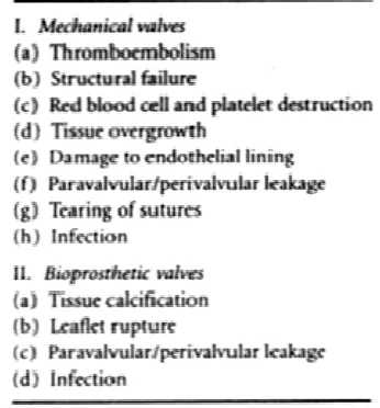
TABLE 44.3 Common Problems with Implanted Prosthetic Heart Valves [Yoganathan et al,. 1979a; Shim and Lenker. 1988; Chandran, 1992]
44.1.3 Total Artificial Hearts (TAH) or Ventricular Assist Devices (VAD)
10/24
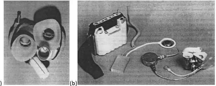
polyurethanes, Biomer, and others). Segmented polyurethane elastomer used in prosthetic ventricles with a thromboresistant additive modifying the polymeric surface have resulted in improved blood compatibility and reduced thromboembolic risk in animal trials [Farrar et al,. 1988]. Design considerations include reduction of regions of stagnation of blood within the blood chamber and minimizing the mechanical stresses induced on the formed elements in blood. Apart from the characteristics of these materials to withstand repetitive high mechanical stresses and minimize failure due to fatigue, surface interaction with blood is also another crucial factor. An electrically powered total artificial heart intended for long term implantation is shown in Figure 44.8b. The details of the design considerations for the circulatory assist devices are included in Rosenberg [1995a] and details of the evaluation of the electrically powered heart is included in Rosenberg et al. [1995b].
(a) (b)
FIGURE 44.8 Typical prototype designs of total artificial hearts: (a) pneumatically powered TAH. The right and left ventricular chambers, inflow and outflow valves, as well as the connector for the pneumatic line are visible in the photograph; (b) electrically powered TAH.
Shown are the external battery pack, transcutaneous energy transmission system (TETS) primary and secondary coils, implanted electronics, energy converter and the blood pumps,
compliance chamber and the subcutaneous access port. (Courtesy of G. Rosenberg, Pennsylvania State University.)
11/24
valves during closure due to the relatively large dp/dt (p is pressure, t is time) at which the TAR was operated. Attempts at reducing the dp/dt during closure of the inflow valves have also been reported with modified designs of the artificial heart driver [Wurzel et al, 1988]. Due to the relatively large dp/dt at which TAHs are operated, there is increased possibility of cavitation bubble initiation and subsequent collapse of the bubbles may also be another important reason for thrombus deposit ion near the mechanical valve at the inflow orifice. Introducing synthetic valves to replace the mechanical valves [Chandran et al., 1991b] may prove to be advantageous with respect to cavitation initiation and may minimize thrombus formation.
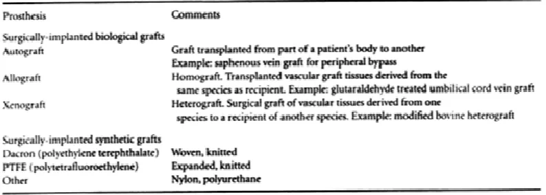
TABLE 44.4 Classification of Vascular Prostheses
44.1.4 Vascular Prostheses
In advanced stages of vascular diseases such as obstructive atherosclerosis and aneurysmal dilatation, when other treatment modalities fail, replacement of diseased segments with vascular prostheses is a common practice. Vascular prostheses can be classified as given in Table 44.4.
44.1.4.1. Surgically Implanted Biological Grafts. Arterial homografts, even though initially used in large scale, resulted in aneurysm formation especially in the proximal suture line [Strandness and Sumner, 1975]. Still, a viable alternative is to use the saphenous vein graft from the same patient. Vein grafts have a failure rate of about 20% in one year and up to 30% in five years after implantation. Vein grafts from the same patients are also unavailable or unsuitable in about 10 to 30% of the patients [Abbott and Bouchier-Hayes, 1978]. Modified bovine heterograft and gluteraldehyde treated umbilical cord vein grafts have also been employed as vascular prostheses with less success compared to autologous vein grafts.
12/24
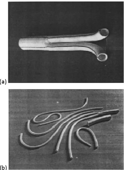
(a)
(b)
FIGURE 44.9 (a) Photograph of a Dacron vascular graft having a bifurcated configuration. (Courtesy of W.L Gore and Associates, Inc., Flagstaff. AZ.) (b) Photographs of expanded
PTFE vascular grafts with straight, straight with external reinforcement rings to resist compression, and bifurcated configurations. (Courtesy of W.L Gore and Associates, Inc.,
Flagstaff. AZ.)
13/24
anastomotic site. At the other end, the graft is attached distal to the occlusion in the host (coronary) vessel to enable perfusion of the vascular bed downstream from the occlusion. Numerous studies analyzing the abnormal flow dynamics within the anastomotic geometry and stress distribution within the vascular material at the junction to the prostheses have been reported in delineating the causes for intimal hyperplasia formation and loss of patency [Kim and Chandran, 1993; Kim et al., 1993; Ojha et al, 1990; Keynton et al., 1991; Chandran et al., 1992; Rodgers et al, 1987; Rhee and Tarbell, 1994] and a detailed discussion on the mechanical aspects of vascular prostheses can be found in Chandran and Kim [1994]. Improvements in the blood-surface interactions are also being attempted in order to improve the functioning capability of vascular grafts. Attempts at seeding the grafts with endothelial cells [Hunter et al, 1983], and modifying the graft material properties by removing the crimping and heat fusing a coil of bendable and dimensionally stable polypropylene at the outer surface to make it kink resistant [Guidoin et al 1983], and employing a compliant and biodegradable graft which will promote regeneration of arterial wall in small caliber vessels [Van der Lei et al., 1985, 1986] are a few examples of such improvements.
44.1.4.3. Transluminally Placed Endovascular Prostheses (Stent.Grafts). Endoluminal approaches to treating vascular disease involve the insertion of a prosthetic device into the vasculature through a small, often percutaneous, access site created in a remote vessel, followed by the intraluminal delivery and deployment of a prosthesis via transcatheter techniques [Veith et al., 1995]. In contrast to conventional surgical therapies for vascular disease, the use of transluminally placed endovascular prostheses are distinguished by their “minimally invasive” nature. Because these techniques do not require extensive surgical intervention, they have the potential to simplify the delivery of vascular therapy, improve procedural outcomes, decrease procedural costs, reduce morbidity, and broaden the patient population that may benefit from treatment. Not surprisingly, endoluminal therapies have generated intense interest within the vascular surgery, interventional radiology, and cardiology communities over recent years.
The feasibility of using transluminally placed endovascular prostheses, or stent-grafts, for the treatment of traumatic vascular injury [Marin et al., 1994], atherosclerotic obstructions [Cragg and Dake, 1993], and aneurysmal vascular disease [Parodi et al., 1991; Yusuf et al., 1994; Dake et al., 1994] has been demonstrated in human beings. Endoluminal scent-grafts continue to evolve to address a number of cardiovascular pathologies at all levels of the arterial tree. Figure 44.l0a depicts endoluminal stent-grafts having a variety of configurations (straight, bifurcated) and functional diameters (peripheral, aortic) that are currently under clinical investigation.
14/24
graft that is externally supported along its entire length by a self-expanding nitinol stent. The implant is radially constrained and attached to the leading end of a dual lumen polyethylene delivery catheter that allows transluminal delivery and deployment. Following introduction into the vascular system, the implant is positioned fluoroscopically within the diseased segment and released from the delivery system.
FIGURE 44.10 (a) Endoluminal stent-grafts of straight and bifurcated configurations and sizes currently under clinical investigation. (Courtesy of W.L Gore and Associates, Inc.,
Flagstaff, AZ.) (b) A stent-graft implant consisting of an expanded PTFE graft that is externally supported by a self-expanding nitinol sent. (Courtesy of W.L Gore and
Associates, Inc.. Flagstaff, AZ.)
15/24
enlarged) to any appreciable degree, accurate sizing of the host vessel is critical. A sizing mismatch resulting in oversizing can cause overcompression of the selfexpanding stent and obstructive invagination of the stent into the lumen. Undersizing, in turn, can result in a poor interference fit, inadequate anchoring, device migration, and/or leakage of blood into the abluminal compartment. In either case, the stent provides a scaffold that structurally supports the graft material. Ongoing work in the field of biomedical engineering is directed at optimizing the biomechanical and biological performance of these devices.
44.1.5 Conclusions
In the last four decades, we have observed significant advances in the development of biocompatible materials to be used in blood interfacing implants. In the case of mechanical heart valve prostheses, pyrolytic carbon has become the material of choice for the occlude’ and the housing. The pyrolytic carbon is chemically inert and exhibits very little wear even after more than 20 years of use. However, thromboernbolic complications still remain significant with mechanical valve implantation. The complex dynamics of valve function and the resulting mechanical stresses on the formed elements of blood appear to be the main cause for initiation of thrombus. More recent reports of structural failure with implanted mechanical valves and pitting and erosion observed on the pyrolytic carbon surfaces have resulted in investigations on cavitation bubble formation during valve closure. Along with further improvements in biomaterials for heart valves, detailed analysis of the closing dynamics and design improvements to minimize the adverse effects of mechanical stresses may be the key to reducing thrombus deposition. Improvements on mechanical heart valves or further developments in durable synthetic leaflet valves may also be vital for the development of TAHs for long-term implantation without neurologie complications.
In the case of vascular grafts. the mismatch of material properties (compliance) between the host artery and the graft, as well as geometric considerations in end-to-side anastomoses, appear to be important for the loss of patency within several months after implantation particularly with medium and small diameter arterial replacement. Most of the vascular grafts are stiffer compared to the host artery and it has been suggested that the mechanical stresses resulting from the discontinuity at the junction is the major cause for neointimal hyperplasia formation and subsequent occlusion of the conduit. Developments with more compliant grafts and in modifying the surface interaction of the graft with blood (endothelialization or other treatment of the graft material) may result in reducing the problems with loss of patency. Recent advances in the use of minimally invasive stent-grafts also show promise in improving the quality of life of patients with vascular disease.
DEFINING TERMS
Acetol: Product of the addition of two moles of alcohol to one of an aldehyde.
Aneurysms: Abnormal bulging or dilatation of a segment of a blood vessel or myocardiuifl.
Artery: Blood vessel transporting blood in a direction away from the heart.
Atherosclerosis: Lipid deposits in the intima of arteries.
ATS valve: A bileaflet mechanical valve made by ATS (Advancing The Standard) Inc.
Autoclaving: Sterilizing by steam under pressure.
Biomer: Segmented polyurethane elastomer.
Bioprostheses: Prosthetic heart valves made of biological tissue.
16/24
Bovine heterograft: Graft material (arterial) transplanted from bovine species.
Calcification: Deposition of insoluble salts of calcium.
Cardiac pacemakers: Prosthesis implanted to stimulate cardiac muscles to contract.
Cardiopulmonary bypass: Connectors bypassing circulation to the heart and the lungs.
Catheters: Hollow cylindrical tubing to be passed through the blood vessels or other canals.
Cavitation bubbles (vapor cavitation): Formation of vapor bubbles due to transient reduction in pressure to below the liquid vapor pressure.
Closing dynamics: Dynamics during the closing phase of heart valves.
Compliance: A measure of ease with which a structure can be deformed; ratio of volumetric strain to increase in unit pressure.
Crimping: Creasing of the synthetic vascular grafts in the longitudinal direction to accommodate the large intermittent flow of blood.
Delrin*: Polyacetal made by Union Carbide.
Dialysers: Devices to filter the blood of waste products taking over the function of the kidney.
dp/dt: Slope of the pressure vs. time curve of the ventricles.
Duramater: A tough fibrous membrane forming the outer cover of the brain and the spinal cord.
Electrohydraulic blood pump: Blood pumps energized by the conversion of electrical to hydraulic energy.
End-to-end configuration: End of the vascular graft anastamosed to the end of the host artery.
End-to-side configuration: End of the vascular graft anastamosed to the side of the host.
Endothelial cells: A layer of flat cells lining the intimai surface of blood vessels.
Erosion: A state of being worn away.
Fascia lata: A sheet of fibrous tissue enveloping the muscles of the thigh.
Fatigue stress: Level of stress below which the material would not undergo fatigue failure (107 cycles is used as the normal limit).
Fibrin: An elastic filamentous protein derived from librinogen in coagulation of the blood.
Fibroblasts: An elongated cell with cytoplasmic processes present in connective tissue capable of forming collagen fibers.
Finite element analysis: Structural analysis with the aid of a computer which divides the structure into finite elements and applies the laws of mechanics on each element.
Formaldehyde: Formic aldehyde, methyl aldehyde, a pungent gas used as antiseptic.
Formed elements in blood: Red blood cells, white blood cells, platelets, and other cells in whole blood.
Haynes 25e: Co—Cr alloy.
Homografts: Transplants (heart valves, arterial segments, etc.) from the same species.
lntra-aortic balloon pumps: A balloon catheter inserted in the descending aorta and alternately inflated and deflated timed to the EKG in order to assist the ventricular pumping.
Laser Doppler anemometry: A velocity measurement device using the principle of Doppler shifted frequency of laser light by particles moving with the fluid.
Leaflets: Occluders on valves which open and close to aid blood flow in one direction.
Left ventricular assist devices: Prosthetic devices to assist the left ventricle in pumping blood.
17/24
LTI: Low temperature (below 1500°C) isotropic pyrolytic carbon.
Mechanical prostheses: Prostheses made of non-biological material.
Negative pressure transients: Reduction in pressure for a short duration.
Neointima: Newly formed intimal surface.
Neointimal hyperplasia: Growth of new intimal surface formed by fibroblasts.
Nylon: Synthetic polymer with condensation polymerization.
Patency: State of being freely open.
Pericardial prostheses: Heart valve prosthesis made with fixed bovine pericardial tissue.
Pitting: Depression or indent on a surface.
Platelet: One of the formed elements of blood responsible for blood coagulation.
Polypropylene: One of the vinyl polymers with good flex life and good environmental stress crack resistance.
Polytetrafluoroethylene (PTFE): A fluorocarbon polymer known as Teflon®.
Pusher plate devices: Artificial heart devices working with pusher plates moving the blood.
Pyrolytic carbon: Carbon deposited onto preformed polycrystalline graphite substrate.
Radiopacity: Being opaque to x-ray.
Sewing rings: Rings surrounding the housing of artificial heart valves used to sew the valve to the tissue orifice with suture.
Solution casting: Casting by pouring molten material on dyes to form a structure.
Stellite 21®: Co—Cr alloy.
Stent: A device used to maintain the bodily orifice or cavity.
Strut: A projection in the structure such as guiding struts in heart valves used to guide the leaflets during opening and closing.
TAH: Total artificial heart replacing a failed natural heart.
Teflon®: See PTFF..
Thrombo-embolic complications: Complications due to breaking away (emboli) of thrombus blocking the distal blood vessels.
Thrombus: A clot in the blood vessels or in the cavities of the heart formed from the constituents of blood.
Tilting disc valves: Valves with a single leaflet tilting open and shut.
Titanium: Highly reactive metal having low density,.good mechanical properties, and biocompatibility due to tenacious oxide layer formation.
Turbulent stresses: Stresses generated in the fluid due to agitated random motion of particles.
Ultra high molecular weight polyethylene: Linear thermoplastics with very high molecular weight (>2 x 10⁶ gl/mol) used for orthopedic devices such as acetabular cup for hip joint replacement.
Umbilical cord vein grafts: Vascular graft made from umbilical cord veins.
Vacuum forming: A manufacturing technique for thermoplastic polymer in which a sheet is heated and formed over a mold while a vacuum is present under the sheet.
Valvular disorders: Diseased states of valves such as stenosis.
Valvular structures: Components of valves such as leaflets, struts, etc.
Vascular grafts: Grafts to replace segments of diseased vessels.
18/24
44.2 NON-BLOOD-INTERFACING IMPLANTS FOR SOFT TISSUES
[K.J.L. Burg and S. W. Shalaby]
Most tissues other than bone and cartilage are of the soft category. Implants do not generally interface directly with blood; the exceptions are located primarily in the cardiovascular systems. Non-blood-interfacing soft tissue implants are used to augnwnt or replace natural tissues or to redirect specific biological functions. The implants can be transient, that is, of short-term functïon and thus made of absorbable materials, or they can be long-term implants which are expected to have prolonged functions and are made of nonabsorbable materials.
Toward the successful development of a new biomedical device or implant, including those used for soft tissues, the following milestones must be achieved: (I) acquire certain biologic and biomechanic data about the implant site and its function to aid in the selection of materials and engineering design of such an implant, to meet carefully developed product requirements; (2) construct a prototype and evaluate its physical and biologic properties both in vitro and in vivo, using the appropriate animal model; and (3) conduct a clinical study following a successful battery of animal safety studies depending on intended application and availability of historical safety and clinical data on the material or design. Extent of the studies associated with any specific milestone can vary considerably. Although different applications require different materials with specific properties, minimum requirements for soft-tissue implants should be met. The implant must (1) exhibit physical properties (e.g., flexibility and texture) which are equivalent or comparable to those called for in the product profile; (2) maintain the expected physical properties after implantation for a specific period; (3) elicit no adverse tissue reaction; (4) display no carcinogenic, toxic, allergenic, and/or immunogenic effect; and (5) achieve assured sterility without compromising the physicochemical properties. In addition to these criteria, a product of potentially broad applications is expected to (1) be easily mass produced at a reasonable cost; (2) have acceptable aesthetic quality; (3) be enclosed in durable, properly labeled, easy-access packaging; and (4) have adequate shelf stability.
The most common types of soft-tissue implants are (1) sutures and allied augmentation devices; (2) percutaneous and cutaneous systems; (3) maxillofacial devices; (4) ear and eye prostheses; (5) space-filling articles: and (6) fluid transfer devices.
44.2.1 Sutures and Allied Augmentation Devices
Sutures and staples are the most common types of augmentation devices. In recent years. interest in using tapes and adhesives has increased and may continue to do so. should new efficacious systems be developed.
44.2.1.1. Sutures and Suture Anchors. Sutures are usually packaged as a thread attached to a metallic needle. Although most needles are made of stainless steel alloys, the thread component can be made of various materials, and the type used determines the class of the entire suture. In fact, it is common to refer to the thread as the suture. Presently, most needles are drilled (mechanically or by laser) at one end for thread insertion. Securing the thread in the needle hole can be achieved by crimping or adhesive attachment. Among the critical physical properties of sutures are their diameter, in vitro knot strength, needle-holding strength, needle penetration force, ease of knotting, knot security, and in vitro strength retention profile.
19/24
and tantalum). Sutures may also be classified according to their physical form, that is, monofilament and twisted or braided multifilament (or simply braids).
The first known suture, the absorbable catgut. is made primarily of collagen derived from sheep intestinal submucosa. It is usually treated with a chrornic salt to increase its in vivo
strength retention and through imparted crosslinking that retards absorption. Such treatment extends the functional performance of catgut suture from 1 to 2 weeks up to about 3 weeks. The catgut sutures are packaged in a specially formulated fluid to prevent drying and maintain necessary compliance for surgical handling and knot formation. The use of synthetic absorbable sutures exceeded that of catgut over the past two decades. This is attributed to many factors including (1) higher initial breaking strength and superior handling characteristics; (2) availability of sutures with a broad range of in vivo strength retention profiles; (3) considerably milder tissue reactions and no immunogenic response; and (4) reproducible properties and highly predictable in vivo
performance. Polyglycolide (PG) was the first synthetic absorbable suture to be introduced, about three decades ago. Because of the high modulus of oriented fibers, PG is made mostly in the braided form. A typical PG suture braid absorbs in about 4 months and retains partial in vivo strength after 3 weeks. However, braids made of the 90/l0 glycolide/l-lactide copolvmer have a comparable or improved strength retention profile and faster absorption rate relative to PG. The copolvmeric sutures absorb in about 3 months and have gained wide acceptance by the surgical community.
As with other types of braided sutures, an absorbable coating which improves suture handling and knot formation has been added to the absorbable braids. To minimize the risk of infection and tissue drag that are sometimes associated with braided sutures, four types of monofilament sutures have been commercialized. The absorbable monofilaments were designed specifically to approach the engineering compliance of braided sutures, by combining appropriate materials to achieve low moduli, for example, polydioxanone and copolymers of glycolide with caprolactone or trimethylene carbonate. Members of the nonabsorbable family of sutures include braided silk (a natural protein), nylon, and polyethylene terephthalate (PET). These braids are used as coated sutures. Although silk sutures have retained wide acceptance by surgeons, nylon and particularly PET sutures are used for critical procedures where high strength and predictable long-term performance are required. Meanwhile, the use of cotton sutures is decreasing constantly because of their low strength and occasional tissue reactivity due to contaminants. Monofilaments are important forms of nonabsorbable sutures and are made primarily of polypropylene, nylon, and stainless steel. An interesting application of monofilament sutures is illustrated in the use of polypropylene loops (or haptics) for intraocular lenses. The polypropylene sutures exhibit not only the desirable properties of monofilaments but also the biologic inertness reflected in the minimal tissue reactions associated with their use in almost all surgical sites. With the exception of its natural tendency to undergo hydrolytic degradation and, hence, continued loss of mechanical strength postoperatively, nylon monofilament has similar attributes to those of polypropylene. Because of their exceptionally high modulus, stainless steel sutures are not used in soft-tissue repair because they can tear these tissues. All sutures can be sterilized by gamma radiation except those made of synthetic absorbable polymers, polypropylene, or cotton, which are sterilized by ethylene oxide.
Related to the suture is the tissue suture anchor, used to attach soft tissue to bone. The anchor is embedded into bone and the suture can be used to reattach the soft tissue. The most common anchor is polylactide-based and is used in shoulder repair.
20/24
similar construction made of multililament PET yarns have been used for repairing tendons and ligaments. Microporous foams of polytetrafluoroethylene (PTFE) are used as pledgets (to aid in anchoring sutures to soft tissues) and in repair of tendons and ligaments. Microporous collagen-based foams are used in wound repair to accelerate healing.
44.2.1.3. Clips, Staples, and Pins. Ligating clips are most commonly used for temporary or long-term management of the flow in tubular t issues. Titanium clips are among the oldest and still-versatile types of clips. Thermoplastic polymers such as nylon can be injection -molded into different forms of ligating clips. These are normally designed to have a latch and living hinge. Absorbable polymers made of lactide/glycolide copolymers and polydioxanone have been successfully converted to ligating dips with different design features for a broad range of applications.
Metallic staples were introduced about three decades ago as strong competitors to sutures for wound augmentation; their use has grown considerably over the past 10 years for everything from skin closure procedures to a multiplicity of internal surgical applications. Major advantages associated with the use of staples are ease of application and minimized tissue trauma. Metallic staples can be made of tantalum, stainless steel, or titanium—nickel alloys. Staples are widely used to facilitate closure of large incisions produced in procedures such as Caesarean sections and intestinal surgery. Many interesting applications of small staples have been discovered for ophthalmic and endoscopic use, a fast-growing area of minimally invasive surgery.
Thermoplaslic materials based on lactide/glycolide copolymers have been used to produce absorbable staples for skin and internal wound closures. These staples consist primarily of two interlocking components, a fastener and receiver. They are advantageous in that they provide a quick means of closure with comparable infection resistance. They are limited to locations which do not have large tensile loads and/or thicker or more sensitive tissue.
A new form of ligating device is the subcutaneous pin. This is designed with a unique applicator to introduce the pin parallel to the axis of the wound. During its application the linear pin acquires a zig-zag-like configuration for stabilized tissue anchoring. The pins are made of lactide/glycolide polymers.
44.2.1.4. Surgical Tapes. Surgical tapes are intended to minimize necrosis, scar tissue formation, problems of stitch abscesses, and weakened tissues. The problems with surgical tapes are similar to those experienced with traditional skin tapes. These include (1) misaligned wound edges, (2) poor adhesion due to moisture or dirty wounds, and (3) late separation of tapes when hematoma or wound drainage occur.
Wound strength and scar formation in skin may depend on the type of incision made. If the subcutaneous muscles in the fatty tissue are cut and the overlying skin is closed with tape, then the muscles retract. This, in turn, increases the scar area. causing poor cosmetic appearance when compared to a suture closure. Tapes also have been used successfully for assembling scraps of donor skin for skin graft.
21/24
healing process; and (5) be easily applied during surgery. The two common types of tissue adhesives currently used are based on alkyl-o-cyanoacrylates and fibrin. The latter is a natural adhesive derived from fibrinogen, which is one of the clotting components of blood. Although fibrin is used in Europe, its use in the United States has not been approved because of the risk of its contamination with hepatitis and/or immune disease viruses. Due to its limited mechanical strength (tensile strength and elastic modulus of 0.1 and 0.15 MPa, respectively), fibrin is used mostly as a sealant and for adjoining delicate tissues as in nerve anastomoses. Meanwhile, two members of the cyanoacrylate family of adhesives, n-butyl- and iso-butyl-cyano-acrylates, are used in a number of countries as sealants, adhesives, and blocking agents. They are yet to be approved for use in the United States because of the lack of sufficient safety data. Due to a fast rate of polymerization and some limited manageability in localizing the adhesive to the specific surgical site, the in vivo performance of cyanoacrylates can be unpredictable. Because of the low strength of the adhesive joints or sealant films produced on in vivo
polymerization of these cyanoacrylates, their applications generally are limited to use in traumatized fragile tissues (such as spleen, liver, and kidney) and after extensive surgery on soft lung tissues. A major safety concern of these alkyl cyanoacrylates is related to their nonabsorbable nature. Hence, a number of investigators have directed their attention to certain alkoxy-alkyl cyanoacrylates which can be converted to polymeric adhesives with acceptable absorbable profiles and rheological properties. Methoxypropyl cyanoacrylate, for example, has demonstrated both the absorbability and high compliance that is advantageous to soft tissue repair.
44.2.2 Percutaneous and Skin Implants
The need for percutaneous (trans or through the skin) implants has been accelerated by the advent of artificial kidneys and hearts and the need for prolonged injection of drugs and nutrients. Artificial skin is urgently needed to maintain the body temperature and prevent infection in severely burned patients. Actual permanent replacement of skin by biomaterials is still a great clinical challenge.
44.2.2.1. Percutaneous Devices. The problem of obtaining a functional and viable interface between the tissue (skin) and an implant (percutaneous device) is primarily due to the following factors. First, although initial attachment of the tissue into the interstices of the implant surface occurs, attachment cannot be maintained for a sustained time since the dermal tissue cells turn over continuously. Downgrowth of epithelium around the implant or overgrowth on the implant leads to extrusion or invagination, respectively. Second, any opening near the implant that is large enough for bacteria to penetrate may result in infection, even though initially there may be a tight seal between skin and implant. Several factors are involved in the development of percutaneous devices:
1. Type of end use — this may deal with transmission of information (biopotentials, temperature, pressure, blood flow rate), energy (electrical stimulation, power for heart-assist devices), transfer of matter (cannula for blood), and load (attachment of a prosthesis);
2. Engineering factors — these may address materials selection (polymers, ceramics, metals, and composites) design variation (button, tube with and without skirt, porous or smooth surface), and mechanical stresses (soft and hard interface, porous or smooth interface);
3. Biologic factors— these are determined by the implant host (human, dog. hog, rabbit, sheep), and implant location (abdominal, dorsal, forearm);
22/24
No percutaneous devices are completely satisfactory Nevertheless, some researchers believe that hydroxyapatite may be part of a successful approach. ¡none experimental trial, a hydroxyapatite-based percutaneous device was associated with less epidermal downgrowth (1 mm after 17 months vs. 4.6 mm after 3 months) when compared with a silicone rubber control specimen in the dorsal skin of canines. Researchers have also investigated coatings such as laniinin-5 which has been shown to enhance epithelial attachment.
44.2.2.2. Artificial Skins. Artificial skin is another example of a percutaneous implant, and the problems arc similar to those described above. Important for this application is a material which can adhere to a large (burned) surface and thus prevent the loss of fluids, electrolytes, and other biomolecules until the wound has healed.
In one study on wound-covering materials with controlled physicochemical properties, an artificial skin was designed with a crosslinked collagen-polysaccharide (chondroitin 6-sulfate) composite membrane. This was specifically chosen to have controlled porosity (5 to 150 pm in diameter), flexibility (by varying crosslink density), and moisture flux rate.
Several polymeric materials and reconstituted collagen have also been examined as burn dressings. Among the synthetic ones are the copolymers of vinyl chloride and vinyl acetate as well as polymethyl cyanoacrylate (applied as a fast-polymerizing monomer). The latter polymer andlor its monomer were found to be too brittle and histotoxic for use as a burn dressing. The ingrowth of tissue into the pores of polyvinyl alcohol sponges and woven fabric (nylon and silicone rubber velour) was also attempted without much success. Nylon mesh bonded to a silicone rubber membrane, another design attempt, prevented water evaporation but has not been found to induce fibrovascular growth.
Rapid epithelial layer growth by culturing cells in vitro from the skin of the burn patient for covering the wound area may offer a practical solution for less severely burned patients. Implantation of an aflogenic tibroblastipolymer construct has proven useful for providing long-term skin replacement. Related to this, temporary tissue engineered replacements are possible alternatives for burns requiring larger area coverage. These can be similar to the synthetic dressing, a nylon mesh and silicone rubber component, but incorporates allogeneic fibrobbsts. This temporary covering hopefully will stimulate or allow fibrovascular growth into the wound bed by providing the appropriate matrix proteins and growth factors.
44.2.3 Maxillofacial Implants
There are two types of maxillofacial implants: extraoral and intraoral. The tormer deals with the use of artificial substitutes for reconstructing defective regions in the maxilla, mandible, and face. Useful polymeric materials for extraoral implants require (I) match of color and texture with those of the patient; (2) mechanical and chemical stability (Le., material should flot creep or change color or irritate skin); and (3) ease of fabrication. Copolymers of vinyl chloride and vinyl acetate (with 5 to 20% acetate), polymethyl methacrylate, silicones, and polyurethane rubbers are currently used. Intraoral implants are used for repairing maxilla, mandibular, and facial bone defects. Material requirements for the intraoral implants are similar to those of the extraoral ones. For the latter group of implants, metallic materials such as tantalum, titanium, and CoCr alloys are commonly used. For soft tissues, such as gum and chin, polymers such as silicone rubber and polymethylmethacrylate are used for augmentation.
44.2.4 Ear and Eye Implants
23/24
(the inflammation of the middle ear which can cause partial or complete impairment of the ossicular chain). A number of prostheses are available for correcting these defects. The porous polyethylene total ossicular implant is used to achieve a firm fixation by tissue ingrowth. The tilt-top implant is designed to retard tissue ingrowth into the section of the shaft which may diminish sound conduction. Materials used in fabricating these implants include polymethyl methacrylate, polytetrafluoroethylene, polyethylene, silicone rubber, stainless steel, and tantalum. More recently, polytetratluoroethylene— carbon composites, porous polyethylene, and pyrolytic carbon have been described as suitable materials for cochlear (inner ear) implants.
Artificial ear implants capable of processing speech have been developed with electrodes to stimulate cochlear nerve cells. Cochlear implants also have a speech processor that transforms sound waves into electrical impulses that can be conducted through coupled external and internal coils. The electrical impulses can be transmitted directly by means of a percutaneous device.
Eye implants are used to restore the functionality of damaged or diseased corneas and lenses. Usually the cornea is transplanted from a suitable donor. In cataracts, eye lenses become cloudy and can be removed surgically. Intraocular lenses (IOL) are implanted surgically to replace the original eye lens and to restore function. IOL are made from transparent acrylics, particularly polymethyl methacrylate, which has excellent optical properties. Infection and fixation of the lens to the tissues are frequent concerns, and a number of measures are being used to address them. Transplantation of retinal pigmented epithelium can be used in the treatment of adult onset blindness; the challenge is developing readily detachable or absorbable materials on which to culture sheets of these cells.
44.2.5 Space-Filling Implants
Breast implants are common space-filling implants. At one time, the enlargement of breasts was done with various materials such as paraffin wax and silicone fluids, by direct injection or by enclosure in a rubber balloon. Several problems have been associated with directly injected implants, including progressive instability and ultimate loss of original shape and texture as well as infection and pain. One of the early efforts in breast augmentation was to implant a sponge made of polyvinyl alcohol. However, soft tissues grew into the pores and then calcified with time, and the so-called marble breast resulted. Although the enlargement or replacement of breasts for cosmetic reasons alone is not recommended, prostheses have been developed for patients who have undergone radical mastectomy or who have nonsymmetrical deformities. The development of the tissue-engineered breast is ongoing, where fat or normal breast tissue may be derived from the patient and combined with an absorbable scaffold for transplantation. A silicone rubber bag filled with silicone gel and backed with polyester mesh to permit tissue ingrowth for fixation is a widely used prosthesis, primarily for psychological reasons. The artificial penis, testicles, and vagina fall into the same category as breast implants, in that they make use of silicones and are implanted for psychological reasons rather than to improve physical health.
44.2.6 Fluid Transfer Implants
24/24
drain into the blood stream through the right internal jugular vein or right atrium of the heart. The simpler peritoneal shunt shows less incidence of infection.
The use of implants for correcting the urinary system has not been successful because of the difficulty of adjoining a prosthesis to the living system for achieving fluid tightness. In addition, blockage of the passage by deposits from urine, salt for example, and constant danger of infection are major concerns. Several materials have been used for producing these implants, with limited long-term success; these include DacronTM, glass, polyvinyl alcohol, polyethylene, rubber, silver, tantalum, TeflonTM, and VitalliumTM. Tissue engineered devices also have application in urinary repair; for examples an alginate-chondrocyte system has been used clinically to treat vesicoureteral reflux. Preliminary results suggest that by transplanting the hydrogel system as a bulking agent below a refluxing ureter, neocartilage gradually develops to correct the reflux. Uroepithelial cells, combined with porous, absorbable polyester matrices show promise in the replacement of urologic tissues.
Drainage tubes, which are impermanent implants for chronic ear infection, can be made from polytetra fluoroethylene (TeflonTM). Glaucoma-related elevated intraocular pressure may be relieved by implanting a tube connecting the anterior eye chamber to the external subconjunctival space in order to direct aqueous humor. Complications can arise with ocdusion of the tube due to wound healing processes; however, researchers are investigating combining this device with an absorbable drug delivery plug to regulate flow and deliver drugs to regulate the wound healing process.
44.2.7 Technologies of Emerging Interest
A new process for uniaxial solid-state orientation, using a range of compressive forces and temperatures, has been developed to produce stock sheets of polyether-ether ketone (PEEK) for machining dental implants with substantially increased strength and modulus as compared to their unoriented counterparts.
Surface treatment of materials is another emerging interest, a process potentially influencing both surface charge, topography, and conductivity. The former has obvious effects on cell adhesion and integration of traditional implants with surrounding tissues, and it has a profound effect on the success or failure of a tissue-engineered device.
Pertinent to the effect of surface chemistry and texture on tissue regeneration/ingrowth, recent studies on surface-phosphonylation to create hydroxyapatite-like substrates show that (1) surface-microtextured polypropylene and polyethylene transcortical implants in goat tibia having phosphonate functionalities (with or without immobilized calcium ions) do induce bone ingrowth, and (2) microtexture and surface phosphonylated (with and without immobilized calcium ions) rods made of PEEK and similarly treated rods of carbon fiber-reinforced PEEK induce bone ingrowth when implanted in the toothless region of the lower jaw of goats. The use of surface-phosphonylated and post-treated (with a bridging agent) UHMW-PE fibers and fabric do produce high strength and modulus composites at exceptionally low filler loading. Among these composites are those based on methyl methacrylate matrices, similar to those used in bone cement.