P-ISSN.2503-0817, E-ISSN.2503-0825
CrossMark
Abstract
Objective: This report discussed about the management of SCC at the tongue with hemiglosectomy and selective neck dissection managed in a 57-year-old male who presented with complaints of a mass on the tongue measuring 0.5 cm that did not heal.
Methods:Squamous Cell Carcinoma (SCC) is a common malignant neoplasma that originates from epithelial cells. It usually involves oral mucosa and underlying tissue of the tongue, hypopharynx and also larynx.
Results: Incision biopsy result confirmed a well-differentiated squamous cell carcinoma on his tongue with enlarged lymph nodes at level I, ipsilateral. The patient was hospitalized and a hemiglosectomy surgery with selective neck dissection was conducted, with the entire edge of the excision boundary becoming free of tumor mass post-surgery.
Conclusion: Hemiglosectomy with selective neck dissection is chosen depending on the size and location of lesion and occurrence of metastases.
Keywords: Hemiglosectomy, Selective neck dissection, Squamous cell carcinoma
Cite this Article: Fauzi A, Hardianto A, Wariz R. 2016. Hemiglosectomy with selective neck dissection in squamous cell carcinoma at the tongue. Journal of Dentomaxillofacial Science 1(2): 132-137. DOI:10.15562/jdmfs.v1i2.13
Intro
duction
Carcinoma is a malignant development of epithelial cells. The most common carcinoma on tongue is termed as Squamous Cell Carcinoma (SCC), which occurs at a rate of 95%.1,2
This malignant tumor originates from squamousepithelium mucosa that is prone to infiltrate and metastases into surround-ing tissue. The incidence of SCC is about 52% of oral cavity carcinoma.3
Other literature found approxi-mately 25–40% of oral cavity carcinoma is related to SCC. SCC on the tongue is the most frequently occurring disease in males and the risk for the disease significantly increases during sixth and seventh decades. If it occurs at an younger age it is commonly associated with substance abuse, such as tobacco and consumption of alcohol. Carcinoma of the tongue is also related to lack of oral hygiene, use of tobacco and alcohol, as well as syphilis glossitis.4
About 45% of the carcinoma lesion on the tongue sites occur on the lateral margin of the middle third of the tongue, about 25% in the posterior third,3 about 20% in the anterior third and about 4% occur on the dorsum.4
Treatment options include surgery, radiation therapy, chemotherapy or a combination of these. Surgical treatment can be either partial or total glossectomy. Decision regarding surgery treatment depends on the size of lesion, location of lesion and occurrence of metastases.1,4
This article discusses a case of a 57-year-old male presenting with a lesion measuring ±0.5 cm in diameter that has been rising at the left edge of the tongue for 2 months and has not healed. Biopsy was
performed under general anesthesia and pathology diagnosis obtained well-differentiated squamous cell carcinoma.
Case Re
port
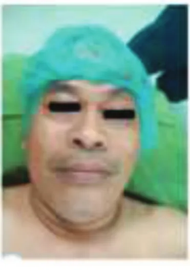
A 57-year-old male was referred to the Dr. Hasan Sadikin Hospital, Department of Oral and Maxillofacial Surgery, Bandung, Indonesia with a complaint of mass on the left edge of his tongue measuring +0.5 cm in diameter for the last two months and the mass did not heal. On physical examination, his general condition seemed compos mentis with blood pressure 130/80 mmHg, temperature 36°C, pulse 80x/minute, no history of shortness of breath or chronic cough. On extra-oral examination showed patient had a symmetrical face and an enlargement of lymph nodes at level I sub-mental group figures 1 and 2. Intra-oral examination on the sinistra on the lateral side of the tongue found a lesion measuring +0.5 cm in diameter with evident border, reddish appearance, easily bleeding, ulceration (+) and T1N1M0 stage
figure 3.
Patient was referred to the Department of Oral and Maxillofacial Surgery Faculty of Den-tistry Padjajaran University, Bandung, Indonesia with an ulcer on the left side of the tongue which was not healing. Patient was scheduled to undergo an incision biopsy under general anesthesia and the results were sent to the Dr. Hasan Sadikin 1Department of Oral and
Maxillofacial Surgery, Faculty of Dentistry, Hasanuddin University, Makassar, Indonesia
3Department of Prostodontic,
Faculty of Dentistry, Hasanuddin University, Makassar, Indonesia
2Department of Oral and
Maxillofacial Surgery, Faculty of Dentistry, Padjadjaran University/ Dr. Hasan Sadikin Hospital, Bandung, Indonesia
*
Correspondence to: Abul Fauzi, Department of Oral and Maxillofacial Surgery, Faculty of Dentistry,
Hemiglosectomy with selective neck dissection in
squamous cell carcinoma at the tongue
Abul Fauzi,1*
Andri Hardianto,2
Hospital, Department of Pathology, Faculty of Medicine, Padjadjaran University, Bandung, Indone-sia.
Results confirmed that the tissue on the lesion of the tongue showed well-differentiated SCC; therefore, it was decided to perform partial glos-sectomy and selective neck dissection surgery
figures 4and5.
Partial glossectomy surgical technique
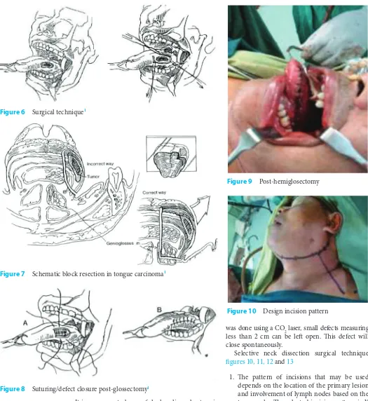
After patient was under general anesthesia and in asepsis condition, a mouth gag was used to open his mouth. Then we marked a margin incision with methylene blue approximately 1.5–2 cm from the ulcer border. We used a retractor on the tip of the tongue to provide good retraction. Ligature of 2–0 silk sutures were placed through the anterior and posterior of the excision lines. Then suture or clamp were used to pull the incision that was made on the tongue. Surgery was performed using the resection block technique, which includes all the muscles below the root of the tongue tumor figures 6 and 7.
Resection block technique prevents the closure of margin on the deeper part of the tumor and includes microscopic lymph duct on specimen. Figure 1 Front view
Figure 2 Lateral view
Figure 3 Lesion on left side of tongue
Figure 4 Biopsy incision
It is necessary to be careful when lingual artery is clamped and ligated with 3–0 silk sutures as well as when the vein on the tongue is clamped, dissected and ligated.
The arising defects after resection of carcinoma of the tongue measuring less than 2 cm in diameter were reconstructed using stitches interrupted with 3-0 sutures on muscle and sub-mucosal layer or interrupted through mucosa figure 8. If dissection
was done using a CO2 laser, small defects measuring less than 2 cm can be left open. This defect will close spontaneously.
Selective neck dissection surgical technique
figures 10, 11, 12 and 13
1. The pattern of incisions that may be used depends on the location of the primary lesion and involvement of lymph nodes based on the topography. The selected incision pattern is Y pattern, it can maintain blood supply to the skin.
2. Vasoconstrictor injecting was used to reduce bleeding on the incision area. The incision is made through the platysma muscle and the flap elevated on sub-platysma plane. After elevating the lateral superior flap, the greater auricular nerve and the external jugular vein Figure 6 Surgical technique1
Figure 8 Suturing/defect closure post-glossectomy1
Figure 9 Post-hemiglosectomy
were identified as overlying the sternocleido-mastoid muscle, with posterior flap retracted to the trapezius muscle.
3. Identification and dissection of the inferior edge of mandible on superior flap. The sub-mental fatty tissue is resected and placed besides the inferior. Then we retracted the mylohyoid muscle anteriorly, exposing the submandibular ganglion, the lingual nerve and the subman-dibular duct. Facial artery is ligated above the digastric muscle. The submandibular duct is then dissected and ligated. Submandibular lymph nodes and submandibular salivary duct are ligated and placed inferiorly.
4. Continued stripping of lymphatic nodes at levels I–III, ipsilateral and biopsy. Histopathological examination confirmed that lymphatic nodes at level I is positive and at level II–III negative. 5. Irrigate the dissected field with isotonic
sodium chloride liquid and secure hemostasis. Furthermore, we put on the drain as much as possible on each side. Layer by layer we covered platysma and then sutured cutis with nylon and applied compressing dressing.
Discussion
Etiology of SCC in oral cavity is multifactorial and consists of extrinsic and intrinsic factors. Extrinsic factors consist of external agents, including tobacco chewing, smoking, betel chewing (50% in Asia), alcohol, syphilis, as well as virus (retrovirus, adenovirus, herpes simplex virus, human papil-loma virus), and other chemical agents. As for the intrinsic factor, it is a systemic condition including malnutrition or iron deficiency, anemia and immunological factors.2,4–6
At the early stage, tongue carcinoma generally does not show any symptom. The pain is often not considered as an early symptom but perceived as neoplasm that directly affects the lingual nerve branch. Pain that spreads to ear is caused by the effect of trigeminal nerve (V3) of lingual nerve that innervates tongue sensation and auricular tempo-ralis nerve that innervates outer ear sensation and tympani membrane. The pain will be more intense with ulcer worsening. Infiltration that spreads to tongue muscles will disturb phonetic and mastica-tion process. If patient has severe lesion there will be smell foul.2
About 30% of the patients experience pain as an early symptom. In about 10% of the cases lesion grows rapidly so that patients experience swelling or mass on tongue as an early symptom, 5% have difficulty swallowing and 6% have enlargement in the cervical lymph gland. Emergence of bleeding
anddifficulty with speaking and swallowing indi-cate the worsening of disease.4
SCC has some clinical representations (morpho-logical growth pattern) that are exopitic, endopitic (infiltratif), ulcerative, leukoplakia and erythroleu-koplacia or erythroplacia. Leukoplakia and eryth-roplacia may not develop into an ulcer or mass at the beginning, and clinical signs resemble a prema-lignant lesion for leukoplakia and erythroplacia. Ectopic lesions tend to arise more toward to the superficial. This takes a shape of a thickened epithel area that shows up as folded on mucosa; later the surface becomes inconsistent, with variance in color starting from normal color, red or white depending on the amount of keratin that is produced. Lesion’s surface goes through induration and lesion feels solid with palpation and is recurrently ulcerative.5,6
Based on histopathological representation, SCC classified as follows: well-differentiated, moderately well-differentiated, moderately differentiated and poorly differentiated. Most of the well-differen-tiated tumor cells resemble normal cells. In some cases, it is commonly found as an epithelial pearl that shows the formation of keratohialin beads beneath the epithelial surface. Keratohialin beads can gather together to form a group of keratins known as keratin pearl. Keratin pearl is a sign for well-differentiated tumor.4,6
In moderately well-differentiated tumor we can see variations in cell size, nucleus size, hiperchro-matism and prominent mitosis activity.Meanwhile, poorly differentiated tumor cells show inconsis-tency and tend to be interpreted as anaplasia which is difficult to recognize. Tumor cells randomly grow in all directions and infiltrate into connective tissues, where the basal layer cannot be identified as they usually disappear.4,6
On histopathological examination of tongue carcinoma a general illustration of broad nucleus, hyperchromatism, and various forms and sizes can be seen. Active mitosis and atypical anaplasia, assorted differentiations, as well as irregular polar-ities can be seen.4,6
Generally, carcinoma occupying two-thirds of anterior is differentiated well and tend to be keratinized without presence of early epithe-lial cells and stroma appears widened. Epitheepithe-lial pearl generally occurs because of the interference of keratohialin grains and other cells and creates the keratin period and a third posterior of the tongue is moderately well-differentiated but depicting no tendency for keratinization but depicts a tendency towards metastases.4,6
lymphogen is the most frequently occurring inci-dence as tongue is richly populated with lympathic vessels.4,6
If the patient presents with an asymme-try and palpable neck lymph nodes in the cervical region, it means the tumor has spread to the neck. Carcinoma spreading to contralateral region can be similar or a continuous region spreading.
The spread through the lymphatic nodes in the middle of the tongue cross to the contralateral or the lymph nodes in the sub-digastric region.The canal/pathway that is often passed through is an afferent lymphatic vessel that has superficial capil-laries and lymph nodes in the anterior midline of the mouth or extends through the midline efferent lymph vessel.Metastases can occur in secondary sites as well, such as mediastinum, axilla, lungs or lever. Direct transplantation may occur because of the continuously direct contact made to surrounding structures. In addition, with release of tumor cells due to trauma, the tumor cells become less cohesive than normal cells.4,6
Diagnosis was obtained with clinical and histo-pathological examination. In the initial stage, most patients do not present any complaints. However, they commonly seek professional advice when the disease becomes severe and presents with various complaints. Usually, patients complain of an inter-ference wound (ulcerations) that do not heal in their tongue. Paresthesias, persistent swelling and unspecific smell occur. Furthermore, with general decline in health due to worsening of diseases and its associated symptoms and complaints, the body becomes weak and patients experience itching in the tongue, persistent burning sensation or pain and sometimes even bleeding.2,4
Patients complain of difficulty in both speaking and swallowing. In the initial stage, mucous membrane is seen to thicken but without erosion on the surface. Later, lesion turn into areas that have visible thickening and hard surface and readily bleed. If the area of indurations thicken, then the central part of the lesion will ulcerate. In about two-thirds of the anterior area of the tongue, carcinoma showed infiltrative growth. Ulceration on the surface or the formation of board ulceration is accompanied by inside infiltra-tion. Secondary infection frequently occurs along with tissue necrosis that causes a specific foul smell.2,4
The third posterior area infiltrates exten-sively into the tongue’s base muscles. Ulceration occurs on one side of the tongue muscles and most frequently in the midline. Ulcerated areas
are surrounded by diffuse groups of tumor tissue and they often bleed.2,4
To ensure accurate diagnosis, a biopsy can be performed by excision that takes a whole tumor out if it is only a small tumor. A biopsy can even be accomplished with sharp V-shaped incision or by puncture biopsy (needle biopsy) or smear technique. On the histopathology examination of tongue carcinoma we obtained a general illustra-tion of broad nucleus, hyperchromatism and the various forms and sizes. Active mitosis, atypic and anaplasia differentiation in assorted stages as well as irregular polarity exist.2,4
Tongue carcinoma lesion can be diagnosed in a manner that is comparable to that of leukopla-kia lesions, Lues, tuberculosis, papilloma or other tumors although it is rare. Carcinoma lesions appear due to ulceration and are surrounded by granu-lation tissues with round border and prominent, painful and ready bleeding, pus, reddish lesions and the lesions become hardened. The lesion has an obscure border or without the capsule and there is presence of adenopathy.3,4
Prognosis of epydermoid carcinoma in oral cavity depends on a number of factors, including size, location of tumor, histopathology, metastases and patient’s age. According to the size, the smaller the size of tumor, the better the prognosis and the larger the size of tumor, the worse the progno-sis is. According to lesion’s location, the closer to posterior, the worse the prognosis will get and the closer to anterior, the better will be the prognosis. A tumor on the anterior surface of the tongue has a better prognosis than carcinoma of the posterior surface of the tongue. Prognosis depends on how early patient comes to treatment as well. Generally, carcinoma of the tongue has a poor prognosis. When carcinoma has metastases to lymphatic gland, survival rate will reduce.4,6
neck dissection.7,8
Supraomohyoid neck dissection is the resection of lymphatic nodes at levels I, II and III figure 4. This surgery resects the soft tissue in the sub-mental triangle, along the submandibular triangle sides, including the submandibular glands and fibro fatty tissues along the internal jugular vein at level II in the upper part. Dissection area includes the fascia layer at the medial part of the sternomas-toid muscle: the muscles retract to lateral and left. Neck content is separated into internal jugular vein and from accessory nerve.8,9
Supraomohyoid neck dissection indicated for prophylaxis treatment in cancer has to address metastases in lymph nodes group, such as small size oral cancer, and when clin-ically regional lymph node (N) neck is considered to be negative. This surgery is performed for the surgical management of oral cavity squamous cell carcinoma stage T2-T4 No or TxN1 if it is clear that the lymphatic nodes identified are less than 3 cm, can be moved and are located at level I or II. This procedure is carried out on both sides of cancer on the anterior tongue and base of the mouth or near the midline. This surgery is also carried out if there is an indication of management of squamous cell carci-noma of the lip or skin in the middle of the face and if the lesions are clinically metastatic and single in sub-mental or submandibular lymphatic nodes.9
The application of this type of neck dissection is for the treatment of lymphatic nodes that are clinically posi-tive still controversial. If surgery is done for N+, the dissection should include the level IV term expanded to supraomuhyoid neck dissection. SOND does not include posterior triangle because the incidence of positive lymph nodes in the area is low. Advantage in this surgery is that it can reduce postoperative complications and disorders of the shoulder.2,8
Conclusion
Hemiglosectomy with selective neck dissection is selected depending on the size of the lesion, location of lesion and metastases occurrence.
Con
flict of Interest
The authors report no conflict of interest
Re
ferences
1. Bailey BJ. Surgery of the oral cavity. London: Year Book Medical Publishers; 2006. p. 45-51.
2. Million RR. Management of the neck for squamous cell carcinoma. In Million RR, Cassisi NJ, editors. Management of head and neck cancer, a multidisciplinary approach. 2nd ed. Philadelphia: JB Lippincoit Company; 1994. p. 113-130.
3. Regezi JA. Oral pathology clinical–pathologic correlations. 2nd ed. Philadelphia: WB Saunders Company; 1993. p. 77-90.
4. Cummings CW. Otolaryngology-head and neck surgery. 2nd ed. St Louis: Mosby Year book; 1993. p. 1248-1270. 5. March, Antonio R. Radical neck dissection. www://http.
eMedicine. Com 2002.
6. Neville BW. Oral & Maxillofacial Pathology. Philadelphia: WB Saunders Company; 1995. p. 295-303.
7. American Joint Committee on Cancer. AJJC Cancer Staging Manual. 5th ed. Philadelphia: Lippincott-Raven; 2002. p. 24-26.
8. Samant, Sandeep. Neck dissection, classification. www:// http. eMedicine. Com 2002.
9. Medina JE, Rigual NM. Neck dissection. In Cummings CW, et al, editors. Otoraryngology head and neck surgery. 2nd ed. St. Louis: Mosby-Year Book; 1993. p. 1649-1671.