M
icroleakage is the movement of bacteria, fluids, molecules or ions, and even air between the prepared cavity wall and the subsequently applied restorative materials.1 Cervical lesions due to caries, erosion, or abrasion often have both enamel and dentin or cemen-tum margins. The longevity of a conventional Class V restoration can be affected by mechanical, thermal, andchemical factors that result in stress in the cervical area.2,3
Bonded composites have been the common choice for the esthetic restoration of Class V lesions. Howev-er, one disadvantage of composites is polymerization shrinkage, which can result in marginal discrepancies leading to microleakage, among other disadvantages.4 This shrinkage has clinical repercussions such as sensi-tivity, marginal discoloration, and secondary caries.2,3
Many new bonding agents and glass ionomer restorative materials have been introduced to bond restorative materials to dentin and cementum margins of cervical lesions,5,6 but microleakage at the dentin (cementum) aspects of restorations remains a problem of clinical significance.1,4,5Glass ionomers are
alterna-restorations
Manuel Toledano, MD, BDS, PhD,aEstrella Osorio, LDS, PhD,bRaquel Osorio, LDS, PhD,cand Franklin García-Godoy, DDS, MSd
University of Granada, Granada, Spain, and University of Texas Health Science Center at San Antonio, San Antonio, Texas
Statement of problem. Resin-modified glass ionomers and polyacid-modified resin composites (com-pomers) have been introduced to provide esthetic restorations. However, there is concern about the margin-al semargin-aling ability of these materimargin-als, especimargin-ally at the dentin (cementum) aspects of restorations.
Purpose. This in vitro study evaluated the microleakage of Class V restorations made with resin-modified glass ionomers or a compomer.
Material and methods. Thirty noncarious human molar teeth were used. Standardized kidney-shaped Class V cavity preparations were placed in the buccal and lingual surfaces at the cementoenamel junction. Teeth were randomly assigned to 3 experimental groups of 10 teeth each and restored as follows: group 1, Fuji II LC; group 2, Vitremer; and group 3, Dyract. In all cases, the manufacturers’ instructions were strict-ly followed. All materials were placed in a single increment. Unfinished restorations were immediatestrict-ly coat-ed with the respective manufacturers’ sealer or varnish and this was either light curcoat-ed for 20 seconds or allowed to air-dry. After 24 hours, teeth were finished to contour and to the cavosurface margins, coated with nail varnish except for 1 mm around the restoration margin, thermocycled (1000×, 5-55°C) and placed in a solution of 2% basic fuchsin dye for 24 hours at room temperature. The staining along the tooth restoration interface was recorded.
Results. Kruskal-Wallis 1-way analysis of variance revealed significant differences among all restorative materials for the overall, occlusal, and gingival scores (P=.03, P=.01, P=.01, respectively). Occlusal and gin-gival scores for each matched pair of restorative materials using the Wilcoxon test showed statistically signifi-cant differences between Fuji II LC glass ionomer cement and Dyract composite, both for the occlusal (P=.005) and gingival (P=.005) margins and also as an overall evaluation (P=.01), with Fuji II LC showing the least dye penetration. Vitremer revealed dye penetration scores not significantly different from Fuji II LC glass ionomer cement or Dyract composite.
Conclusion. Resin-modified glass ionomers showed less or similar microleakage than the polyacid-modi-fied composite resin tested. (J Prosthet Dent 1999;81:610-5.)
aProfessor, Department of Dental Materials, University of Granada,
Spain.
bAssistant Professor, Department of Dental Materials, University of
Granada.
cProfessor, Department of Dental Materials, University of Granada. dProfessor and Director of Clinical Materials Research, Department
of Restorative Dentistry, Dental School, University of Texas Health Science Center at San Antonio.
CLINICAL IMPLICATIONS
tive materials to composites for the conservative restoration of these lesions because of their adhesion to tooth structure, fluoride release, biocompatibility, lower shrinkage values, reduced microleakage, and acceptable esthetics.7-11 Light-cured resin-modified glass ionomer cements were developed to improve the handling and working characteristics of the original glass ionomer formulation.12,13 Improved adhesion to dentin is probably caused by both a chemical bond from the polyacrylic acid component and formation of a hybrid layer from the hydrophilic HEMA.14-21
Favorable adhesive and fluoride-releasing properties of glass ionomer cements have lead to their widespread use as restorative, lining, and luting materials. To over-come the problems of moisture sensitivity and low early mechanical strengths associated with the conventional glass ionomer cements (GICs) and at the same time maintain their clinical advantages, some hybrid versions of GIC were introduced that are light-cured, because of their small quantity of resin components such as HEMA or BIS-GMA. In some situations, the polyacid also has been modified with side chains that can be polymerized by light-curing mechanisms. The actual formulations vary between manufacturers, but the amount of resin in the final set restoration is between 4.5% to 6%, such as for Fuji II LC and Vitremer glass ionomer cements. The addition of a resin component to GIC and its effects on the development of the ionic crosslink and the subsequent marginal seal against the tooth structure needs further evaluation.
To overcome technique-sensitive mixing and han-dling properties of the resin-modified glass ionomer cements, new materials containing acid-decomposable glass and acidic polymerizable monomers substituting the polyalkenoic acid polymer were developed. These materials were termed polyacid-modified resin compos-ites,22-26 commonly called compomers. Dyract poly-acid-modified resin composite belongs to the new materials that have either been marketed as multipur-pose materials, or contain both of the essential compo-nents of a glass-ionomer cement but at levels that are insufficient to produce an acid-base reaction.13 With this material, the resin content is approximately 28%.
The purpose of this study was to compare the microleakage of Class V restorations produced with the 3 materials, which differ in their resin content, to test the hypothesis that resin content affects microleakage.
MATERIAL AND METHODS
Thirty noncarious human molars, which were stored in a solution of 1% sodium hypochlorite for up to 4 months at room temperature, were test specimens. After surface debridement with a hand-scaling instru-ment and cleaning with a rubber cup and slurry of pumice, a standardized Class V cavity preparation was placed in the buccal and lingual surface at the
cemen-toenamel junction. Preparations were made with a no. 329 carbide bur in a high-speed handpiece and a template to a uniform kidney-shaped outline. Prepara-tions measured 5 mm long, 3 mm wide, and 2 mm deep with the occlusal margin in enamel and the gingi-val margin in dentin or cementum.
Subsequently, teeth were randomly assigned to 3 experimental groups of 10 teeth each. Buccal and lin-gual preparations of group 1 were restored with Fuji II LC (GC Corp, Tokyo, Japan) resin-modified glass ionomer cement; group 2 with Vitremer (3M, St Paul, Minn.) resin-modified glass ionomer cement; and group 3 with Dyract (De Trey Dentsply, Konstanz, Germany) polyacid-modified resin composite.
In all cases, the manufacturers’ instructions for dentin conditioning, powder/liquid proportioning and mixing were strictly followed. For Fuji II LC glass ionomer cement, the cavity wall was conditioned for 20 seconds with dentin conditioner (GC Dental Corp). For Vitremer glass ionomer cement, Vitremer primer (3M Dental Products) was applied on the cavity wall for 20 seconds, gently air dried and light cured for 30 seconds. For Dyract composite restorations, the cav-ity wall was treated with PAS primer/adhesive (DeTrey Dentsply) for 30 seconds, excess was removed with a blast from an air syringe and the adhesive was cured for 20 seconds. A second coat of the primer/adhesive was applied and immediately light cured for 20 seconds. Dyract composite was placed in 1 increment. The teeth were prevented from dehydration by remaining in deionized water storage at room temperature when not being prepared for restoration.
Teeth were prepared for microleakage evaluation by coating the entire tooth with 1 application of nail var-nish, except for 1 mm around the restoration margins. These specimens were then subjected to 1000 temper-ature cycles as suggested in a previous study.27 Each cycle consisted of 30 seconds at 6°C and 30 seconds at 60°C. After thermocycling, teeth were placed in a solu-tion of 2% basic fuchsin dye (Fisher Scientific, Fair Lawn, N.J.) for 24 hours at room temperature.
After removal of the specimens from the dye solu-tion, the superficial dye was removed with a pumice slurry and rubber cup. Teeth were then mounted in a light-curing 1-component methacrylate-based resin (Technovit 7200 VLC, Kulzer, Norderstedt, Germany) to facilitate handling during sectioning. The resin was cured for 24 hours (Histolux, EXAKT, Norderstedt, Germany), then teeth were sectioned longitudinally with a hard tissue microtome (Exakt-apparerteban, Otto Herrman, Norderstedt, Germany) in 0.6-mm thick sections to evaluate the dye penetration.28The sections were then separated, and the cut surfaces cor-responding to the most mesial, central (mesial and dis-tal), and most distal portion of the tooth restoration interface were examined at the occlusal and gingival margins with a stereomicroscope (Olympus Co, Tokyo, Japan) at ×16 magnification. Examination of the speci-mens was undertaken at random, and the investigators were unaware of the exact nature of the restorative material.
Staining along the tooth restoration interface was recorded by 2 evaluators, according to the following criteria: 0 = no dye penetration; 1 = partial dye pene-tration; 2 = dye penetration along the occlusal or gingival wall, but not including the axial wall; and 3 = dye penetration to and along the axial wall. If disagree-ment occurred between the evaluators, a consensus was obtained after reexamination of the specimen by both investigators. Occlusal, gingival, and overall scores for each group of restoration were compared with the Kruskal-Wallis 1-way analysis of variance (ANOVA) nonparametric statistical test to identify any statistical
significant differences between the materials, and the Wilcoxon test was performed to compare each matched pair of restorative materials. Significance was consid-ered at the .05 level.
RESULTS
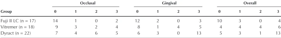
Microleakage scores for the occlusal, gingival, and overall walls are presented in Table I. Kruskal-Wallis 1-way ANOVA indicated significant differences between the restorative materials for overall, occlusal, and gingival scores (P=.03; P=.01; P=.01, respectively). Further matched analysis by Wilcoxon test was under-taken to compare occlusal, gingival, and overall scores of each material, which revealed statistically significant differences between Fuji II LC glass ionomer cement and Dyract resin composite, both for the occlusal (P=.005) and gingival (P=.005) margins and also as an overall evaluation (P=.01) (combining the occlusal and gingival margins scores) with Fuji II LC demonstrating the least dye penetration between these 2 products. Vitremer glass ionomer cement revealed dye penetra-tion scores between Fuji II LC glass ionomer cement and Dyract resin composite, with no statistically signif-icant differences between Vitremer glass ionomer cement and the other 2 products.
DISCUSSION
Polymerization shrinkage of resin-containing restora-tive materials may result in marginal discrepancies that lead to microleakage, marginal discoloration, and sensi-tivity.2-4 Hygroscopic expansion can compensate, to some degree, for polymerization shrinkage. Water sorp-tion can help to reduce marginal gaps3; for this reason, glass ionomer cements, which absorb the most water during the first 24 hours after placement,6 can display less microleakage than resins. Attin et al7reported that Fuji II LC glass ionomer cement expanded after curing and immersion in water, whereas Dyract resin composite and Vitremer glass ionomer cement revealed a total vol-umetric loss. Thus, they concluded that water expansion is 1 factor that reduces the leakage.
Table I. Microleakage of the different groups
Occlusal Gingival Overall
Group 0 1 2 3 0 1 2 3 0 1 2 3
Fuji II LC (n = 17) 14 1 0 2 12 2 0 3 10 3 0 4
Vitremer (n = 18) 9 3 2 4 8 1 4 5 4 4 4 6
Dyract (n = 22) 7 4 6 5 6 3 0 13 5 3 1 13
Kruskal-Wallis 1-way analysis of variance indicated significant differences between all the restorative materials for both overall, occlusal, and gingival scores (P=.03; P=.01; P=.01, respectively).
Our results disagree with those of Yap et al8 who compared the microleakage of Dyract resin composite and Fuji II LC glass ionomer cement and reported no statistically significant differences in microleakage scores. In their study, they reported a significant differ-ence between enamel and dentin; in our study, even if microleakage was less common in enamel, the differ-ence was not significantly different. These differdiffer-ences between the studies could be because Yap et al8stored their specimens in a saline solution for 1 week before testing. This storage time allows hygroscopic expansion of the material,7 which may compensate the original polymerization shrinkage of the material, which allows less microleakage. In our study, specimens were ther-mally cycled for approximately 2 days, and the material may not have expanded completely. Yap et al8also sug-gested that 1 of the unique features of the resin that releases fluoride to enamel is the omission of acid etch-ing, which is a critical step in most resin composite and adhesive systems. The manufacturers have claimed that this is achieved through the use of a specially formulat-ed coupling agent with hydrophilic phosphate groups that is thought to form ionic bonds with the calcium of hydroxyapatite. Dyract resin composite also aims to be self-adhesive because of hydrophilic carboxylic groups present in its patent tetrachlorobiohenyl (TCB) resin. These questions need further investigation.
No restorative material evaluated in our study com-pletely resisted microleakage at the occlusal or gingi-val walls of the tooth. Of the 3 products egingi-valuated, Fuji II LC glass ionomer cement exhibited the least dye penetration, at both the occlusal and gingival margins, and when evaluated as overall values (enam-el and gingival scores pooled together). However, only with the overall evaluation did Fuji II LC glass ionomer cement reveal a statistically significant differ-ence with Dyract resin composite. The lack of statisti-cally significant difference in microleakage between resin-modified glass ionomers has also been previous-ly reported.15Uno et al16concluded that the superior adaptation of Fuji II LC glass ionomer cement to the cavity walls was responsible for the lower dye penetra-tion, which may be a result of the glass ionomer cement undergoing minimal setting shrinkage over a longer period and approximately one half that of resins.17 Because the resin component is responsible for the polymerization shrinkage, and Dyract resin composite has more resin than Fuji II LC glass ionomer cement in its composition, it is possible that this is the reason for the greater microleakage scores observed with Dyract resin composite. Another reason that could explain the results is the resin component of Fuji II LC glass ionomer cement undergoing dif-ferent rates of polymerization shrinkage during light curing (as it is a dual-cure material) compared with Dyract resin composite.
The microleakage scores for Vitremer glass ionomer cement fell between those recorded for Dyract resin composite and Fuji II LC glass ionomer cement, which could be due to 2 reasons. Fuji II LC is a resin-modified glass ionomer in which the HEMA content is merely blended with a polyalkenoic acid liquid, whereas Vit-remer, in addition to being a simple mixture of HEMA with polyalkenoic acid, is also modified by the attach-ment of polymerizable methacrylate side groups.13It is possible that Vitremer has more polymerizable resin than Fuji II LC, but less than Dyract; its microleakage values fell in between these 2 materials. The better adap-tation of Fuji II LC glass ionomer cement compared with Dyract resin composite could be also due to the 15-second dentin conditioning performed with the 10% polyacrylic acid. This dentin treatment produces a close relation between the ionomer and dentin struc-tures as it removes the smear layer, leaving the surface clean and theoretically better able to accept a glass ionomer.18 Moreover, the Fuji II LC liquid contains approximately 40% HEMA (manufacturer’s data) and primers that contain similar hydrophilic monomers than resin-containing materials, facilitating the bonding between dentin and these type of materials.19
Although the PAS adhesive of the Dyract resin com-posite tested had orthophosphoric acid to condition the dentin, it also contained TGDMA and elastomeric resins, which have chemical affinity with the resin con-tained in the material. When these resins shrink during polymerization, they could generate a gap where microleakage could be detected. The extent of the cur-ing shrinkage determines the formation of marginal gaps if the restorative material does not adhere enough to tooth structure or it can cause cohesive failures in the material.23
However, this bonding is not so strong and does not produce an adequate marginal sealing.20,21
The increase in leakage of Dyract resin composite also could be attributed to thermal expansion mismatch with tooth substance, which is reported to be signifi-cantly higher than that of conventional cements and also less than that of composites,9,10 perhaps due to their different chemical composition. Leakage of com-posite resin restorations may be attributed to a con-traction gap produced by polymerization shrinkage and expansion and contraction with temperature changes, because the coefficient of thermal expansion of com-posites is different from that of the dental hard tissues. Glass ionomer cements exhibit limited shrinkage dur-ing settdur-ing and their coefficient of thermal expansion is similar to that of dentin.4Mitra and Conway9reported that Fuji II LC and Vitremer materials had coefficients of thermal expansion of 31.5 and 11.5 ppm/°C, respectively, and Silux Plus microfilled composite 56.6 ppm/°C 7 days after curing. Dyract has a composition closely related to the microfilled composites and has a coefficient of thermal expansion of 40.52 ppm/°C (P Hammesfahr, verbal communication, 1998). This may explain why Dyract resin composite is more sus-ceptible to thermal stresses than the other materials. Also, because the resin component of the material adheres poorly to the cervical dentin than to enamel, this justifies, in part, that the Dyract resin composite revealed more leakage at the gingival margin than at the enamel margin.
Although Vitremer glass ionomer cement displayed microleakage values between those of Fuji II LC and Dyract materials, there was no statistically significant difference among the 3 materials. Some authors have pointed out that significant dimensional changes and surface hardening can occur after initial light curing of the resin component of resin-modified glass ionomers, and further contraction continues for the first 24 hours as the material matures.10,11 Because both Vitremer and Fuji II LC glass ionomer cements contain approx-imately the same percentage of resin, which is less than that for Dyract composite, it could be thought that this is another reason to explain the different microleakage patterns.10,11 Uno et al16 considered that the differ-ences observed between Vitremer and Fuji II LC glass ionomer cements might be due to differences in matu-ration of setting reactions.
Although the results obtained from this study may not be directly extrapolated to the clinical situation, they provide some information regarding the perfor-mance of the restorative system evaluated. Independent long-term clinical data are still required.
CONCLUSIONS
Within the limits of this study, the following conclu-sions were drawn:
1. The resin-modified glass ionomers showed less or similar microleakage than the polyacid-modified com-posite resin tested.
2. The amount of resin content and filler particles of the materials may influence the degree of microleakage.
REFERENCES
1. Kidd EA. Microleakage: a review. J Dent 1976;4:199-206.
2. Davidson CL. Resisting the curing contraction with adhesive composites. J Prosthet Dent 1986;55:446-7.
3. Feilzer AJ, de Gee AJ, Davidson CL. Relaxation of polymerization con-traction shear stress by hygroscopic expansion. J Dent Res 1990;69:36-9. 4. Kaplan I, Mincer HH, Harris EF, Cloyd JS. Microleakage of composite resin and glass ionomer cement restorations in retentive and nonretentive cervical cavity preparations. J Prosthet Dent 1992;68:616-23.
5. Gordon M, Plasschaert AJM, Stark MM. Microleakage of several tooth-colored restorative materials in cervical cavities. A comparative study in vitro. Dent Mater 1986;2:228-31.
6. Mount GJ. Atlas práctico de cementos de ionómero de vidrio. Barcelona: Salvat; 1990. p. 128.
7. Attin T, Buchalla W, Kielbassa AM, Helwig E. Curing shrinkage and volu-metric changes of resin-modified glass ionomer restorative materials. Dent Mater 1995;11:359-62.
8. Yap AU, Lim CC, Neo JC. Marginal sealing ability of three cervical restora-tive systems. Quintessence Int 1995;26:817-20.
9. Mitra SB, Conway WT. Coefficient of thermal expansion of some methacrylate-modified glass ionomers. J Dent Res 1994;73:219 (abstr 944).
10. Cárdenas HL, Burgess JO. Thermal expansion of glass ionomers. J Dent Res 1994;73:220 (abstr 946).
11. Hallett KB, García-Godoy F. Microleakage of resin-modified glass ionomer cement restorations: an in vitro study. Dent Mater 1993;9: 306-11.
12. Antonucci JM, Mckinney JE, Stansbury JW. Resin-modified glass-ionomer cement restoration. US Patent 160856, 1988.
13. Gladys S, Van Meerbeek B, Braem M, Lambrechts P, Vanherle G. Com-parative physico-mechanical characterization of new hybrid restorative materials with conventional glass-ionomer and resin composite restora-tive materials. J Dent Res 1997;76:883-94.
14. Ferrari M, Davidson CL. Interdiffusion of a traditional glass ionomer cement into conditioned dentin. Am J Dent 1997;10:295-7.
15. Sidhu SK. Sealing effectiveness of light-cured glass ionomer cement lin-ers. J Prosthet Dent 1992;68:891-4.
16. Uno S, Finger WJ, Fritz UB. Effect of cavity design on microleakage of resin-modified glass ionomer restorations. Am J Dent 1997;10:32-5. 17. Trushkowsky RD, Gwinnett AJ. Microleakage of Class V composite, resin
sandwich, and resin-modified glass ionomers. Am J Dent 1996;9:96-9. 18. Pachuta SM, Meiers JC. Dentin surface treatments and glass ionomer
microleakage. Am J Dent 1995;8:187-90.
19. Van Meerbeek B, Inokoshi S, Braem M, Lambrechts P, Vanherle G. Mor-phological aspects of the resin-dentin interdiffusion zone with different dentin adhesive systems. J Dent Res 1992;71:1530-40.
20. Charlton DG, Haveman CW. Dentin surface treatment and bond strength of glass ionomers. Am J Dent 1994;7:47-9.
21. Zyskind D, Frenkel A, Fuks A, Hirschfeld Z. Marginal leakage around V-shaped cavities restored with glass-ionomer cements: an in vitro study. Quintessence Int 1991;22:41-5.
22. McLean JW, Nicholson JW, Wilson AD. Proposed nomenclature for glass-ionomer dental cements and related materials. Quintessence Int 1994;25: 587-9.
23. Van Dijken JW. 3-year clinical evaluation of a compomer, a resin-modi-fied glass ionomer and a resin composite in Class III restorations. Am J Dent 1996;9:195-8.
24. Abdalla AI, Alhadainy HA, García-Godoy F. Clinical evaluation of glass ionomers and compomers in Class V carious lesions. Am J Dent 1997;10: 18-20.
25. García-Godoy F, Hosoya Y. Bonding mechanism of Compoglass to dentin in primary teeth. J Clin Pediatr Dent 1998;22:217-20.
27. Crim GA, García-Godoy F. Microleakage: the effect of storage and cycling duration. J Prosthet Dent 1987;57:574-6.
28. Mixson JM, Eick JD, Chappell RP, Tira DE, Moore DL. Comparison of two-surface and multiple-two-surface scoring methodologies for in vitro microleakage studies. Dent Mater 1991;7:191-6.
Reprint requests to: DR. FRANKLINGARCIA-GODOY
DEPARTMENT OFRESTORATIVEDENTISTRY
UNIVERSITY OFTEXASHEALTHSCIENCECENTER
7703 FLOYDCURLDR
SANANTONIO, TX 78284-7850 FAX: (210) 567-3522 E-MAIL: [email protected]
Copyright © 1999 by The Editorial Council of The Journal of Prosthetic Dentistry.
0022-3913/99/$8.00 + 0. 10/1/96694
Shear stresses in the adhesive layer under porcelain veneers Troedson M, Derand T. Acta Odontol Scand 1998;56:257-62.
Purpose. In vitro studies into which part of the enamel-resin–composite-porcelain laminate sys-tem breaks have shown that the luting interface is the weakest part of the lamination and that it will fail due to sheer stresses. This study calculated sheer stress in the composite cement and enamel bond with the facing loaded in the incisal area under different angles and adhesive con-ditions.
Material and methods. Two-dimensional finite element models of veneers on teeth with an intermediate layer of resin were designed according to the size of an average maxillary central incisor. The abutment was considered to be homogenous, and the remaining enamel layer under the buccal surface of the veneer and the pulp were treated as dentin with regard to material prop-erties. Three models of the tooth were created with different margin designs, while all designs had preparations that covered the incisal edge. Porcelain facings were made to be 0.5 mm thick: composite cement layer, 25 µm; enamel bond layer, 1 µm. Three adhesive conditions were test-ed: (1) lack of polymerization in the facing’s periphery, (2) lack of polymerization in the middle, and (3) total bonding of the facing. All models were loaded at 0, 30, and 60 degrees to the long axis of the tooth.
Results. Rather extensive tables presented the numeric results of the study. Maximum sheer stresses did not exceed the stress level for debonding, but great differences in maximum shear stress appeared with varying loss of bond and different loading angles. Fully laminated facing showed stress levels in the composite cement to be only 1⁄
5 of those in the facing with a lack of
adhesion in the periphery and 1⁄
15of those in the enamel bond. Maximum stresses increased about
4 times when the load angle was 30 degrees compared with 0 degrees, and increased 1.5 times from 30 to 60 degrees.
Conclusions. A porcelain veneer that is kept inside the enamel, with full lamination, demon-strated fairly low shear stresses in the enamel bond and composite cement and thus should indi-cate good long-term prognosis. 13 References.—ME Razzoog