Lithium Increases
N
-Acetyl-Aspartate in the Human
Brain: In Vivo Evidence in Support of bcl-2’s
Neurotrophic Effects?
Gregory J. Moore, Joseph M. Bebchuk, Khondakar Hasanat, Guang Chen,
Navid Seraji-Bozorgzad, Ian B. Wilds, Michael W. Faulk, Susanne Koch,
Debra A. Glitz, Libby Jolkovsky, and Husseini K. Manji
Background:
Recent preclinical studies have shown that
lithium (Li) robustly increases the levels of the major
neuroprotective protein, bcl-2, in rat brain and in cells of
human neuronal origin. These effects are accompanied by
striking neuroprotective effects in vitro and in the rodent
central nervous system in vivo. We have undertaken the
present study to determine if lithium exerts neurotrophic/
neuroprotective effects in the human brain in vivo.
Methods:
Using quantitative proton magnetic resonance
spectroscopy,
N
-acetyl-aspartate (NAA) levels (a putative
marker of neuronal viability and function) were
investi-gated longitudinally in 21 adult subjects (12
medication-free bipolar affective disorder patients and 9 healthy
volunteers). Regional brain NAA levels were measured at
baseline and following 4 weeks of lithium (administered in
a blinded manner).
Results:
A significant increase in total brain NAA
con-centration was documented (
p
,
.0217). NAA
concentra-tion increased in all brain regions investigated, including
the frontal, temporal, parietal, and occipital lobes.
Conclusions:
This study demonstrates for the first time
that Li administration at therapeutic doses increases brain
NAA concentration. These findings provide intriguing
indirect support for the contention that chronic lithium
increases neuronal viability/function in the human brain,
and suggests that some of Li’s long-term beneficial effects
may be mediated by neurotrophic/neuroprotective events.
Biol Psychiatry 2000;48:1– 8 ©
2000 Society of
Biologi-cal Psychiatry
Key Words:
bcl-2, lithium, bipolar mood disorder,
mag-netic resonance spectroscopy, neuroprotection,
N
-acetyl-aspartate
Introduction
B
ipolar affective disorder (BD, manic-depressive
ill-ness) is a common, severe, chronic, and
life-threaten-ing illness (Goodwin and Jamison 1990). Despite the
devastating impact that this illness has on the lives of
millions, there is still a dearth of knowledge concerning its
etiology and pathophysiology. The discovery of lithium’s
efficacy as a mood-stabilizing agent revolutionized the
treatment of patients with BD. Indeed, it is likely that the
remarkable efficacy of lithium served to spark a revolution
that has reshaped not only medical and scientific but also
popular concepts of severe mental illnesses. After more
than three decades of use in North America, lithium
continues to be the mainstay of treatment for this illness,
both for the acute manic phase and as prophylaxis for
recurrent manic and depressive episodes (Goodwin and
Jamison 1990). Interestingly, long-term lithium treatment
not only reduces the excessive mortality from suicide
observed in the illness but also reduces cardiovascular
mortality (Baldessarini et al 1999). The effect on the
broader community is highlighted by one estimation that
the use of lithium saved the United States $4 billion in the
period 1969 –1979, by reducing associated medical costs
and restoring productivity (Reifman and Wyatt 1980).
Despite its role as one of psychiatry’s most important
treatments (Goodwin and Jamison 1990), the biochemical
basis for lithium’s therapeutic effects remains to be fully
elucidated (Jope 1999; Manji et al 1995). It has been
increasingly appreciated in recent years that the long-term
treatment of complex neuropsychiatric disorders likely
involves the regulation of gene expression in critical
neuronal circuits. In this context it is noteworthy that a
recent mRNA reverse transcription polymerase chain
re-action differential display study has shown that chronic
(4-week) lithium administration robustly increases the
levels of the neuroprotective protein bcl-2 in rat frontal
cortex (Chen et al 1999). Subsequent studies have shown
that chronic lithium also increases the levels of bcl-2 in
areas of hippocampus and striatum in vivo, as well as in
From the Departments of Psychiatry and Behavioral Neurosciences (GJM, JMB, KH, GC, NS-B, IBW, MWF, SK, DAG, LJ, HKM), Radiology (GJM), and Pharmacology (HKM) and the Cellular and Clinical Neurobiology Program (GJM, HKM), Wayne State University School of Medicine, Detroit, Michigan. Address reprint requests to Gregory J. Moore, Ph.D., Wayne State University School of Medicine, Dept. of Psychiatry and Behavioral Neurosciences, 4201 St. Antoine UHC 9B-28, Detroit MI 48201.
Received July 13, 1999; revised December 28, 1999; accepted December 31, 1999.
© 2000 Society of Biological Psychiatry 0006-3223/00/$20.00
rodent and human neuronal cells in culture (Chen and
Chuang 1999; Manji et al 1999).
Bcl-2 is the acronym for the B-cell
lymphoma/leuke-mia-2 gene; this gene was first discovered because of its
involvement in B-cell malignancies, where chromosomal
translocations activate the gene in the majority of
follicu-lar non-Hodgkin’s B-cell lymphomas (Adams and Cory
1998; Bruckheimer et al 1998 and references therein;
Merry and Korsmeyer 1997). A role for bcl-2 in protecting
neurons from cell death is now supported by abundant
evidence; thus, bcl-2 has been shown to protect neurons
from a variety of insults in vitro, including growth factor
deprivation, glucocorticoids, ionizing radiation, and
oxi-dant stressors, such as hydrogen peroxide,
tert
-butylhy-droperoxide, reactive oxygen species, and buthionine
sul-foxamine (Adams and Cory 1998; Bruckheimer et al
1998). In addition to these potent in vitro effects, bcl-2 has
been shown to prevent cell death in vivo in a variety of
experimental paradigms. In transgenic models, bcl-2
over-expression has been shown to prevent motor neuron death
induced by facial nerve axotomy and sciatic nerve
axo-tomy, and to protect from the deleterious effects of
1-methyl-4-phenyl-1,2,3,6-tetrahydropyridine
or
focal
ischemia; interestingly, neurons that survive ischemic
lesions or traumatic brain injury in vivo show upregulation
of bcl-2 (Chen et al 1997; Lawrence et al 1996; Merry and
Korsmeyer 1997; Yang et al 1998, and references therein).
Overexpression of bcl-2 has also recently been shown to
prolong survival and attenuate motor neuron degeneration
in a transgenic animal model of amyotrophic lateral
sclerosis (Kostic et al 1997). Most recently, it has been
clearly demonstrated that not only does bcl-2
overexpres-sion protect against apoptotic and certain types of necrotic
cell death, it can also promote
regeneration
of axons in the
mammalian central nervous system (CNS), leading to the
intriguing postulate that bcl-2 acts as a major regulatory
switch for a genetic program that controls the
growth
of
CNS axons (Chen et al 1997).
Consistent with the robust increases in bcl-2 levels,
lithium has recently been shown to protect neurons from
the deleterious effects of a variety of insults both in vitro
and in vivo. Thus, lithium robustly protects against the
toxic effects of glutamate,
N
-methyl-
D-aspartate (NMDA)
receptor activation, low potassium, serum/nerve growth
factor deprivation, thapsigargin, and
1-methyl-4-phe-nylpyridinium–induced cell death (reviewed in Chen and
Chuang 1999; Manji et al 1999 and references therein;
Nonaka et al 1998a). Lithium’s protective effects against
the deleterious effects of glutamate and NMDA receptor
activation have also been demonstrated to occur in
hip-pocampal and cortical neurons in culture, and in addition
to these “harsh insults,” lithium has been shown to exert
protective effects in a more “naturalistic” paradigm,
age-induced cerebellar granule cell death (Nonaka et al
1998b). Most recently, chronic lithium was shown to exert
dramatic protective effects against middle cerebral artery
occlusion, reducing not only the infarct size (56%), but
also the neurological deficits (abnormal posture and
hemiplegia)(Nonaka and Chuang 1998).
Our study was undertaken to determine if lithium may
also exert neurotrophic/neuroprotective effects in the
hu-man brain in vivo. Proton magnetic resonance
spectros-copy (MRS) is a tool which provides a noninvasive
window to functional brain neurochemistry.
N
-Acetyl-aspartate (NAA) is one of the many neurochemical
com-pounds that can be quantitatively assessed via MRS. NAA
is the predominant resonance in the proton MRS spectrum
of the normal adult human brain, and although the
func-tional role of this amino acid has not been definitively
determined, NAA is a putative neuronal marker (Birken
and Oldendorf 1989), localized to mature neurons and not
found in mature glial cells, cerebrospinal fluid (CSF), or
blood. A relative decrease in this compound may reflect
decreased neuronal viability, neuronal function, or
neuro-nal loss (for an excellent recent review of NAA see Tsai
and Coyle 1995). In this prospective longitudinal study,
we have utilized quantitative in vivo proton MRS to test
the hypothesis that similar to the preclinical findings in the
rodent brain and in human neuronal cells in culture,
chronic lithium increases neuronal viability/function in the
human brain in vivo, as evidenced by increased CNS
levels of NAA in both medication-free BD patients and
healthy subjects.
Methods and Materials
Subjects
BIPOLAR DISORDER PATIENTS (N512). Adult patients
who gave written informed consent as approved by the institu-tional review board were eligible for this study. In addition, all eligible patients were required to meet diagnostic criteria for Bipolar Mood Disorder Most Recent Episode Depressed. The diagnosis was determined using the structured interview for DSM-IV (SCID). Patients were excluded if they met diagnostic criteria for any other DSM-IV Axis I disorder during the 21⁄2
years preceding the index episode. In addition, patients with Psychoactive Substance Abuse or Dependence within 1 year of the index episode were excluded. Patients were also excluded from the study if they had any of the following medical conditions, which may put them at greater risk for side effects from lithium: 1) renal disease; 2) hepatic disease; 3) hematolog-ical disease; or the MRS procedure: 1) a cardiac pacemaker; 2) brain surgery for an aneurysm; 3) recent major surgery; 4) the presence of ferromagnetic implanted devices, such as neuro-stimulators; or 5) metal fragments in or near the eye or brain.
years, seven female, five male) who met the criteria outlined above. Eleven of these patients had a diagnosis of Bipolar I (history of Major Depression plus Mania) and one had a diagnosis of Bipolar II (history of Major Depression and Hypo-mania). Upon admission to the inpatient research unit, the subjects were administered blinded research capsules four times per day, tapered off any previous medications (through these capsules), and underwent a minimum 14-day drug washout period (depending on the half-lives of the previous treatment medications). On completion of the washout period, the patients had their affective symptomatology reassessed with the Hamilton Depression Rating Scale (HAM-D; Hamilton 1967) by trained blinded raters. All patients remained depressed following the washout period (HAM-D mean 18.75, range 11–29). The patients then underwent a baseline MRS scan (see neuroimaging methods described below) prior to the initiation of lithium treatment again through blinded research capsules. Lithium treatment was initi-ated and titriniti-ated to obtain a therapeutic plasma level (;0.8meq/L) over the first week of treatment. Quantitative proton (1H) MRS was utilized to measure brain NAA levels in
the patients at baseline and after chronic (;4 weeks) blinded lithium administration.
HEALTHY VOLUNTEERS (N59). Healthy adult subjects who gave written informed consent as approved by the institu-tional review board were eligible for this study. Subjects were free of any personal or family history of psychiatric disorders, including psychoactive substance abuse or dependence. In addi-tion, they were healthy and free of any significant neurological, cardiovascular, respiratory, or endocrine disorders and had no history of head trauma. The subjects were free of all medications (including health food supplements) for at least 2 weeks before beginning the study and had no factors (described above) that would preclude lithium administration or MR scanning. A total of nine healthy adults (mean age 27.1 years, range 18 – 48 years, 6 female, 3 male) completed the identical lithium administration and MRS scanning protocol with the timetable described above.
MRS Protocol
Quantitative single voxel1H MRS exams were performed using
a 1.5T clinical scanner (GE Signa/Horizon 5.7, Milwaukee, WI). A Stimulated Echo Acquisition Mode (STEAM) pulse sequence (Frahm et al 1987) was used to acquire spectra using the following acquisition parameters and also included unsuppressed water reference scans for neurochemical quantitation: an echo time of 30 msec, a modulation time of 13.7 msec, a repetition time of 2 sec, 8 step phase cycle, 2048 points, a spectral width of 2500 Hz, and 128 averages for a total acquisition time of approximately 5 min. Spectra were acquired from approximately 8 mL regions of interest (ROIs) in the right frontal, left temporal, central occipital, and left parietal lobes (see Figure 1). Special care was taken to place the ROIs in identical locations for the exams (baseline and 4 weeks of lithium) using a systematic approach that referenced voxel position to readily identifiable anatomical gyral landmarks within the brain.
Additional procedures were undertaken to evaluate the preci-sion of the voxel placement from scan to scan in this longitudinal
study and to control for potential partial voluming effects, which may potentially confound any MRS findings. We utilized a simple robust semi-automated image segmentation approach to determine the relative percentage of the various components, namely gray matter (GM), white matter (WM), and CSF making up the voxel (Moore et al, in press).
Quantitative MRS Anaylsis
The NAA resonance at 2.02 ppm, which also includes a minor contribution from N-acetyl-aspartyl-glutamate (Koller et al 1984), was identified in each of the spectra. The area under the resonance is proportional to the concentration of the compound. Individual peak areas were fit using time domain analysis software (deBeer et al 1992; van den Boogaart et al 1994) and the concentration of NAA is reported in arbitrary quantitative units as a ratio to brain water concentration (3104/water). This water
referencing method has been used in the field for over a decade and has been validated by a number of research groups (Barker et al 1993; Christiansen et al 1993; Frahm et al 1990; Hennig et al 1992; Hetherington et al 1996; Klose 1990; Soher et al 1996; Thulborn and Ackerman 1983). The analysis software is publicly available (http://carbon.uab.es/mruiwww) and eliminates much of the subjectivity previously involved in determining spectral peak areas.
of 2 sec to minimize T1 error resulting from collecting spectra under less than fully relaxed conditions. This is a common trade-off in clinical research studies.
Two research assistants trained in nuclear magnetic resonance spectral analysis using this protocol evaluated the data in this study. The individuals were blind to the study information and to each others’ results. Intra-class correlation coefficient analysis revealed an inter-rater reliability of greater than 98% for in vivo quantitative measurement of brain NAA concentration.
Data Analysis
Changes in the NAA concentration with lithium administration were assessed using a within-subjects repeated measures analysis of variance design (RM-ANOVA). Reportedpvalues are two tailed.N-Acetyl-aspartate values were normalized to baseline to control for individual differences among subjects and diagnostic groups and the known regional variability of NAA in the different brain regions (Kreis et al 1993). N-Acetyl-aspartate changes are reported as a percent difference compared to baseline values for all analyses. Comparisons among groups were performed usingttests.
Results
Of the 168 potential in vivo proton brain spectra (four
brain regions at two time points for each of the 21
subjects), 146 (87%) were available and judged to be of
adequate quality to undergo quantitative analysis. Two
subjects (both BD patients) failed to complete the chronic
time point scans (loss of eight spectra), and 14 other
spectra were discarded because of poor quality due to
inadequate water suppression, subject motion during the
scan, or magnetic susceptibility artifacts. The output of the
spectral analysis program and a typical MR spectrum is
shown in Figure 2. Voxel content within each ROI
remained stable across the two time points, repeated
measures analysis of variance revealed there was no
significant differences over time for GM, WM, or CSF.
Individual analysis of the combined data for voxel tissue
content over time demonstrated that voxel content was
highly correlated and highly significant (baseline vs.
chronic
r
5
0.93,
p
,
.0001).
No significant differences in baseline NAA
tion were found between the healthy volunteer and patient
groups (using unnormalized NAA concentrations), so data
from both groups were combined to increase the power of
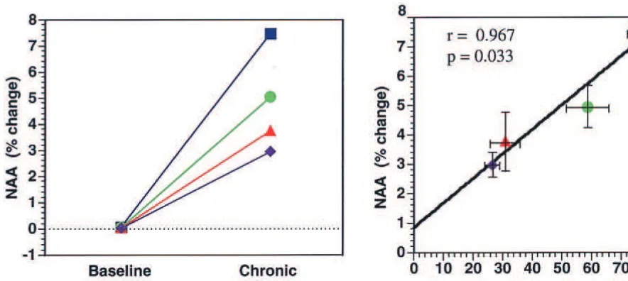
the study. Four weeks of lithium administration resulted in
a small (
;
5%) but significant increase of total brain NAA
concentration (RM-ANOVA,
F
5
5.528,
p
5
.0217; see
Figure 3). There was no correlation between lithium levels
and NAA increases. All brain regions investigated
dem-onstrated an increase in NAA over the course of the study
(Figure 4).
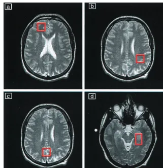
When regional brain NAA increases were examined
together with the regional voxel image segmentation data,
we noted a significant positive correlation between
in-creased NAA and the voxel gray matter content (
r
5
0.967,
p
5
.033; Figure 4), indicating that NAA increases
were occurring primarily in CNS gray matter. The
per-centage of the NAA increase with lithium administration
was not significantly different between the volunteer and
patient groups either on a total brain or regional basis. Of
the 19 completing both the acute and chronic scans, 14
(74%) had increased NAA levels, 1 (5%) showed no
change (
6
1%), and 4 (21%) showed a small decrease.
Examining data on a regional basis again together with
voxel gray matter content segmentation results, revealed
an interesting finding that the percentage of NAA increase
per voxel gray matter content was highest in the frontal
lobe region. Furthermore, examining the BD patient data
separately revealed an NAA increase per voxel gray
matter content two-fold higher in both the frontal and
temporal lobe regions compared to the parietal and
occip-ital regions; however, these regional findings should be
considered speculative as they failed to reach statistical
significance in this small sample.
Discussion
This study demonstrates for the first time that chronic Li
administration at therapeutic doses increases NAA
con-centration in the human brain in vivo. These findings
provide intriguing indirect support for the contention that,
similar to the findings observed in the rodent brain and in
human neuronal cells in culture, chronic lithium increases
neuronal viability/function in the human brain, and
sug-gests that some of Li’s long-term beneficial effects may be
mediated by neurotrophic/neuroprotective events.
Although the magnitude of the increase in brain NAA
with lithium administration over the course of this 4-week
trial is small (
;
5%), a recent in-depth study by Brooks
and colleagues (Brooks et al 1999) investigating the
reproducibility of in vivo proton MRS in repeated scans
convincingly documented that with 20 subjects, in vivo
proton MRS can reliably detect a mean change in NAA
concentration of 3% between two scans. The acquisition
protocol and data processing used in the Brooks study are
identical to the protocols used in this study, and we have
obtained similar or greater reliability measures in our own
Figure 2. Typical frontal lobe proton magnetic resonance spec-troscopy spectrum from a bipolar patient and example of quan-titative analysis method. The bottom trace is the original proton magnetic resonance spectrum. The spectral analysis software models the entire spectrum (next trace up), followed by a fit of the individual peaks (next trace up). The uppermost trace in each panel shows the residual component, which remains after the peak fitting procedure is completed. Ideally this should consist only of unstructured noise to achieve optimal quantitative results. MI, myo-inositol; Cho, choline compounds; Cr, creatine/phos-phocreatine; Glx, glutamate/glutamine/g-aminobutyric acid; NAA,N-acetyl-aspartate.
test-retest quality assurance measurements both in
phan-toms and in normal volunteers.
A number of studies have now shown that initial
abnormally low brain NAA measures may increase and
even normalize with remission of CNS symptoms in
disorders such as demyelinating disease, amyotrophic
lateral sclerosis, mitochondrial encephalopathies, and
hu-man immunodeficiency virus (HIV) dementia. (Davie et al
1994; De Stefano et al 1995a, 1995b; Ellis et al 1997;
Holshouser et al 1995; Kalra et al 1998; Pavlakis et al
1998; Salvan et al 1997; Takanashi et al 1997; Vion-Dury
et al 1995). In the case of temporal lobe epilepsy in which
proton MRS often reveals abnormal NAA measures in the
contralateral hippocampus, several studies have shown
normalization of these measures following successful
neurosurgical removal of the ipsilateral anterior temporal
lobe to control seizures (Cendes et al 1997; Da Silva et al,
unpublished data, 1999; Hugg et al 1996;). In the majority
of these studies, the magnitude of increase in NAA
reported is quite similar to that reported in our study.
There is now considerable literature support from a variety
of sources demonstrating significant reductions in regional
CNS volume and cell numbers (both neurons and glia)
associated with mood disorders. Thus, volumetric
neuroim-aging studies have demonstrated reduced basal ganglia
vol-ume and reduced temporal lobe volvol-ume (including the
hippocampus) in mood disorders (reviewed in Ketter et al
1997; Soares and Mann 1997). Within the frontal lobe,
volumetric neuroimaging studies have also consistently
shown reduced volumes in mood disorders. In particular,
recent volumetric MRI studies in familial bipolar depressives
and familial unipolar depressives have demonstrated
reduc-tions in the mean gray matter volume of approximately 40%
in the prefrontal cortex ventral to the genu of the corpus
callosum (Drevets et al 1997). Intriguingly, lithium-treated
subjects demonstrate smaller reductions in prefrontal cortical
volumes (personal communication from W. Drevets to H.K.
Manji, March 1999). In addition to the accumulating
neuro-imaging evidence, recent postmortem studies of the
prefron-tal cortex have also demonstrated reduced CNS volume and
cell numbers in mood disorders. Rajkowska (1997) has used
three-dimensional cell counting and morphological
tech-niques to demonstrate significantly reduced sizes and
densi-ties of both neurons and glia in several distinct areas in mood
disorder subjects (Rajkowska et al 1999). Intriguingly,
neu-ronal dimunition was especially pronounced in layer II of the
rostral orbitofrontal region (Rajkowska et al 1999), an area
where we have observed among the largest lithium-induced
increases in bcl-2 immunoreactivity (Chen et al 1999; Manji
et al 1999). Also in the prefrontal cortex, Ongur and
col-leagues (Ongur et al 1998) have recently reported a
histolog-ical study examining the cellular composition of area sg24
located in the subgenual prefrontal cortex. They found
striking reductions in glial cell numbers in patients with
familial major depression (24% reductions) and BD (41%
reductions), compared to controls. This is a particularly
striking finding, as it is consistent with neuroimaging
find-ings showing cortical volume loss in this same region on
volumetric magnetic resonance imaging in a similar
diagnos-tic group (Drevets et al 1997). Together, the preponderance
of the data from the neuroimaging studies and the growing
body of postmortem evidence presents a convincing case that
there is indeed a reduction in regional CNS volume,
accom-panied by cell atrophy/loss, in mood disorders; these
find-ings, along with the results of our study, raise the possibility
that chronic lithium may exert some of its long term benefits
via hitherto underappreciated neurotrophic/neuroprotective
effects.
Interestingly, recent findings from Duman and
associ-ates have demonstrated that chronic administration of a
variety of antidepressants increases the expression of
BDNF (brain derived neurotrophic factor), and its receptor
trkB in certain populations of neurons in the hippocampus
(Duman et al 1997). Moreover, Smith and associates
(Smith et al 1995) have also recently demonstrated that
antidepressants increase the locus coeruleus expression of
another neurotrophic factor, neurotrophin 3.
In conclusion, we report here the novel observation that
chronic lithium administration increases brain NAA levels.
Clearly a larger longitudinal study with a follow-up of BD
patients over a period of years will be required to determine
if lithium indeed has a long term
neurotrophic/neuroprotec-tive effect in BD patients. The increases in NAA levels, the
robust increases in bcl-2 levels, as well as the clear evidence
for neuroprotective effects in preclinical studies also suggest
that lithium may have utility in the long term treatment of
certain neurodegenerative disorders.
Supported in part by a NARSAD Young Investigator grant to GJM and grants from the Theodore and Vada Stanley Foundation, the State of Michigan (Joseph Young, Sr.), and the National Institutes of Health (NIMH MH-59107) to HKM and GJM.
The authors acknowledge the invaluable assistance of the Nursing and Research Staff of the Neuropsychiatric Research Unit, and Ms. Caroline Zajac Benitez for outstanding editorial assistance.
References
Adams JM, Cory S (1998): The Bcl-2 protein family: Arbiters of cell survival.Science281:1322–1326.
Baldessarini RJ, Tondo L, Hennen J (1999): Effects of lithium treatment and its discontinuation in bipolar manic-depressive disorders.J Clin Psychiatry Suppl60:77– 84.
Barker P, Breiter S, Soher B, Chatham J, Forder J, Samphilipo M, et al (1994): Quantitative proton spectroscopy of canine brain: In vivo and in vitro correlations.Magn Reson Med32:157–163. Barker PB, Soher BJ, Blackband SJ, Chatham JC, Mathews VP,
Ryan RN (1993): Quantitation of proton NMR spectra of the human brain using tissue water as an internal concentration reference.NMR Biomed6:89 –94.
Birken DL, Oldendorf WH (1989): N-acetyl-L-aspartic acid: A literature review of a compound prominent in 1H-NMR spec-troscopic studies of brain.Neurosci Biobehav Rev13:23–31. Brooks WM, Friedman SD, Stidley CA (1999): Reproducibility
of 1H-MRS in vivo.Magn Reson Med41:193–197. Bruckheimer EM, Cho SH, Sarkiss M, Herrmann J, McDonnell
TJ (1998): The Bcl-2 gene family and apoptosis. Adv Bio-chem Eng Biotechnol62:75–105.
Cendes F, Andermann F, Dubeau F, Mathews PM, Arnold DL (1997): Normalization of neuronal metabolic dysfunction after surgery for temporal lobe epilepsy. Evidence from proton MR spectroscopic imaging.Neurology49:1525–1533.
Chen DF, Schneider GE, Martinou JC, Tonegawa S (1997): Bcl-2 promotes regeneration of severed axons in mammalian CNS.Nature385:434 – 439.
Chen G, Zeng WZ, Jiang L, Yuan PX, Zhao J, Manji HK(1999): The mood stabilizing agents lithium and valproate robustly increase the expression of the neuroprotective protein bcl-2 in the CNS.J Neurochem72:879 – 882.
Chen RW, Chuang DM (1999): Long term lithium treatment suppresses p53 and Bax expression but increases bcl-2 expression. A prominent role in neuroprotection against excitotoxicity.J Biol Chem274:6039 – 6042.
Christiansen P, Henriksen O, Stubgaard M, Gideon P, Larsson H (1993): In vivo quantification of brain metabolites by 1H-MRS using water as an internal standard. Magn Reson Imaging11:107–118.
Davie CA, Hawkins CP, Barker GJ, Brennan A, Tofts PS, Miller DH, et al (1994): Serial proton magnetic resonance spectros-copy in acute multiple sclerosis lesions.Brain117:49 –58. deBeer R, van den Boogaart A, van Ormondt D, Pijnappel WW,
den Hollander JA, Marien AJ, et al (1992): Application of time-domain fitting in the quantification of in vivo 1H spectroscopic imaging data sets.NMR Biomed5:171–178. De Stefano N, Mathews PM, Arnold DL (1995a): Reversible
decreases in N-acetylaspartate after acute brain injury.Magn Reson Med34:721–727.
De Stefano N, Matthews PM, Ford B, Genge A, Karpati G, Arnold DL (1995b): Short-term dichloroacetate treatment improves indices of cerebral metabolism in patients with mitochondrial disorders.Neurology45:1193–1198.
Drevets WC, Price JL, Simpson JR Jr, Todd RD, Reich T, Vannier M, et al (1997): Subgenual prefrontal cortex abnor-malities in mood disorders.Nature386:824 – 827.
Duman RS, Heninger GR, Nestler EJ (1997): A molecular and cellular theory of depression.Arch Gen Psychiatry54:597– 606. Ellis CM, Lemmens G, Williams SC, Simmons A, Dawson J, Leigh PN, et al (1997): Changes in putamen N-acetylaspartate and choline ratios in untreated and levodopa-treated Parkin-son’s disease: A proton magnetic resonance spectroscopy study.Neurology49:438 – 444.
Frahm J, Bruhn H, Gyngell ML, Merboldt KD, Hanicke W, Sauter R (1989): Localized high-resolution proton NMR spectroscopy using stimulated echoes: Initial applications to human brain in vivo.Magn Reson Med9:79 –93.
Frahm J, Merboldt KD, Hanicke W (1987): Localized proton spectroscopy using stimulated echoes.J Magn Reson72:502– 508.
Frahm J, Michaelis T, Merboldt KD, Bruhn H, Gyngell ML, Hanicke W (1990): Improvements in localized 1H NMR spectroscopy of human brain: Water suppression, short echo times and 1 ml resolution.J Magn Reson90:464 – 473. Goodwin FK, Jamison KR (1990): Manic-Depressive Illness.
New York: Oxford University Press.
Hamilton M (1967): Development of a rating scale for primary depressive illness.Br J Soc Clin Psychol6:278 –296. Hennig J, Pfister H, Ernst T, Ott D (1992): Direct absolute
quantification of metabolites in the human brain with in vivo localized proton spectroscopyNMR Biomed5:193–199. Hetherington HP, Pan JW, Mason GF, Adams D, Vaughn MJ,
imaging of human brain at 4.IT using image segmentation. Magn Reson Med36:21–29.
Holshouser BA, Komu M, Moller HE, Zijlmans J, Kolem H, Hinshaw DB Jr (1995): Localized proton NMR spectroscopy in the striatum of patients with idiopathic Parkinson’s disease: A multicenter pilot study.Magn Reson Med33:589 –594. Hugg JW, Kuzniecky RI, Gilliam FG, Morawetz RB, Faught RE,
Hetherington HP (1996): Normalization of contralateral meta-bolic function following temporal lobectomy demonstrated by
1H magnetic resonance spectroscopic imaging. Ann Neurol
40:236 –239.
Jope RS (1999): Anti-bipolar therapy: Mechanism of action of lithium.Mol Psychiatry4:117–128.
Kalra S, Cashman NR, Genge A, Arnold DL (1998): Recovery of N-acetylaspartate in corticomotor neurons of patients with ALS after riluzole therapy.Neuroreport9:1757–1761. Ketter TA, George MS, Kimbrell TA, Willis MW, Benson BE,
Post RM (1997): Neuroanatomical models and brain imaging studies in bipolar disorder: Biological models and their Clinical Application. In: Joffe RT, Young LT, editors. Bipo-lar Disorder: Biological Models and Their Clinical Applica-tion.New York: Marcel Dekker, 179 –218.
Klose U (1990): In vivo proton spectroscopy in presence of eddy currents.Magn Reson Med14:26 –30.
Koller KJ, Zaczek R, Coyle JT (1984): N-acetyl-aspartyl-gluta-mate: Regional levels in rat brain and the effects of brain lesions as determined by a new HPLC method.J Neurochem 43:1136 –1142.
Kostic V, Jackson-Lewis V, de Bilbao F, Dubois-Dauphin M, Przedborski S (1997): Bcl-2: Prolonging life in a transgenic mouse model of familial amyotrophic lateral sclerosis. Sci-ence277:559 –562.
Kreis R, Ernst T, Ross B (1993): Absolute quantitation of water and metabolites in the human brain. II metabolite concentra-tions.J Magn ResonB102:9 –19.
Lawrence MS, Ho DY, Sun GH, Steinberg GK, Sapolsky RM (1996): Overexpression of Bcl-2 with herpes simplex virus vectors protects CNS neurons against neurological insults in vitro and in vivo.J Neurosci16:486 – 496.
Manji HK, Moore GJ, Chen G (1999): Lithium at 50: Have the neuroprotective effects of this unique cation been over-looked?Biol Psychiatry46:929 –940.
Manji HK, Potter WZ, Lenox RH (1995): Signal transduction pathways. Molecular targets for lithium’s actions.Arch Gen Psychiatry52:531–543.
Merry DE, Korsmeyer SJ (1997): Bcl-2 gene family in the nervous system.Annu Rev Neurosci20:245–267.
Michaelis T, Merboldt KD, Bruhn H, Hanicke W, Frahm J (1993): Absolute concentrations of metabolites in the adult human brain in vivo: Quantification of localized proton MR spectra.Radiology187:219 –227.
Moore GJ, Seraji-Bozorgzad N, Wilds IB, Manji HK (in press): Assesment of MRS voxel placement reliability using a simple semi-automated voxel segmentation approach.Med Phys. Narayana PA, Fotedar LK, Jackson EF, Bohaw ID, Butler IJ,
Wolinsky JS (1989): Regional in vivo proton magnetic resonance spectroscopy of brain.J Magn Reson83:44 –52. Nonaka S, Chuang DM (1998): Neuroprotective effects of
chronic lithium on focal cerebral ischemia in rats. Neurore-port9:2081–2084.
Nonaka S, Hough CJ, Chuang DM (1998a): Chronic lithium treatment robustly protects neurons in the central nervous system against excitotoxicity by inhibitingN-methyl-D -aspar-tate receptor-mediated calcium influx.Proc Natl Acad Sci U S A95:2642–2647.
Nonaka S, Katsube N, Chuang DM (1998b): Lithium protects rat cerebellar granule cells against apoptosis induced by anticon-vulsants, phenytoin and carbamazepine. J Pharmacol Exp Ther286:539 –547.
Ongur D, Drevets WC, Price JL (1998): Glial reduction in the subgenual prefrontal cortex in mood disorders. Proc Natl Acad Sci U S A95:13290 –13295.
Pavlakis SG, Lu D, Frank Y, Wiznia A, Eidelberg D, Barnett T, et al (1998): Brain lactate and N-acetylaspartate in pediatric AIDS encephalopathy.AJNR Am J Neuroradiol19:383–385. Rajkowska G (1997): Morphometric methods for studying the prefrontal cortex in suicide victims and psychiatric patients. Ann N Y Acad Sci836:253–268.
Rajkowska G, Miguel-Hidalgo JJ, Wei J, Dilley G, Pittman SD, Meltzer HY, et al (1999): Morphometric evidence for neuro-nal and glial prefrontal cell pathology in major depression. Biol Psychiatry45:1085–1098.
Reifman A, Wyatt RJ (1980): Lithium: A brake in the rising cost of mental illness.Arch Gen Psychiatry37:385–388. Salvan AM, Vion-Dury J, Confort-Gouny S, Nicoli F,
Lamou-reux S, Cozzone PJ (1997): Brain proton magnetic resonance spectroscopy in HIV-related encephalopathy: Identification of evolving metabolic patterns in relation to dementia and therapy.AIDS Res Hum Retroviruses13:1055–1066. Smith MA, Makino S, Altemus M, Michelson D, Hong SK,
Kvetnansky R, et al (1995): Stress and antidepressants dif-ferentially regulate neurotrophin 3 mRNA expression in the locus coeruleus.Proc Natl Acad Sci U S A92:8788 – 8792. Soares JC, Mann JJ (1997): The anatomy of mood disorders—
review of structural neuroimaging studies.Biol Psychiatry41: 86 –106.
Soher BJ, Hurd RE, Sailasuta N, Barker PB (1996): Quantitation of automated single-voxel proton MRS using cerebral water at an internal reference.Magn Reson Med36:335–339. Takanashi J, Sugita K, Ishii M, Aoyagi M, Niimi H (1997):
Longitudinal MR imaging and proton MR spectroscopy in herpes simplex encephalitis.J Neurol Sci149:99 –102. Thulborn KR, Ackerman JH (1983): Absolute molar
concentra-tions by NMR in inhomogeneous B1. A scheme for analysis
of in vivo metabolites.J Magn Reson55:357–371.
Tsai G, Coyle JT (1995): N-acetylaspartate in neuropsychiatric disorders.Prog Neurobiol46:531–540.
van den Boogaart A, Ala-Korpela M, Jokisaari J, Griffiths JR (1994): Time and frequency domain analysis of NMR data compared: an application to 1D 1H spectra of lipoproteins. Magn Reson Med31:347–358.
Vion-Dury J, Nicoli F, Salvan AM, Confort-Gouny S, Dhiver C, Cozzone PJ (1995): Reversal of brain metabolic alterations with zidovudine detected by proton localized magnetic reso-nance spectroscopy.Lancet345:60 – 61
![Figure 3. Total brain N-acetyl-aspartate (NAA) concentration isplotted for the two time points (baseline and chronic lithiumtreatment [�4 weeks])](https://thumb-ap.123doks.com/thumbv2/123dok/3144300.1383706/5.612.68.288.79.330/figure-total-aspartate-concentration-isplotted-baseline-chronic-lithiumtreatment.webp)