RENAL REGULATION OF ACID-BASE BALANCE
Bruce M. Koeppen
Depa rtm ents of Medicine a nd Physiology, University of Connecticut School of Medicine, Fa rm ington, Connecticut 06030
T
his article reviews the role of the kidneys in the regulation of acid-base balance. It is intended as a guide for those who teach this aspect of renal physiology to health professions students. An approach is described, which begins with an overview of acid-base balance and then proceeds to describe the details of renal H1 transport and the important role the production and excretion of NH41plays in the abilityof the kidneys to generate new HCO32. In the overview, the role of the kidneys in
acid-base balance is placed in context for the student by examining the impact of diet and cellular metabolism on acid-base balance. Also, the interactions between the kidneys and lungs to maintain extracellular HCO32concentration within a narrow range are described.
This is followed by a detailed look at the cellular mechanisms of H1secretion along the nephron, how these mechanisms are regulated, and how they result in the reabsorption of the filtered load of HCO32. Finally, the important role of NH41production and excretion
in the generation of new HCO32is reviewed and highlighted.
AM. J. PHYSIOL. 275 (ADV. PHYSIOL. EDUC. 20): S132–S141, 1998.
Key words:urine acidification; renal ammoniagenesis
Our knowledge of renal acid-base physiology has progressed over the years as we have been able to study and understand the mechanisms of H1 and
HCO32 transport in increasing detail (i.e., from the
level of specific nephron segments to single renal tubule cells, individual cell membranes, and, most recently, the membrane transporters themselves). In general, the teaching of renal acid-base physiology has paralleled this progression of knowledge and has focused on the mechanisms and regulation of H1
secretion in the various portions of the nephron.
Typically, students are taught that the kidneys reab-sorb the filtered load of HCO32and in addition excrete
acid by titrating urinary buffers, with both processes being the result of specific H1secretory mechanisms.
Traditionally, and for simplicity of presentation to students, the processes of HCO32 reabsorption and
acid excretion are ascribed to different portions of the nephron. Accordingly, the proximal tubule is the
primary site in which the filtered load of HCO32 is
reabsorbed, and the distal portions of the nephron are involved in acid excretion. Acid excretion assumes central importance in this scheme because it results in the generation of ‘‘new HCO32,’’ which is returned to
the body to replenish that lost during the titration of metabolically produced acids. The principal urinary buffers used for acid excretion are usually said to be phosphate and NH3.
Although much of this simplified scheme of renal acid-base physiology is essentially correct, our under-standing of the mechanisms involved in the produc-tion and excreproduc-tion of NH41have changed dramatically
in recent years, and it is now clear that NH3 cannot
simply be viewed as a urinary buffer. Consequently, the teaching of renal acid-base physiology must empha-size our new understanding of the role of NH3/NH41in
function of other organs that also influence acid-base balance (e.g., lungs and liver).
In this article the role of the kidneys in the mainte-nance of acid-base balance is reviewed from the perspective of teaching this material to health profes-sions students. First, an overview of the role of the kidneys in acid-base balance is presented. The cellular mechanisms of H1 secretion along the nephron are
then briefly reviewed, with recent discoveries from molecular biological studies highlighted. This is fol-lowed by a description of our current understanding of renal NH41 production and excretion. Finally, the
integrated function of the kidneys and lungs in the setting of acid-base disturbances (i.e., compensation) is considered.
OVERVIEW
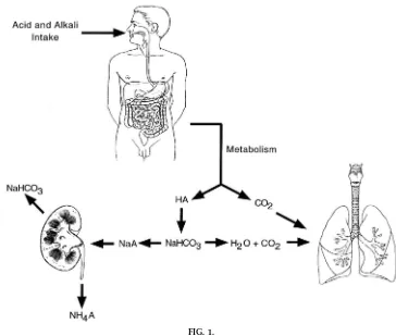
Figure 1 provides a general overview of acid-base balance and the role of the kidneys. Also depicted is the role of the lungs in the excretion of metabolically
produced CO2. The interrelationships between H1,
CO2, and HCO32are central to understanding acid-base
balance and reflect the physiological importance of the CO2/HCO32buffer system.
CO2 1H2O&H2CO3&H11HCO3
2 (1)
The CO2/HCO32buffer system is not only of
quantita-tive importance in acid-base balance [extracellular fluid (ECF) contains 350–400 meq of HCO32], but the
H1 concentration ([H1])/pH of the body fluids is
influenced by both PCO2 and HCO32 concentration
([HCO32]). Thus acid-base balance can be effected by
both the lungs and the kidneys, the lungs through control of PCO2 and the kidneys through control of
[HCO32]. This dual impact of CO
2and [HCO3
2] on pH
has led to the distinction between CO2-derived or
‘‘volatile acid’’ and ‘‘nonvolatile acid’’ such as lactic acid. Although this distinction is in widespread use, it is important to recognize that CO2itself is not an acid,
and under normal conditions the production and
FIG. 1.
excretion of CO2 does not impact acid-base balance
(3). However, as indicated byrea ction 1, retention of CO2 (production . excretion) will produce an
in-crease in [H1] and thus the development of acidosis.
Conversely, if CO2 production is less than excretion,
there will be a decrease in [H1], and alkalosis results.
Acid-base disorders resulting from primary alterations in the PCO2are termed ‘‘respiratory’’ disorders.
As depicted in Fig. 1, nonvolatile acids are buffered by HCO32 (other non-HCO32 buffers are also involved in
this process). Thus, if nonvolatile acid production exceeds the excretion of acid from the body, [HCO32]
decreases and [H1] increases (see rea ction 1), and
acidosis results. Conversely, if nonvolatile acid produc-tion is less than the excreproduc-tion of acid from the body, then [HCO32] increases and [H1] decreases, and
alkalo-sis results. Acid-base disorders resulting from nonvola-tile acid or alkali are termed ‘‘metabolic’’ disorders. They are readily detected by the associated change in [HCO32].
Each day, acid and alkali are ingested in the diet. In addition, c ellular metabolism produc es ac id-base equivalents. The majority of energy (i.e., the largest source of calories) is derived from the metabolism of dietary carbohydrates and fats. When tissue perfusion is adequate, and insulin is present at normal levels, cellular metabolism of carbohydrates and fats results in the production of CO2and H2O. On a typical diet,
,15–20 mol of CO2are produced. Normally, this large
quantity of CO2 is effectively eliminated by the lungs,
and there is no impact of this metabolically derived CO2on whole body acid-base status.
With inadequate tissue perfusion, hypoxia, or in the absence of insulin, the cellular metabolism of carbohy-drates and fats does not yield CO2and H2O but instead
results in the production of significant quantities of nonvolatile acids such as lactic acid and ketoacids. With restoration of tissue perfusion (i.e., delivery of adequate amounts of O2) or treatment with insulin,
many of these nonvolatile acids are then further metabolized to CO2 and H2O. In this later process,
much of the HCO32lost during titration of the
nonvola-tile acids is regenerated.
Cellular metabolism of other dietary constituents also impacts acid-base balance. Amino acid metabolism
results in the addition of either acid or alkali to the body. For example, the sulfur-containing (e.g., methio-nine) and cationic (e.g., argimethio-nine) amino acids result in acid production on metabolism, whereas alkali results from the metabolism of anionic (e.g., aspartate) amino acids. Given the mix of amino acids in the typical diet, acid production exceeds alkali production. Organic anions (e.g., citrate), when metabolized, result in the generation of alkali.
Several points must be considered when one is trying to determine the impact of nonvolatile acid and alkali on acid-base balance.
1) The source of the nonvolatile acid and alkali is multifactorial and not limited to cellular metabolism. Direct ingestion of acid or alkali can and does occur and, depending on diet, can have a significant impact on acid-base balance.
2) Acid and alkali can be and are lost from the body. For example, vomiting results in the loss of gastric acid, which from an acid-base perspective is equiva-lent to adding alkali to the body. As a result, vomiting can result in the development of a metabolic alkalosis. Conversely, diarrhea results in the loss of alkali (equiva-lent to addition of acid), and can result in metabolic acidosis.
3) The impact on acid-base balance of nonvolatile acid or alkali derived from cellular metabolism is highly variable and critically dependent on diet. The inges-tion of a vegetarian diet, for example, results in a much reduced acid load to the body, and in some instances may even impart a net alkali load.
Most textbooks state that the direct intake of acid and alkali in a typical diet, the normal loss of some HCO32
in the feces, and the production of nonvolatile acid and alkali from metabolism result in the net addition of acid to the body. Collectively, these processes are referred to as nonvolatile acid production and as-cribed a value of ,1 meq.kg body wt21.day21 (70
there is net addition of this amount of nonvolatile acid to the body on a daily basis.
Nonvolatile acids are quickly buffered throughout the body. This buffering occurs in both the intracellular fluid (ICF) and the ECF. As already noted, HCO32is a
major ECF buffer, and in this titration process it is consumed producing the sodium salts of the nonvola-tile acids. To maintain acid-base balance, the kidneys must excrete the anions of the nonvolatile acids and replenish the HCO32lost during the titration process.
This later process, frequently referred to as ‘‘new HCO32 generation,’’ results from the excretion of
titratable acid (i.e., the excretion of H1 with urine
buffers) and from the production and excretion of NH41. In addition, the kidneys must reabsorb the
filtered load of HCO32to prevent its loss in the urine,
because any lost in the urine would be equivalent to the addition of acid to the body. This overall process is termed ‘‘net acid excretion’’ (NAE) and is quantitated as
NAE5[(UNH413V)1(UTA3V)2(UHCO323V)] (2)
where U is the urine concentration, V is the urine flow rate, UNH413V is the amount of NH4
1excreted, U
TA3V
is the amounted of titratable acid excreted, and UHCO32 3 V is the amount of HCO3
2 excreted. To
maintain acid-base balance, net acid excretion must equal nonvolatile acid production. If nonvolatile acid production exceeds net acid excretion, metabolic acidosis results (serum [HCO32] and pH decrease).
Conversely, if nonvolatile acid production is less than net acid excretion metabolic alkalosis results (serum [HCO32] and pH increase).
Several important points regarding net acid excretion by the kidneys require comment and emphasis.
1) NH41excretion, titratable acid excretion, and HCO32
reabsorption all result from H1 secretion along the
nephron.
2) Very little acid is excreted by the kidneys as ‘‘free H1.’’ Even with urine of pH 4.0, only 0.1 meq/l of H1
is excreted in this form.
3) Titratable acid represents H1excreted with urinary
buffers, with the principal urinary buffer being phos-phate.
4) When urine pH is ,6.5, very little HCO32 is
excreted, and therefore NAE is simply equal to the sum of titratable acid and NH41excretion.
5) The production and excretion of NH41is critically
important in this process, because it is regulated by the kidneys in response to alterations in acid-base balance. The role of NH41excretion in renal acid-base
physiology is emphasized in Fig. 1, in which the anions of the nonvolatile acids are shown as being excreted with NH41. Importantly, for every NH41
ex-creted in the urine an HCO32is returned to the body.
H1TRANSPORT ALONG NEPHRON
H1 secretion by the cells of the nephron serves to
reabsorb the filtered load of HCO32, lower the pH of
the urine, titrate urinary buffers, and cause the excre-tion of NH41. Of these processes, the reabsorption of
the filtered load of HCO32 is quantitatively the most
important, because the filtered load of HCO32 is
,4,500 meq/day, whereas the amount of H1required
for NH41 excretion plus the amount excreted with
urine buffers is generally,100 meq/day.
Figure 2 summarizes H1 secretion (HCO
3
2
reabsorp-tion) along the nephron. The proximal tubule reab-sorbs ,80% of the filtered load of HCO3
2, and an
additional 15% is reabsorbed by the thick ascending limb of Henle’s loop. The cellular mechanisms in-volved are essentially the same in these segments. H1
secretion occurs by two apical membrane transport-ers, Na1/H1antiporter and H1-ATPase. Of these
trans-porters the Na1/H1 antiporter is the predominant
pathway for H1 secretion. Thus H1 secretion is
dependent on the lumen-to-cell Na1gradient. Because
of this coupling, factors that regulate Na1transport in
these segments will secondarily effect H1 secretion
(see below).
Recent studies on the molecular biology of Na1/H1
antiporters found that the Na1/H1exchanger 3 (NHE-3)
isoform is present in the apical membrane of both the proximal tubule and thick ascending limb cells and is the physiologically important antiporter for H1
secre-tion in these segments (14). The H1-ATPase provides a
parallel pathway for H1 secretion across the apical
intercalated cells of the collecting duct (Refs. 4, 5; see below).
Carbonic anhydrase plays an important role in H1
secretion by the cells of the proximal tubule and thick ascending limb. It is found in the cytoplasm of these cells, in which it catalyzes the production of H1and
HCO32from CO2and H2O. In the proximal tubule, but
not the thick ascending limb, carbonic anhydrase is also found in the apical membrane. The isoforms for the cytoplasmic (CA-II) and apical membrane enzymes (CA-IV) differ (12).
HCO32generated in the cell from the hydration of CO2
exits the cell across the basolateral membrane by a symporter that couples the movement of 3 HCO32
with 1 Na1. An additional portion of basolateral HCO
3
2
exit may also occur in exchange for Cl2.
The distal tubule and collecting duct reabsorb the portion of the filtered load of HCO32 that escapes
reabsorption by the proximal tubule and thick ascend-ing limb of Henle’s loop (,5% of the filtered load).
HCO32is reabsorbed as a result of H1secretion by the
intercalated cells found in this region of the nephron. H1secretion occurs by two transporters, H1-ATPase
and H1-K1-ATPase. The H1-ATPase, as already noted,
is a distinct isoform from that found in the proximal tubule. The H1-K1-ATPase is similar to, but distinct
from, the isoform found in the gastric parietal cells (15). As in the proximal tubule and thick ascending limb cells, CA-II catalyzes the intracellular production of H1 and HCO
3
2. The predominant mechanism for
HCO32 exit across the basolateral membrane is via a
Cl2/HCO
3
2 antiporter similar to that found in red
blood cells (i.e., Band-3).
In addition to the H1-secreting intercalated cell, there
is a second intercalated cell subtype that secretes HCO32(see Fig. 2). Because of nonvolatile acid
produc-tion, and thus the need to excrete acid, H1secretion
predominates in the collecting duct. However, HCO32 FIG. 2.
Summary of cellular mechanisms of H1secr etion (HCO
3
2r eabsorption) along nephr on. Appr ox imately
80% of filter ed load of HCO32is r eabsorbed by pr ox imal tubule and 15% by thick ascending limb of Henle’s loop. Collecting duct contains both H1-secr eting and HCO
3
secretion is important in states of metabolic alkalosis, when renal HCO32excretion must be enhanced.
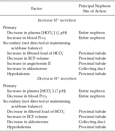
H1secretion by the cells of the nephron is regulated
by a number of factors (see Table 1). From a cellular perspective, an important factor regulating the secre-tion of H1across the apical membrane is the
cell-to-tubular fluid gradient for H1. This gradient depends on
the pH of the tubular fluid relative to the pH within the tubular cells. Acidosis, whether of metabolic (decreased [HCO32] and pH) or respiratory (increased
PCO2) origin, decreases intracellular pH, creating a more favorable cell-to-tubular fluid H1 gradient, and
thus stimulates H1secretion along the entire nephron.
Alternatively, metabolic (increased [HCO32] and pH)
and respiratory (increased PCO2) alkalosis inhibit H1
secretion by their effect to increase intracellular pH. Although changes in intracellular pH can directly influence the cell-to-tubular fluid H1 gradient and
thereby H1secretion across the apical membrane of
the cell, there is also evidence that changes in intracel-lular pH, perhaps mediated by other intracelintracel-lular messengers, also alter the activity and expression of key H1 and HCO
3
2 transporters in the cell (1, 4, 5,
9–11, 13, 14). For example, H1-secreting intercalated
cells in the collecting duct respond to acidosis by exocytotically inserting more H1-ATPase into the
apical membrane (4, 5, 11, 13). Also, the abundance of Na1/H1 antiporter in proximal tubule cells is
in-creased in chronic metabolic acidosis (1, 9, 10, 14).
Other factors may alter renal H1excretion, but their
influence is not directed at the maintenance of acid-base balance (see Table 1). Because, as noted, H1
secretion is linked to Na1 reabsorption in both the
proximal tubule and thick ascending limb of Henle’s loop, factors that are primarily related to Na1
reabsorp-tion also influence renal H1secretion. These include
alterations in the filtered load (i.e., glomerulotubular balance) and changes in ECF volume. The effect of alterations in ECF volume are mediated by the renin-angiotensin-aldosterone system, with angiotensin II acting on the cells of the proximal tubule to stimulate the Na1/H1antiporter and aldosterone acting on the
intercalated cells of the collecting duct to stimulate the H1-ATPase (7, 8, 10). Alterations in peritubular
Starling forces that occur with changes in ECF volume also are involved in enhancing proximal tubule fluid (and HCO32) reabsorption in volume depletion and
decreasing reabsorption during volume expansion.
PRODUCTION AND EXCRETION OF NH41
Although the reabsorption of the filtered load of HCO32 is quantitatively an important process, simply
preventing the loss of HCO32 in the urine does not
replenish the HCO32lost during the titration of
non-volatile acid. This later process is accomplished through the excretion of H1with urine buffers
(titrat-able acid) and by the production and excretion of NH41. It should be emphasized that the availability and
thus excretion of urinary buffers is not regulated to meet the requirements for acid-base balance. For example, the most abundant urinary buffer is phos-phate, the excretion of which is regulated not to effect acid-base balance but in response to phosphate bal-ance needs. In contrast, NH41production and
excre-tion by the kidneys is regulated to effect acid-base balance. Thus understanding how the kidneys pro-duce and excrete NH41is critical to understanding the
role of the kidneys in acid-base balance.
Traditionally, the excretion of NH41 has been taught
from the perspective of urinary buffering. Specifically, NH3 was viewed as a urinary buffer that could accept
TABLE 1
Factors influencing H1secr etion by the nephr on
Factor Principal Nephron
Site of Action
Increa se H1secretion
Primary
Decrease in plasma [HCO32] (<pH) Entire nephron
Increase in blood PCO2 Entire nephron
Secondary (not directed at maintaining acid-base balance)
Increase in filtered load of HCO32 Proximal tubule
Decrease in ECF volume Proximal tubule Increase in angiotensin II Proximal tubule Increase in aldosterone Collecting duct
Hypokalemia Proximal tubule
Decrea se H1secretion
Primary
Increase in plasma [HCO32] (>pH) Entire nephron
Decrease in blood PCO2 Entire nephron
Secondary (not directed at maintaining acid-base balance)
Decrease in filtered load of HCO32 Proximal tubule
Increase in ECF volume Proximal tubule Decrease in aldosterone Collecting duct
H1. HCO
3
2 was generated in this process from the
hydration of CO2within the intercalated cell (i.e., the
H1was secreted into the tubular fluid and the HCO
3
2
returned to the blood). However, new knowledge regarding the production and excretion of NH41makes
it clear that NH3cannot be viewed simply as a urinary
buffer (2).
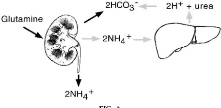
The essential features of NH41production and
excre-tion are summarized in Fig. 3. Glutamine is metabo-lized by the kidneys to produce 2NH41 and 2HCO32.
The NH41is excreted in the urine, and the HCO32 is
returned to the body to replenish that which was lost earlier during the titration of nonvolatile acids. For every equivalent of NH41 excreted in the urine, an
equivalent of HCO32is returned to the body. Figure 3
also illustrates what happens if the kidneys are unable to excrete NH41. When this occurs, NH41returns to the
liver, where it is metabolized to urea. The net result of this process is that 2 NH41are converted to urea with
the production of 2 H1. These 2 H1are then titrated
by 2 HCO32, thus negating the efforts of the kidneys to
generate HCO32from the metabolism of glutamine.
The details of NH41 production and excretion are
summarized in Fig. 4. The cells of the proximal tubule are the site of ammoniagenesis. Here glutamine is metabolized to 2 NH41and the tricarboxylic acid cycle
intermediate 2-oxoglutarate5, which is then further
metabolized to 2HCO32(2). The HCO
3
2is returned to
the body, and the NH41is secreted by the cell into the
tubular fluid.
NH41secretion by the proximal tubule cells occurs by
two mechanisms. The majority is exchanged for Na1
via the Na1/H1antiporter (NH
4
1substituting for H1).
An additional small portion leaves the cell as NH3and
is reprotonated in the lumen. At this point the process of generating HCO32is complete (i.e., NH
4
1 has been
secreted into the tubular fluid and HCO32returned to
the blood). However, NH41 must still be eliminated
from the body, because as already noted, if any NH41is
reabsorbed by the nephron it will be metabolized to urea by the liver and in that process consume the HCO32 produced from ammoniagenesis (see Fig. 3).
Unfortunately, significant amounts of NH41 are
reab-sorbed by the thick ascending limb of Henle’s loop. Unlike the other portions of the nephron, which are highly permeable to NH3 but not NH41, the thick
ascending limb has the opposite characteristics (low permeability to NH3 and high permeability to NH41).
The reabsorption of NH41by the thick ascending limb
occurs via transcellular and paracellular routes. Trans-cellular reabsorption involves uptake into the cell across the apical membrane via the Na1-K1-2Cl2
symporter (NH41substituting for K1) and movement
across the basolateral membrane via K1 channels.
Paracellular reabsorption of NH41 is driven by the
lumen positive potential difference. The reabsorbed NH41 accumulates in the interstitial fluid of the
me-dulla by the processes of countercurrent multiplica-tion and countercurrent exchange. As a result, this accumulated NH41, which is in chemical equilibrium
with NH3 (pKa59), is available for secretion into the
tubular fluid by the cells of the collecting duct.
The secretion of NH41by the collecting duct is indirect
and involves nonionic diffusion of NH3 and diffusion
trapping of the NH41 in the acidic tubular fluid. As
indicated in Fig. 4, the secretion of NH41 by the
collecting duct is critically dependent on H1
secre-tion. If H1secretion is impaired in any way, reduced
amounts of NH41will also be secreted and more NH
4
1
will be returned to the body. It should be emphasized that even though the secretion of NH41by the
collect-ing duct requires H1secretion, no additional HCO
3
2is
generated in this process (i.e., the HCO32generated in
the intercalated cell titrates the H1generated in the FIG. 3.
Overview of NH41 pr oduction and ex cr etion by kid-neys. Glutamine is metabolized by kidneys to pr oduce 2 NH41and 2 HCO32. NH41is ex cr eted in urine, and HCO32 is r etur ned to body (‘‘new HCO32’’). Shaded arr ows illustrate what happens to NH41if it is not ex cr eted in urine. When this occurs 2 NH41ar e converted to ur ea by liver, in a pr ocess yielding 2 H1. This pr oduction of
interstitial fluid from the dissociation of NH41to NH
3).
All the HCO32 derived from ammoniagenesis was
generated during the process of glutamine metabo-lism in the proximal tubule. The H1 secreted by the
collecting duct in the process of NH41secretion simply
prevents the NH41from being returned to the liver and
converted to urea (see Fig. 3).
Importantly, NH41 production and excretion is
regu-lated by the kidneys. With acidosis ammoniagenesis is enhanced, and as already noted, H1secretion by the
nephron is increased. Thus more NH41is excreted, and
more HCO32 is generated and returned to the body.
This response to acidosis, frequently termed renal compensation (see COMPENSATION DURING ACID-BASE DIS
-TURBANCES), involves upregulation of the enzymes
involved in proximal tubule glutamine metabolism. Therefore, hours to days are required for the full response.
Assessing NH41 excretion by the kidneys is done
indirectly, because assays of urine NH41 are not
rou-tinely available. Consider, for example, the situation of metabolic acidosis. In the setting of metabolic acido-sis, the appropriate renal response is to increase net acid excretion. Accordingly, little or no HCO32 will
appear in the urine, the urine will be acidic, and NH41
excretion will be increased. To assess this, and espe-cially the amount of NH41 excreted, the ‘‘urinary net
charge’’ or ‘‘urine anion gap’’ can be calculated by
FIG. 4.
NH41pr oduction and ex cr etion. Two NH41and two HCO32 ar e pr oduced in pr ox imal tubule fr om glutamine. HCO32 is r etur ned to body as new HCO32, and NH41is secr eted into tubular fluid. A significant amount of NH41is r eabsorbed by thick ascending limb, in which it accumulates in interstitial fluid of r enal medulla. Some of this NH4
1is secr eted into tubular fluid
by collecting duct. This secr etion pr ocess is indir ect and involves nonionic diffusion and diffusion trapping. Shaded arr ows emphasize pathway for NH4
measuring the urinary concentrations of Na1, K1, and
Cl2(6)
urine anion gap5[Na1]1[K1]2[Cl2] (3)
The concept of urine anion gap assumes that the major cations in the urine are Na1, K1, and NH
4
1, and
that the major anion is Cl2 (with urine pH , 6.5,
virtually no HCO32is present). As a result, the urine
anion gap will yield a negative value when adequate amounts of NH41 are being excreted. Indeed, the
absence of a urine anion gap or the existence of a positive value indicates a renal defect in NH41
produc-tion and excreproduc-tion.
COMPENSATION DURING ACID-BASE DISTURBANCES
When there is a disturbance of acid-base balance the body uses several mechanisms to minimize the impact on the pH of the body fluids. These mechanisms are compensatory in that they do not correct the underly-ing disorder and include bufferunderly-ing (intracellular and extracellular), respiratory compensation, and renal compensation.
Buffering is the first line of defense because of the rapidity at which it occurs. Extracellular buffering is virtually instantaneous and involves titration of H1by
HCO32, phosphate, and serum proteins (histidine
groups). Intracellular buffering can take several min-utes and utilizes the same buffering species. Although it is difficult to estimate,,50% of nonvolatile acid and
70% of nonvolatile alkali is buffered in the extracellu-lar fluid and the remainder is buffered inside cells.
With metabolic acidosis or alkalosis there is respira-tory compensation. This compensarespira-tory response is mediated by changes in ventilatory rate, which in turn occur in response to H1. With metabolic acidosis
there is an increase in the ventilatory rate, which drives down PCO2. Given the mechanics of breathing, PCO2can be reduced to 10–15 mmHg in a young adult. However, lesser degrees of hypoc apnia c an be achieved in elderly or weakened patients. Conversely, there is a decrease in ventilatory rate with metabolic alkalosis. The concomitant hypoxia that develops as a result of hypoventilation limits the range of this response, and in general PCO2 cannot be maintained
above 60 mmHg. The respiratory compensation to metabolic acidosis and alkalosis is not as fast as the intracellular and extracellular buffers. However, an appropriate response can be developed in several minutes to hours.
As already noted, the kidneys respond to acidosis (metabolic and respiratory) by increasing H1
secre-tion by the nephron segments and by increasing the production and excretion of NH41. Both responses are
necessary to eliminate all HCO32from the urine and to
generate HCO32. If the kidneys are responding to a
metabolic acidosis, the serum [HCO32] will still be less
than the normal value but not as low as would be the case if the renal compensatory response had not occurred. In contrast, the serum [HCO32] is increased
above normal in response to respiratory acidosis, because additional HCO32is added to the normal levels
present before the development of the respiratory acidosis. Because of the need for upregulation of acid-base transporters and the enzymes involved in ammoniagenesis, the renal compensatory response can take a day or more to become fully developed.
The renal response to alkalosis is more complicated and can differ for metabolic and respiratory disorders. In general, it is expected that renal H1secretion and
ammoniagenesis are reduced, resulting in loss of HCO32in the urine and reduced generation of HCO
3
2.
Enhanced HCO32secretion by the collecting duct also
contributes to enhancing HCO32 excretion. These
mechanisms typify the response seen in respiratory alkalosis and many cases of metabolic alkalosis. How-ever, metabolic alkalosis can be seen in a setting of volume depletion. When this occurs, it is difficult for the kidneys to increase HCO32excretion, because of
the overriding need to reduce NaCl excretion. For example, loss of gastric contents produces a meta-bolic alkalosis and reduces extracellular fluid volume (volume depletion). The volume depletion in turn results in a decrease in the glomerular filtration rate, which limits the filtered load of HCO32. In addition,
proximal tubule Na1reabsorption is stimulated,
result-ing in enhanced HCO32 reabsorption because H1
secretion and Na1 reabsorption are linked via the
Na1/H1antiporter (seeH1TRANSPORT ALONG NEPHRON).
Finally, collecting duct H1secretion is also stimulated
of volume depletion. As a result, the kidneys cannot increase the excretion of HCO32until the ECF volume
is restored and the stimuli for enhancing renal NaCl reabsorption are turned off.
SUMMARY
The role of the kidneys in acid-base balance is to excrete acid in an amount equal to nonvolatile acid production. In this way HCO32 is generated and
returned to the body to replenish that lost during the titration of nonvolatile acids. The process of acid excretion involves the secretion of H1by cells of the
nephron. The secreted H1 serve to reabsorb the
filtered load of HCO32, acidify the urine, titrate urine
buffers, and excrete NH41. Because the production and
excretion of NH41can be regulated by the kidney, it
assumes central importance in understanding the physiology of renal acid-base balance.
Suggested Reading for Students
Halperin, M. L., and M. B. Goldstein.Fluid, Electrolyte, a nd Acid-Ba se Physiology(2nd ed.). Philadelphia, PA: Saunders, 1994, chapt. 1.
Koeppen, B. M., and B. A. Stanton.Rena l Physiology(2nd ed.). St. Louis, MO: Mosby Year Book, 1997, chapt. 8.
Rose, B. D., and H. G. Rennke.Rena l Pa thophysiology—The Essentia ls. Baltimore, MD: Williams and Wilkins, 1994, chapt. 6.
Address reprint requests to the author at Univ. of Connecticut Health Ctr., MC-1915, Farmington, CT 06030.
Refer ences
1. Ambu¨ hl, P. M., M. Amemiya, M. Danczkay, M. Lotscher, B. Kaissling, O. W. Moe, P. A. Pr eisig, and R. J. Alper n. Chronic metabolic acidosis increases NHE3 protein abundance in rat kidney. Am . J. Physiol. 271 (Rena l Fluid Electrolyte Physiol.40): F917–F925, 1996.
2. Br osnan, J. T., M. Lowry, P. Vinay, A. Gougoux , and M. L. Halperin. Renal ammonium production—une vue canadi-enne.Ca n. J. Physiol. Pha rm a col.65: 489–498, 1987.
3. Gamble, J. L., Jr. Moving more closely to acid-base relation-ships in the body as a whole.Perspect. Biol. Med.39: 593–600, 1996.
4. Gluck, S. L., M. Iyori, L. S. Holliday, T. Kostr ominova, and B. S. Lee.Distal urinary acidification from Homer Smith to the present.Kidney Int.49: 1660–1664, 1996.
5. Gluck, S. L., D. M. Underhill, M. Iyori, L. S. Holliday, T. Y. Kostr ominova, and B. S. Lee.Physiology and biochemistry of the kidney vacuolar H1-ATPase.Annu. Rev. Physiol.58: 427–
445, 1996.
6. Goldstein, M. B., R. Bear, R. M. A. Richar dson, P. Marsden, and M. Halperin.The urine anion gap: a clinically useful index of ammonium excretion.Am . J. Med. Sci.292: 198–202, 1986. 7. Hays, S. R.Mineralocorticoid deficiency inhibits apical
mem-brane H pump and basolateral memmem-brane Cl/HCO3exchange in
parallel in the inner stripe of the outer medullary collecting duct (OMCDi) (Abstract).J. Am . Soc. Nephrol.2: 702, 1991.
8. Hays, S. R.Mineralocorticoid modulation of apical and basolat-eral membrane H1/OH2/HCO
3
2 transport processes in the
rabbit inner stripe of outer medullary collecting duct.J. Clin. Invest.90: 180–187, 1992.
9. Laghmani, K., P. Bor ensztein, P. Ambu¨ hl, M. Fr oissart, M. Bichara, O. W. Moe, R. J. Alper n, and M. Paillar d.Chronic metabolic acidosis enhances NHE-3 protein abundance and transport activity in the rat thick ascending limb by increasing NHE-3 mRNA.J. Clin. Invest.99: 24–30, 1997.
10. Moe, O. W. Sodium-hydrogen exchange in renal epithelia: mechanisms of acute regulation.Curr. Opin. Nephrol. Hyper-tens.6: 440–446, 1997.
11. Sabolic, I., D. Br own, S. L. Gluck, and S. L. Alper. Regula-tion of AE1 anion exchanger and H1-ATPase in rat cortex by
acute metabolic acidosis and alkalosis. Kidney Int.51: 125– 137, 1997.
12. Sly, W. S., and P. Y. Hu.Human carbonic anhydrases and carbonic anhydrase deficiencies. Annu. Rev. Biochem . 64: 375–401, 1995.
13. Tsuruoka, S. and G. J. Schwartz.Metabolic acidosis stimu-lates H1secretion in the rabbit outer medullary collecting duct
(inner stripe) of the kidney. J. Clin. Invest.99: 1420–1431, 1997.
14. Wakabayashi, S., M. Shigekawa, and J. Pouyssegur. Molecu-lar physiology of vertebrate Na1/H1exchangers.Physiol. Rev.
77: 51–74, 1997.