‘‘VIRTUAL RAT’’: A TOOL FOR UNDERSTANDING HORMONAL
REGULATION OF GASTROINTESTINAL FUNCTION
Christopher T. Hsu, Cynthia M. Bailey, and Stephen E. DiCarlo
Depa rtm ent of Physiology, Wa yne Sta te University School of Medicine, Detroit, Michiga n 48201
T
his manuscript describes a ‘‘dry laboratory’’ using the ‘‘virtual rat’’ to help students understand the hormonal regulation of gastrointestinal function. The laboratory was modeled after a recent exercise that used the virtual rat to teach basic endocrine physiology. The virtual rat concept avoids the many obstacles associated with animal experimentation (for example, lack of adequate animal facilities, expense, equipment, and limited teacher experience). Our goal was to create a fun and educational experience while avoiding the complications associated with laboratory experimentation. No additional materials are required to complete this exercise. After finishing this laboratory, the students should have a greater understanding and appreciation for experimental design and the collection, analysis, and interpretation of data.AM. J. PHYSIOL. 276 (ADV. PHYSIOL. EDUC. 21): S23–S38, 1999.
Key words:science education; dry laboratory; endocrine physiology; analytical thinking
Animal experimentation is a useful means of teaching experimental design, data collection, analysis, and interpretation. Furthermore, laboratory experiments promote analytical thinking, small group discussion, and problem-solving skills. However, animal experi-mentation is rapidly becoming an ‘‘endangered tool’’ because of the expense and ethical issues surrounding the care and use of animals. For these reasons, we created a ‘‘dry laboratory’’ using the ‘‘virtual rat’’ to aid students in understanding the hormonal regulation of gastrointestinal (GI) function. GI physiology, an often neglected area of study, was selected because of its complex and specialized function.
The idea for this project was modeled after a similar laboratory that used the virtual rat to help teach endocrine physiology (2). Using the virtual rat avoids the complications associated with the use of live animals for scientific experimentation. In this labora-tory, the students are presented with an experimental design that requires the identification of unknown agents on the basis of measurements obtained from
the virtual rats. To correctly identify the unknown agents, the students must use the knowledge gained from the background material to interpret the changes seen in the virtual rat.
Our goal for the students is to instill an appreciation for experimental design, data collection, and analysis. We hope to encourage students to continue their pursuit of science by presenting the material in an exciting, challenging, and inexpensive manner.
LABORATORY EXERCISE
Objectives
A. To review GI physiology
B. To review endocrine physiology
D. To explain the various effects of the hormones on GI function
E. To promote small group discussion and enhance problem-solving skills
F. To have the student use analytical thinking to identify an unknown agent from an experimental situation
Materials Needed
None.
Backgr ound to GI Physiology
Questions are integrated throughout the text to help focus thinking and to test comprehension of the material. Arrowed questions (=) are used to review the previous text, whereas asterisked questions (*) are used to provoke thought on upcoming passages.
The GI tract consists of the mouth, salivary glands, esophagus, stomach, small intestine, large intestine (colon), pancreas, liver, and gallbladder. Its main function is to transport food from the mouth to the anus at a rate that is slow enough for digestion and absorption to take place but fast enough to provide the nutrients needed by the body for energy. For this transport to occur, coordinated smooth muscle con-tractions must push food through the GI tract. This movement is known as motility.
The role of the mouth is to begin mechanical digestion through a process known as mastication (chewing). The salivary glands, an accessory organ, are located in the mouth and primarily serve to produce saliva. Saliva provides lubrication for food as it passes through the esophagus and is important for the hygiene and comfort of the teeth and mouth. The most important component of saliva is bicarbonate ( HCO3
2
), which neutralizes the acid produced by bacteria in the mouth, thus preventing the formation of cavities. The salivary glands also producea-amylase, which aids in preliminary carbohydrate digestion.
The only function of the esophagus is to transport food from the mouth to the stomach. Its inner surface is coated with mucus to help lubricate food as it travels to the stomach. Ringlike bands of muscle called
sphincters close off the esophagus at its upper and lower ends. The upper esophageal sphincter prevents air from entering into the stomach, and the lower esophageal sphincter prevents reflux of acid and food into the esophagus.
There are two functional divisions of the stomach, the orad (proximal stomach) and the caudad (distal stom-ach). When food enters the stomach, the orad ex-pands to accommodate and store the meal. The food is then gently pushed toward the caudad (an example of motility), where it is mechanically broken down and mixed with gastric juices. The mixture of food and digestive juices is collectively called chyme, and it is propelled toward the small intestine.
The small intestine consists of three sections, duode-num, ileum, and jejunum. The small intestine opti-mizes the processes of digestion and absorption by mixing the chyme with digestive secretions, repeat-edly exposes the contents to digestive and absorptive surfaces, and moves the chyme toward the large intestine.
The pancreas, liver, and gallbladder are all accessory organs to the digestive tract. They release their secre-tions into the duodenum of the small intestine to aid in the chemical breakdown and absorption of food. The pancreas is responsible for the release of digestive enzymes, HCO3
2
, and hormones, whereas the liver produces bile that is stored and concentrated in the gallbladder. Bile is necessary for the absorption of fats. From the small intestine, chyme enters the large intestine, in which fecal matter is stored and large volumes of water are absorbed.
=1. Name the accessory organs of the GI tract.
=2. In what section of the GI tract does the majority of digestion and absorption occur?
*3. Discuss the importance of the portal vein.
of the parasympathetic nervous system) acts to in-crease motility and glandular secretions throughout the GI tract. Conversely, the sympathetic nervous system acts to inhibit GI motility and glandular secre-tions.
Three major arteries supply blood to the GI tract, the celiac, superior mesenteric, and inferior mesenteric arteries. Blood leaving the GI organs drains into the portal vein, which then transports blood and nutrients through the liver (for the removal of any toxins and processing of the constituents of the meal) and on to the heart for distribution to the rest of the body. The circulation through the digestive system is important for the distribution of hormones, as will be discussed.
Backgr ound to Hor mone Regulation
The primary objective of every living organism is to maintain a constant internal environment, also known as homeostasis. The importance of homeostasis is to allow the individual (organism) to experience a vari-ety of stressors without having large changes in the internal environment. For example, if a person be-comes dehydrated during exercise, mechanisms exist to compensate for the loss of fluids. Specifically, enhanced reabsorption of water from the kidneys and large intestine occurs to compensate for the loss of fluid during exercise. To achieve homeostasis, the organ systems must balance and coordinate their functions so that each system works in an integrative fashion to regulate physiological activities. In the body, there are two communication networks that
control and regulate homeostasis, the nervous system and the endocrine system.
=4. Why is homeostasis important?
*5. Describe the general differences between the nervous and endocrine systems.
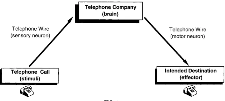
The anatomy and physiology of the nervous system can be compared with a telephone network. Each call that is made must pass through a telephone wire (sensory neuron) to the telephone company (brain). In the telephone company, the call is processed and a decision is made before the call is sent to its intended destination. Subsequently, the call must travel down specific telephone wires (motor neurons) to reach the intended destination (effectors). The information is transmitted from one neuron to another or from one neuron to the effector cell by electrochemical signals known as neurotransmitters. The nervous system is fast and direct and can allow an immediate response to occur, just like the telephone system. Figure 1 illustrates a neural transmission as it compares with a phone call.
In contrast, the endocrine system can be compared with the postal service. Like bulk mailings, the mes-sage reaches its destination more slowly than the aforementioned telephone call and is more diffuse (reaches a greater area). The endocrine system uses chemical substances known as hormones to send information throughout the body. These hormones
FIG. 1.
are released from tissues into the circulation (blood-stream), where they travel to other cells and regulate organ and cell function. The release of these hor-mones is pulsatile and may occur during daily rhythms (circadian rhythms). The physiological effects of hor-mones persist longer than those of neurotransmitters, even though the method of communication is slower for the endocrine system than the nervous system.
Although the nervous and endocrine systems are anatomically separate, their functions are highly coor-dinated and interrelated. Table 1 compares the differ-ences between the nervous and endocrine systems.
=6. Compare and contrast the similarities and differ-ences between neurotransmitters and hormones.
*7. What is the role of a feedback system? Name two types of feedback systems.
For a hormone to regulate an activity, it must be able to recognize and bind to a specific receptor. This process is traditionally described as a lock and key mechanism. Specifically, for a response to occur, the hormone (key) must fit exactly into the receptor (lock). Once inserted, the key can influence the lock to regulate ongoing activities and cause a number of intracellular events. Specific receptors are only found on certain cells. A hormone cannot regulate a cell’s activity if the specific receptor is not present.
The endocrine system can regulate the release of hormones through feedback mechanisms. Negative feedback is the most common form of regulation and can be compared with how temperature is regulated in your home. For example, if the thermostat in your home is set at 72°, a decrease in temperature will stimulate the thermostat to cause the heater to release warm air. When the room returns to 72°, the
thermo-stat detects the rise in temperature and turns off the heater. Conversely, if room temperature rises above 72°, the thermostat will stimulate the air conditioner to cool down the room. The temperature of the room is regulated through the thermostat, which assesses the situation and reacts accordingly to maintain a constant temperature.
Similarly, the endocrine system regulates the release of hormones in a negative feedback function. For example, when released fromcell A,horm one Aacts oncell Bto increase the production ofhorm one B. If blood levels ofhorm one Bincrease above a set range, horm one B will communicate back to cell A to prevent further release of horm one A. Figure 2A illustrates a negative feedback loop.
A positive feedback loop can also occur but is less common than negative feedback. During positive feedback, the amount of hormone released can fur-ther stimulate the releasing organ to continue secre-tions. This occurs through a limited range. The posi-tive feedback mechanism stops once the range has been surpassed. For example, when released fromcell A, horm one Aacts on cell Bto increase the produc-tion ofhorm one B. However, instead of feeding back to inhibitcell A, horm one B will enhance the secre-tions ofcell A. Note, however, that positive feedback TABLE 1
Comparison between the nervous and endocrine systems
Nervous System
Endocrine System
Speed of response fast slow Route of action neurons blood stream Messenger neurotransmitters hormones Duration of effects short long
FIG. 2.
loops naturally occur only in unique situations. For example, during childbirth, stretching of the cervix causes a neuroendocrine reflex that enhances uterine contractions until the baby is born. Figure 2B illus-trates a positive feedback loop.
All hormones of the GI tract are peptides, but not all peptides in the GI tract are hormones. There are four major types of peptides in the GI system, autocrine, paracrine, endocrine, and neurocrine. The peptides are defined by the route and distance that they must travel to reach their target organs. Autocrines are peptides that are released into the extracellular fluid, and they act on the same cell from which they are released. Paracrines are peptides released into the extracellular fluid, but they diffuse short distances to reach and regulate neighboring cells. Endocrines are peptides (hormones) released into the blood stream, and they travel long distances to reach their target cells. Neurocrines are peptides released from nerve endings, and they diffuse across short synapses to affect secretory cells. A schematic illustration of the types of peptide actions found in the GI tract is presented in Fig. 3.
=8. Where are autocrine agents secreted? Where are their effects seen?
=9. Where are paracrine agents secreted? Where are their effects seen?
=10. Where are neurocrine agents secreted? Where are their effects seen?
*11. Name the four main GI hormones.
There are four main hormones found in the GI system, gastrin, secretin, cholecystokinin (CCK), and gastric inhibitory peptide (GIP). The GI hormones are stored within endocrine cells that are dispersed throughout the mucosa of the stomach and small intestine. Vagal (parasympathetic) nerve activity, distension of the GI wall, and chemical stimulation corresponding to the intake of food can release the hormones into the bloodstream. Once in the portal circulation, the GI hormones pass through the liver to the heart and back to the digestive system to regulate its movements and secretions. These hormones also help to regulate the growth of the stomach, small intestine, and pan-creas (1).
*12. Of the four hormones, which one is released from cells found in the stomach? Where are the other three hormones mainly found?
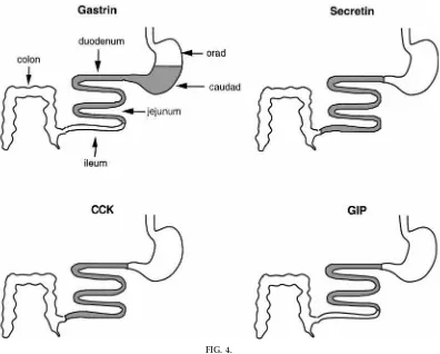
The cells that release these hormones are not evenly distributed throughout the GI tract. Gastrin is primar-ily released from cells located in the caudad and duodenum, but it can be present in the jejunum. Secretin is discharged from cells primarily found in the duodenum, but it can also be present in the jejunum and ileum, whereas CCK is released through-out the small intestine. Finally, GIP is emitted from cells found in the duodenum and jejunum. Figure 4 shows the sites of distribution of the hormones found in the GI tract.
Gastrin, the first of the four hormones to be discussed, is secreted from G cells found in the lower third of the stomach. Distension of the stomach (expansion of the walls of the stomach because of food intake), prod-FIG. 3.
ucts of protein digestion, and activation of the vagus nerve are all stimuli that can cause the release of gastrin. Gastrin is the only hormone demonstrated to be strongly released because of neural stimulation. In addition to releasing gastrin, the vagus nerve also causes an increase in motility of the stomach and small intestine. Once secreted into the bloodstream, gastrin travels through the circulation up to the heart and returns to the stomach to stimulate the parietal cells (found in the middle third of the stomach) that secrete hydrochloric acid (HCl) and intrinsic factor. A sche-matic representation of the activities of gastrin is seen in Fig. 5.
The gastric juices found in the stomach consist of HCl, intrinsic factor, pepsinogen, and mucus. HCl main-tains a low bacterial level in the stomach. HCl also
denatures proteins and allows pepsinogen to be converted into its active form, pepsin. Pepsin begins the preliminary digestion of proteins. Intrinsic factor is essential for the absorption of vitamin B12, which is important in blood formation. A thick mucus lining protects the stomach from the low pH caused by HCl.
The second hormone discussed is secretin (nature’s antac id). Onc e ac idic gastric juic es and c hyme enter the duodenum of the small intestine and lower the pH to 4.5, the S cells release secretin. Secretin travels through the blood to the pancreas to stimulate pancreatic HCO3
2
to be released. HCO3
2
neutralizes the acidic gastric juices so that the small intestine is not harmed; hence, the name ‘‘nature’s antacid.’’ The neutral pH is also necessary for the conversion of FIG. 4.
digestive enzymes from their precursor to their active forms. Figure 6 schematically depicts the actions of secretin.
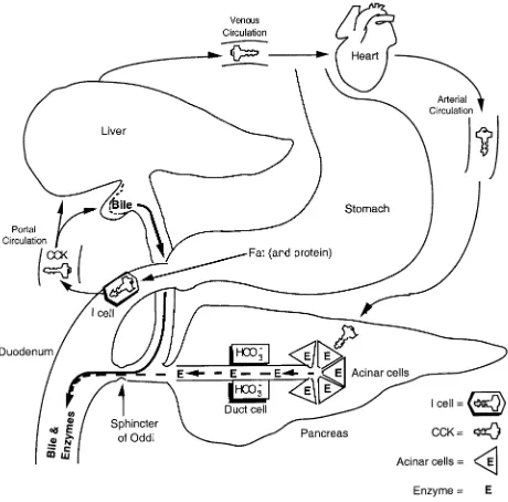
The third hormone discussed is CCK. Fat, and to a lesser extent protein, in the small intestine will stimulate the I cells in the duodenum to release CCK. CCK travels through the blood to the gallbladder and pancreas. As a result, the gallbladder contracts to release bile, and pancreatic digestive enzymes are released from the pancreas. CCK also causes the sphincter of Oddi to relax so that both the pancreatic enzymes and bile can flow into the duodenum. The activities of CCK are presented in Fig. 7.
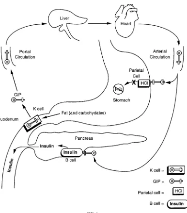
GIP, released from the K cells, is secreted mainly in response to fat, and to a lesser extent to carbohy-drates, present in the duodenum. GIP decreases motor and secretory activity of the stomach, but it mainly causes insulin release from thebcells of the pancreas (hence it can also be called ‘‘glucose-dependent
insulinotropic peptide’’). Insulin is essential for the uptake of sugar (glucose) into the cells. GIP’s actions and mechanisms are presented in Fig. 8.
=13. Why is hydrochloric acid (HCl) important? What cells release HCl?
=14. What is the primary role of bic arbonate (HCO3
2
)?
=15. Which hormone(s) can be released by vagal stimulation? Which hormone(s) can be released by fat?
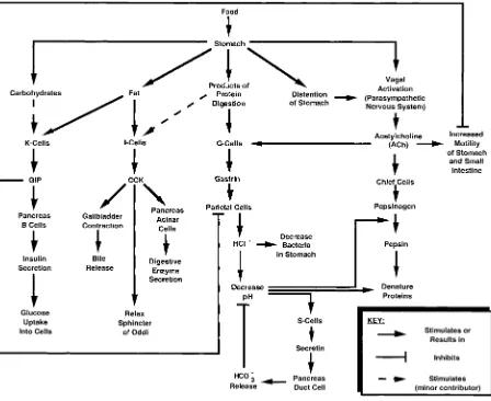
Figure 9 presents a concept map that students may find useful to organize the material and review the GI hormones. The following example helps to illus-trate how the concept map may be used. Food entering the stomach (stimuli) causes the G cells (releasing cells) to release gastrin (hormone). Gastrin FIG. 5.
stimulates the parietal cells of the stomach (target cells) to secrete HCl (function), which then begins preliminary digestion.
Answers to Tex t Questions
1. The accessory organs of the GI tract include the salivary glands (which produce saliva for protec-tion, lubricaprotec-tion, and preliminary carbohydrate
diges-tion), pancreas (produces the digestive enzymes and HCO32), liver (produces bile salts), and gallbladder
(stores and concentrates bile salts).
2. The majority of digestion and absorption occurs in the small intestine.
3. The portal vein is important because it directs all of the blood from the GI system to the liver for FIG. 6.
Summary of secr etin mechanisms. An incr ease in HCl in small intestine causes S cells (of duodenum) to r elease secr etin into bloodstr eam. Secr etin then travels thr ough venous cir culation to heart and r etur ns thr ough arterial cir culation to stimulate duct cells of pancr eas to r elease HCO32. HCO
3
2neutralizes HCl so that
processing. This is especially important when toxins are ingested. When this occurs, the toxins travel first to the liver, where they can be detoxified (neutral-ized) before reaching the rest of the body.
4. Homeostasis is important in maintaining a relatively constant internal environment. This is essential for sustaining vital organ function when the organism is faced with everyday stressors.
FIG. 7.
5. The nervous system uses neurons to transmit information quickly from one area of the body to another. Neurotransmitters are the messengers of the nervous system. In contrast, the endocrine system uses hormones as messengers and sends them through the bloodstream to reach distant organs where they regulate function.
6. Neurotransmitters are chemical messengers sent from neurons to a specific target cell. Their effects usually do not last very long after transmission has stopped. Hormones are also chemical messengers, but they are sent through the bloodstream to act on distant groups of cells. The effects of hormones persist longer than those of the neurotransmitters.
FIG. 8.
7. The role of a feedback system is to regulate body function so that homeostasis is maintained. Negative feedback and positive feedback are two types of regulatory mechanisms found in the body. Negative feedback is much more common than positive feed-back.
8. Autocrine agents are secreted into the extracellular fluid. They exert their effects on cells from which they were released.
9. Paracrine agents are also secreted into the extracel-lular fluid. They travel short distances to affect neigh-boring cells.
10. Neurocrine agents are secreted from nerve end-ings. They diffuse across short synapses to affect target cells.
11. The four main GI hormones are gastrin (secreted in response to distension of the stomach, products of protein digestion, or vagal stimulation), secretin (se-creted in response to acid), CCK (se(se-creted mainly in response to fat), and GIP (secreted in response to fats and carbohydrates).
12. Gastrin is the only hormone that is secreted by the stomach. Secretin, CCK, and GIP are mainly found in the duodenum and jejunum of the small intestine. FIG. 9.
Secretin and CCK are also seen in the ileum of the small intestine.
13. HCl is important for three reasons.1) HCl main-tains a low level of bacteria in the stomach. 2) HCl denatures proteins to begin protein digestion.3) HCl allows pepsinogen to be converted to pepsin. Parietal cells in the stomach release HCl in response to the hormone gastrin.
14. The small intestine is sensitive to acid. When acidic chyme enters the duodenum, it stimulates the release of HCO32. HCO32, released from duct cells of the pancreas, enters the duodenum to neutralize the acidic chyme emptying from the stomach.
15. Gastrin is the only hormone released in response to vagal stimulation. GIP and CCK are both released when fat enters the duodenum.
LABORATORY
Pr esenting the Laboratory Ex perience
We have developed two different options for present-ing the laboratory material.
Option 1.The students should form groups of three or four members to enhance small group discussion. Each student should be given a copy of the data presented in Table 2 and a copy of Table 3. The students should complete Table 3 using the data presented in Table 2. Once Table 3 has been com-pleted, the instructor will assign an unknown agent to each group. The laboratory group will be responsible for identifying the unknown agent and presenting the solution and rationale for their choice at the end of class. The students are encouraged to determine the solution and rationale for the remaining unknown agents as well, time permitting.
Option 2.The students should form groups of two or three members to enhance small group discussion. Each student should be given a copy of the data presented in Table 2 and a copy of Table 3. The students should complete Table 3 using the data presented in Table 2. Once Table 3 has been com-pleted, the students will determine the solution and rationale for all five unknown agents. That night for
homework or during class the next day, the students will write up a two-page laboratory report about their laboratory experience that includes their solutions and rationales for each of the unknown agents.
TABLE 2
Ex perimental data fr om the ‘‘virtual rats’’
Ex perimental Data Contr ol
Unknowns
1 2 3 4 5
Drops of fluid from
salivary duct, min21 10 10 10 70 12 9 pH of the stomach 2 1.9 1.8 1 1 4 Drops of fluid from
pancreatic duct,
min21 8 77 30 9.5 9 7
pH of fluid from a main
pancreatic duct 7 7.2 10 7.6 7.2 7 Drops of fluid from
common bile duct,
min21 2 64 2 2.3 2.4 2.4 Motility of stomach, no.
of contractions/min 3 3.6 3 15 3.4 1.0 Motility of small
intes-tine, no. of
contrac-tions/min 15 17 18 30 17 13 Blood glucose level,
mg/dl 100 101 100 104 102 60 Strength of contraction,
mmHg 10 12 12 50 12 7
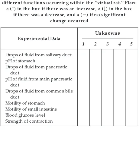
TABLE 3
Comparison of the effects of the unknown agents on the differ ent functions occurring within the ‘‘virtual rat.’’ Place
a (>) in the box if ther e was an incr ease, a (<) in the box
Drops of fluid from salivary duct pH of stomach
Drops of fluid from pancreatic duct
pH of fluid from main pancreatic duct
Drops of fluid from common bile duct
To allow more time for class discussion, the teacher may elect to have each student complete Table 3 before class. In addition, the number of students per group and time allotted may differ from class to class. The formats presented above are only sug-gestions. The teacher may develop other ideas for presenting the material. We estimate that the labora-tory itself should take no longer than one class period (,1 h).
Ex perimental Pr ocedur e
The data for this laboratory were compiled from six male laboratory rats. After an overnight fast, the rats were anesthetized and carefully monitored during the procedure to insure that no pain or discomfort was experienced by the animals. Catheters (tubes) were then inserted into the salivary duct, stomach, main pancreatic duct, and common bile duct. This process is referred to as cannulation. The catheters allowed the student to measure the volume of fluid secreted from the salivary gland, pancreas, and gallbladder. Once the fluid was collected, further tests were performed to determine the pH and content of the fluids secreted from each organ. Balloons, attached to pressure transducers, were placed in the stomach and small intestine to monitor pressure changes in gastro-intestinal motility. Contractions (motility) in the GI tract squeezed the balloon and sent a signal to the pressure transducer. The transducer then recorded both the frequency and strength of the contractions. Figure 10 illustrates the instrumentation of the virtual experimental animal.
Once the animals were prepared, each laboratory rat was given a 1-ml injection of one of the following: gastrin, secretin, CCK, GIP, or acetylcholine (ACh). ACh mimics the actions of the parasympathetic ner-vous system (i.e., vagus nerve). One rat was injected with 1 ml saline; this rat served as the control. Ten minutes after injection, the following data were ob-tained from the rats: drops of fluid from the salivary, main pancreatic, and common bile ducts (per minute), pH of the stomach and main pancreatic duct, motility of the stomach and small intestine (number of contrac-tions/min), strength of smooth muscle contraction (mmHg), and blood glucose level (mg/dl). The data obtained from the control rat (baseline data) and
each rat receiving an unknown were recorded in Table 2.
For this experiment, a significant difference is defined as any change of .20% from the control values. A change of,20% is attributed to experimental error or biological variability. Experimental errors may include small errors in calibration procedures, measurements, or instrumentation. Biological variability is the natu-rally occurring differences between animals.
Before beginning their analysis of the unknown, students are encouraged to complete Table 3 using the data presented in Table 2.
ANALYSIS
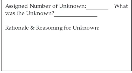
Complete the box below to use in your small group discussion and presentation at the end of class.
Assigned Number of Unknown: What
was the Unknown?
Rationale & Reasoning for Unknown:
Be sure to answer the following questions, either in your small group (if time permits), or on your own outside of class.
1. What wasunk nown 1? Explain your answer.
2. What wasunk nown 2? Explain your answer.
3. What wasunk nown 3? Explain your answer.
4. What wasunk nown 4? Explain your answer.
5. What wasunk nown 5? Explain your answer.
SOLUTIONS TO LABORATORY EXERCISE
Hor mone 1: CCK
CCK acts to cause gallbladder contraction and pancre-atic enzyme secretion. Fluid flow from both the common bile duct and the main pancreatic duct is
substantially increased. CCK does not greatly affect the salivary glands, blood glucose levels, pH of the stomach, pH of the pancreatic duct, or motility of the GI tract.
FIG. 10.
Hor mone 2: Secr etin
Secretin acts to increase bicarbonate secretion from the duct cells lining the pancreas. Therefore, the pH of the main pancreatic duct increases (becomes basic) and the volume of fluid secreted will increase. No other significant differences are seen compared with the control rat.
Hor mone 3: ACh
ACh mimics the effects of the parasympathetic ner-vous system on the GI tract. Therefore, in general, motility and secretory activity will increase. This can be observed by the increase of fluid from the salivary duct as well as the increased motility of the stomach and small intestine. Note that the only hormone that will cause gallbladder contraction is CCK (thus no significant change is observed). However, strong con-tractions in the GI wall are seen. ACh does not greatly affect blood glucose levels.
Hor mone 4: Gastrin
Gastrin acts on the parietal cells of the stomach to increase HCl and intrinsic factor secretion. Because of increased HCl levels, the pH of the stomach will decrease (become more acidic). In addition, a small increase of motility in the stomach and small intestine occurs. No other measurements are significantly af-fected.
Hor mone 5: GIP
GIP acts to inhibit gastric motility, strength of contrac-tion, and the release of HCl from the parietal cells of the stomach. Because of decreased HCl levels, the pH of the stomach becomes less acidic. More importantly, however, GIP stimulates the release of insulin. Nor-mally, insulin is released into the bloodstream after a meal to enhance glucose absorption into cells. As a result, blood glucose levels are lower than in the control rat.
SUMMARY
We wanted to develop a laboratory experiment that would provide teachers with the appropriate educa-tional materials required for teaching GI physiology. We provided background information on the con-cepts of GI physiology and the hormonal regulation of GI function. Thought-provoking questions were distrib-uted throughout the text to enhance the students’ learning and retention of GI physiology. Subsequently, the students conducted a dry laboratory experiment to reinforce the background material. The student was challenged to identify an unknown hormone or drug given to the virtual rat on the basis of the material previously presented.
Through the use of the virtual rat, the students were provided with an opportunity to analyze and interpret laboratory data without the complications associated with using live animals. On completion of this labora-tory, we hope that the students will have a basic comprehension of the hormones involved in GI physi-ology and will be encouraged to continue their pursuit in scientific education.
Suggested Readings
1. DiCarlo, S. E., E. Sipe, J. P. Layshock, and R. L. Rosian.
Experim ents a nd Dem onstra tions in Physiology. Upper Saddle River, NJ: Prentice Hall, 1998.
2. Granger, D. N., J. A. Barr owman, and P. R. Kvietys.Clinica l Ga strointestina l Physiology. Philadelphia: Saunders, 1985. 3. Johnson, L. R. Ga strointestina l Physiology. St. Louis, MO:
Mosby-Year Book, 1991.
4. Solcia, E., C. Capella, R. Buffa, L. Usellini, R. Fiocca, and F. Sessa.Endocrine cells of the digestive system. In:Physiology of the Ga strointestina l Tra ct(2nd ed.), edited by L. R. Johnson. New York: Raven, 1987.
TABLE 4
Answers to Table 3. Place a (>) in the box if ther e was an incr ease, a (<) in the box if ther e was a decr ease, and a (2) if
no significant change occurr ed
Ex perimental Data
Unknowns
1 2 3 4 5
Drops of fluid from salivary duct 2 2 > 2 2 pH of stomach 2 2 < < >
Drops of fluid from pancreatic
duct > > 2 2 2
pH of fluid from main pancreatic
duct 2 > 2 2 2
Drops of fluid from common bile
duct > 2 2 2 2
Motility of stomach 2 2 > 2 <
Motility of small intestine 2 2 > 2 2 Blood glucose level, mg/dl 2 2 2 2 <
5. Walsh, J. H.Gastrointestinal hormones. In:Physiology of the Ga strointestina l Tra ct(2nd ed.), edited by L. R. Johnson. New York: Raven, 1987.
The authors thank David W. Rodenbaugh for assisting with the preparation of this manuscript.
C. T. Hsu was supported by the Summer Fellowship Program at Northeastern Ohio Universities College of Medicine.
Address for reprint requests and other correspondence: S. E. DiCarlo, Department of Physiology, Wayne State University School
of Medicine, 540 E. Canfield Ave., Detroit, MI 48201 (E-mail: [email protected]).
Received 4 September 1997; accepted in final form 29 March 1999.
Refer ences
1. Johnson, L. R.Ga strointestina l Physiology. St. Louis, Mosby-Year Book, 1991.
2. Odenweller, C. M., C. T. Hsu, E. Sipe, J. P. Layshock, S. Varyani, R. L. Rosian, and S. E. DiCarlo.A laboratory exercise using virtual rats to teach endocrine physiology.Am . J. Physiol.