* Corresponding author. Tel: +62-21-64713850 ext 123 Fax: 021-64711948; Email: [email protected]
THE ANTIBACTERIAL COMPOUND COLLISMICIN A DERIVED FROM
MARINE
Streptomyces
sp Q-629K
Tutik Murniasih
1and Kyoko Adachi
2 1Research Center for Oceanography Indonesian Institute of Sciences Jl. Pasir putih No. 1 Ancol Timur Jakarta 14430 2
Marine Biotechnology Institute Co. Ltd., Kamaishi Lab.,3-75-1 Heita, Iwate 026-0001, Kamaishi City, Japan
Received 9 June 2008; Accepted 17 September 2008
ABSTRACT
In our course of screening for secondary metabolite derived from marine bacterial, we isolate the antimicrobial compound collysmicin A from the ethyl acetate extract of Streptomyces sp Q-629K. Separation of this compound was carried out by silica gel open column chromatography. Purification of an active compound was done using HPLC C18 with acetonitril-water system. Determination of chemical structure was done by 1H, 13C NMR and LC-MS analysis. Collysmicin A was contained in fraction 3, fraction 7.2 and fraction 8.7. The antimicrobial assayed from purified compound Fr.8.7 gave diameter inhibition approximately 13 mm against S. aureus and 12 mm against B. subtilis .
Keywords: antimicrobial, collismycin A and marine Streptomyces sp.
INTRODUCTION
The discovery of penicillin in 1929 heralded the era of antibiotics and realization that microorganism are a rich source of clinically useful natural products. Since that time, between 30,000 and 50,000 natural products have been discovered from microorganisms. Of these substances, more than 10,000 are biologically active and more than 8,000 are antibiotics [1,2]. Due to this potential, over 100 microbial products are in use today as antibiotics, antitumor agents, and agrichemicals. Most antibiotics of microbial origin come from terrestrial bacteria belonging to one taxonomic group, the order Actinomycetales. Unfortunately, the rate of discovering novel metabolites from terrestrial actinomycetes is decreasing and that new sources of bioactive natural products must be explored. One such resource can be found in marine microorganisms.
Two serious problems which prevent development of drugs from marine natural products were the majority of bioactive compounds have complex structures and the limiting of samples to let on chemical synthesis. Many researchers believed that these bioactive compounds are actually produced by ‘symbiotic’ microorganisms. Yet bacteria and fungi which produce highly promising metabolites have not been isolated from parent organisms. However Faulkner and co-workers have obtained fascinating results with sponge or bacteria association. If such symbiotic bacteria are culturable, we can expect a more promising future in ‘Drugs from the Sea’. Strictly speaking, the discovery of new structures is very important. Marine microbes are relatively new targets for such research. Unculturable microbes should be interesting to be explored. [3].
Marine bacteria are the best studied chemically of any of the marine microorganisms. This field is growing rapidly and there are frequent additions to growing literature. The first documented identification of a bioactive marine bacterial metabolite was the highly brominated pyrrole antibiotic isolated by Burkholder and co-workers from a bacterium obtained from surface of Caribbean sea grass Thalassia. [4] The Tokyo group at the Institute of Microbial Chemistry has been an innovator in search for new metabolites from marine actinomycetes. An isolate identified as Chainia purpurgena SS-228, found in mud samples obtained from Sagami Bay, was found to produce the antibiotic benzanthraquinone [5,6]. The antibiotic showed selective inhibition of Gram-positive bacteria, with MIC
values of between 1 and 2 μg/ml, and was an active
antitumor agent in vivo against Ehrlich carcinoma in mice, showing significant life span extension at low doses. Most interestingly, the production of this antibiotic was only observed in selected seawater media containing the unique Japanese seaweed product “Kobu cha.” Kobu Cha is a dried and pulverized powder produced in Japan from the brown seaweed Laminaria. The observation clearly shows that marine microorganisms have specific nutrients adaptations which relate to their natural nutrient sources in the ocean. The other bioactive compounds that isolated by Tokyo group were aplasmomycins A-C. This compounds produced by a marine actinomycete identified as Streptomyces griseus SS-20 propagating by media containing Kobu Cha under conditions (27 °C and very low nutrient) [7-10].
to investigate on useful substance for drug or agrichemical. This paper was conducted to investigate antibiotics target from marine bacterial Streptomyces sp.
Q-629K.
EXPERIMENTAL SECTION
Material
The bacterial strain of Streptomyces sp. Q-629K was obtained from the MBI culture collection. Media culture of KBG containing Glucose 0.3%, CaCO3 0.2 %,
Bouillon 0.1% and tangle weed powder was purchase from Wako pure chemicals. Organic solvent, ethyl acetate, chloroform, methanol and acetonitrile were purchase from Merck. Antimicrobial assays was conduct against Staphylococcus aureus IFO 12732 and Bacillus subtilis IFO 3134. Antibiotic standard Penicillin G (Sigma Co. Ltd). were used as positive control.
Instruments
Instruments that used in this work are Nano space S1-2 HPLC (Shiseido) with PDA (Diode Array) detector and column X Terra C18, LC-MS (PDA detector) LCQ Advantage ion trap mass spectrometer (Thermo Electron Co.Ltd), NMR 500MHz NMR (Varian Co. Ltd.) for structural determination. Autoclave, clean bench, Shimadzu UV/Vis spectrometer, shaker incubator were used for microbial preparation.
Procedure
Cultivation
Propagating of semi-large scale strain
Streptomyces sp. Q629K was done in the 3 x 1 L KBG media containing Glucose 0.3%, CaCO3 0.2%, Bouillon
0.1% and tangle weed powder (Kobu cha) diluted by 50% sea water. Cultivation was carried on the rotary shaker incubator during 6 days, at 30 °C. Measuring pH during cultivation was done to prevent an extreme acidity as the effect of decomposing carbon source.
Antibacterial assays
Antibacterial assays against S. aureus and B. subtilis was done using agar diffusion method. All about
15 μL bacterial extracts, fractions and isolated
compounds were applied into paper disc (diameter 6 mm), then put on the agar surface of the bacterial culture. After one day incubation at 30 °C, the diameter of inhibition was determined. The same procedure was applied to the antibiotic Penicillin G as a positive control.
Isolation of active compound
After 6 days cultivation, the bacterial culture was centrifuged for separating cell and secondary metabolite. The supernatant was extracted using ethyl acetate to get
organic phase substances. The pellet was extracted with ethanol 70%. All of extract were evaporated and applied for bioassay.
The active ethyl acetate phase was led to the separation by column silica gel developing using chloroform-acetonitrile by gradient method. About 8 fractions was collected by this open column separation. All of fractions were applied to the antimicrobial assays. There are 3 active fractions (Fr.3, Fr.7 and Fr.8), but only fraction Fr.7 and Fr.8 could be continued to the purification processes.
The potent open column fractions (Fr.7 and 8) was chromatographed using HPLC/PDA detector by column X Terra C18, 4.6 x 250 mm with mobile phase acetonitrile-water containing 0.1% TFA (Triflouroacetic Acid).
Structural Determination of the potent compound
Structural determination was done toward all of active antimicrobial compounds. The LC-MS (PDA detector) with Inertsil ODS-2 column diameter 1.5 x 250 mm, eluted with 70% Water containing 0,1% TFA and 30% methanol containing 0,1% TFA and ESI mode for the MS analysis was applied to detect the molecular weight of potent compound.
Analysis of 1H, 13C, COSY and HMBC were done using 500 MHz NMR (Varian co.ltd).
RESULT AND DISCUSSION
Cultivation of Streptomyces sp Q629K
The pH on zero day cultivation of Streptomyces
sp was 7.11, and after harvesting was 7.10 for
Streptomyces sp. These value indicated that the bacterial growth on pH neutral (around 7.1 – 7.5). Thus the reason why in media containing glucose must be added calcium carbonate for prevent decreasing pH because of carbohydrate hydrolysis.
Antimicrobial Assay
Tabel 2. Anti-bacterial assay of column fractions Streptomyces sp Q-629K.
Q-629K-EtOAc Fr.3 (CHCl3/CH3CN) 11/1 17 13
Q629K-EtOAc Fr.4 (CHCl3/CH3CN) 4/1 16 13
Q629K-EtOAc Fr.5 (CHCl3/CH3CN) 4/1 -
-Q629K-EtOAc Fr.6 (CHCl3/CH3CN) 1/1 - 9
Q629K-EtOAc Fr.7 (CH3CN) 100% 15 15
Q629K-EtOAc Fr.8 (MeOH) 100% 28 28
* the hallow was not clear
Fig 1. Inhibition effect against S. aureus by column fractions of ethyl acetate extract Q-629K strain
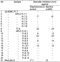
Table 3. Anti-gram positive bacterial assay by HPLC fractions of Streptomyces sp (Q-629K)
Diameter Inhibition (mm) antibacterial activity compare with ethanol and water extract. We can consider that the active compound has intermediate polarity. Base from this data then we
continued for separation process of ethyl acetate extract using open column method. The table 2 showed the antibacterial activity of separated fraction using silica gel open column with chloroform and acetonitrile solvent system. potent fraction with the strong activity was indicated by fraction 3, 4, 7 and 8. The figure 1 was present the clear zone inhibition of Staphylococcus aureus growth by these column fraction.
Otherwise the fraction 3 and 4 have strong activity, we could’t continued to the later purification due to the small quantity of both fraction. We decided to purify the fraction 7 and 8. The reverse phase HPLC method was applied to the fraction 7 and 8. The table 3 showed the result of antibacterial assay by HPLC column of both fraction.
HPLC fractions of Streptomyces sp Q-629K strain produced active fractions Fr. 7.2, Fr. 8.2, Fr.8.3, Fr. 8.5 and Fr. 8.7. Even though very weak there is only one fraction (Fr. 7.2) from purification fraction 7 that gave a positive result against both of the pathogenic bacteria. The most potent fraction from purification fraction 8 was fraction Fr. 8.7, otherwise this activity was lower than a positive control penicillin G.
Result of Separation Active Substance
Separation active compounds of Streptomyces sp (Q-629K)
N
N
O C H3
S
N O H H
H
H
H
H
H
C H3
5 ,8 . 6 3 8 ,7 . 4 4
7 ,7 . 9 2
1 0 ,8 . 5 3
1 2 3 . 2 7
3 ,1 5 6 4 5 1 4 9 . 8 5
1 2 5 . 6 1 1 3 8 . 5 2
1 0 4 . 8 6
1 1 ,8 . 0 1 4
1 2 ,4 . 0 9 9 5 6 . 8 6 1 ,1 6 8 7 5
9 ,1 2 3 . 4 2 4 ,1 5 3 . 9 6 2 ,1 5 8 . 2 1
6 ,8 . 9 2 4
1 4 8 . 7 0
1 3 ,2 . 3 7 6 1 8 . 3 4 Fig 3. HPLC chromatogram of Streptomyces sp (
Q-629K) Fr.8
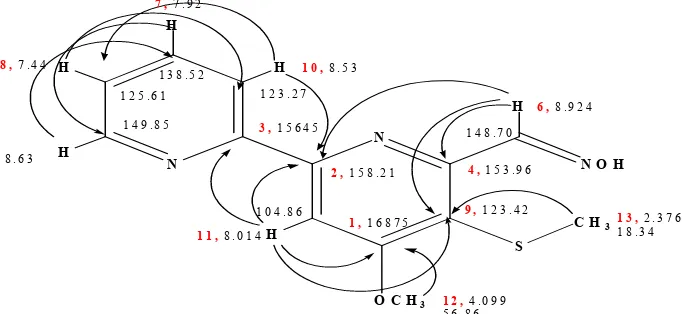
Table 4. The chemical shif of 13C and 1H NMR of Collismycin in CDCl3
Position C H (multiplicity ,J=Hz)
1 168.75 C
2 158.21 C
3 156.45 C
4 153.96 C
5 149.85 CH 8.63(d, 5.0)
6 148.70 CH=NOH 8.924(s)
7 138.52 CH 7.92 (t ,8.0)
8 125.61 CH 7,44(dd,8.0, 5.0 )
9 123.42 C
10 123.27 CH 8.53(d, 8.0)
11 104.86 CH 8.014 (s)
12 56.86 CH3 4.099 (s)
13 18.34 CH3 2.376 (s)
NOH 10.19 (br s)
Solvent : CDCl3
Fig 3. The structure of Collismycin A with HMBC correlation
trile containing 0.1% TFA by gradient system. Separation using HPLC method of open column fraction Fr.7 gave 6 HPLC fraction (Fr.7.1…Fr7.6). The twelve fractions (Fr8.1….Fr8.12) were collected from HPLC separation of open column 8 The HPLC chromatogram profile of open column 8 separation could be seen in the figure 2.
There are four potent fractions Fr.8.2, Fr.8.3, Fr8.5 and Fr.8.7 that appeared on the retention time 3, 4, 7 and 10 min. The available second purification was done to the fraction Fr.8.7. This fraction was chosen to the structural determination analysis.
Chemical structural determination
As a mention before, open column
chromatographic separation of ethyl acetate phase of Q-629K strain led to the active fraction Fr.3, Fr.4, Fr.7 and Fr8. Chemical structural determination of Fr.3, Fr.7.2, and Fr.8.7 gave an active compound collismycin A. The
detail of chemical structural analysis would be explained in the next paragraph.
The LC-MS data of Q-629K Fr.3, Fr.7.2, Fr.8.7. gave [M+H]+ 276.2 at retention time 7.6 min. Elucidation structure of these active fractions was carried out by proton and carbon NMR analysis. Proton and carbon chemical shift could be shown in the table 4.
The 1H NMR spectra showed there were
aromatically proton at position 5 H 8.63; position 7,H
7.92; position 8 ,H 7,44, position 10, H 8.53 and
position 11, H 8.014 respectively. The duplet peaks at
position 6, H 8.924 considered to the proton that
attached carbon – nitrogen bond (HC-N~). The singlet peak at position 12 corresponded to the proton on ~O-CH3 bond. The last position at shift,H 2.376 indicated
proton methyl that attach sulphuric such as ~S-CH3.
compound Fr.3 was, Collismycin A (Figure 3). The presence of shift C-13 at C 18.34 indicated signal of
~S-CH3 bond. This data supported to the HMBC spectral signed that only one correlation between proton position 13 at shift,H 2.376 with carbon C-9, C 123.27.
Literature study proved that the spectral data of isolated compound was very similar with antibacterial & antitumor compound collismycin A that already isolated by Shindo, S. et.al [10] from Streptomyces sp. MQ22. This compound had been reported active against S. aureus, B. subtilis, E. coli, C. albicans, S. cerevisiae and
A. niger [11]. GOMI et. al. [12] informed that this compound has antitumor activity and showed cytotoxic against L1210 murine leukemia cells ( IC50 0.08 μg/mL
and 0.12 μg/mL,respectively).
CONCLUSION
Base to explanation mentioned in this paper, We could conclude that the antibacterial compound contained in ethyl acetate extract of marine bacterium
Streptomyces sp (Q-629K) was collismycin A. In this study, this compound was inhibit S. aureus and B. subtillis.
ACKNOWLEDGEMENT
We appreciated to Dr. Yoshikazu Shizuri for his guidance in structural determination. I would also thank to JICA for financial support in this training program.
REFERENCES
1. Berdy, J., 1982, Search and Discovery Methods for Novel Antimicrobials in: Bioactive Metabolites from Microorganisms (M.E. Bushell and U. Grafe, eds), Elsevier, Amsterdam, pp. 3-25.
2. Beetina, V., 1983, The Chemistry and Biology of Antibiotics, Elsevier, Amsterdam.
3. Fusetani, N., 2000, Drugs from The Sea. Karger, Basel Switzerland, pp.1-5
4. Burkholder, P.R., Pfister, R.M., and Leitz, F.P., 1966, Appl. Microbiol. 15:85-89.
5. Kitahara, T., Naganawa, H., Okazaki, T., Okami, Y., and Umezawa, H., 1975, J. Antibiot. 28:280. 6. Okazaki, T., Kitahara, T., and Okami, Y., 1975, J.
Antibiot. 28:176-184.
7. Hotta, K., Yoshida, T., Hamada, M., and Okami, Y., 1980, J. Antibiot. 33:1515.
8. Okami, Y., Okazaki,T., Kitahara, T., and Umezawa, H., 1976, J. Antibiot. 29:1019-1025.
9. Sato, K., Okazaki, T., Maeda, K., and Okami, Y., 1978, J. antibiot. 31:632
10. Shindo, K., Yamagishi, Y., Okada, Y., Kawai, H.,1994,The Journal of Antibiotics vol.47 no.9. 1072-1074.