www.elsevier.com/locate/jinsphys
Apocrine secretion of amylase and exocytosis of trypsin along the
midgut of Tenebrio molitor larvae
Plinio T. Cristofoletti
a, Alberto F. Ribeiro
b, Walter R. Terra
a,*aDepartamento de Bioquı´mica, Instituto de Quı´mica, Universidade de Sa˜o Paulo, C.P. 26077, 05513-970, Sao Paulo, Brazil bDepartamento de Biologia, Instituto de Biocieˆncias, Universidade de Sa˜o Paulo, C.P. 11461, 05422-970, Sao Paulo, Brazil
Received 12 December 1999; accepted 10 May 2000
Abstract
Amylase and trypsin were purified from Tenebrio molitor midgut larvae and used to raise antibodies in a rabbit. A Western blot of T. molitor midgut homogenates, after sodium dodecyl sulfate–polyacrylamide gel electrophoresis using amylase and trypsin antisera, showed only bands co-migrating with the purified enzymes. The antisera were used to localize the enzymes by immunofluo-rescence and immunogold labeling. Amylase occurs in a few regularly disposed anterior midgut cells. Non-amylase-secreting anterior midgut cells are proposed to be water-absorbing cells based on morphology and dye movements. Amylase is found inside vesicles originating from Golgi areas that seem to fuse together before their release along with the now disorganized apical cytoplasm (apocrine secretion). Trypsin precursors are observed inside small vesicles near the apical plasma membrane of posterior midgut cells, suggesting an exocytic mechanism of secretion, followed by putative trypsin activation. Apocrine secretion is thought to be an adaptation to enhance the dispersion of secretory vesicle contents released from a water-absorbing epithelium, whereas exocytosis is an efficient secretory mechanism in a water-secreting epithelium. 2000 Elsevier Science Ltd. All rights reserved.
Keywords: Apocrine secretion; Microapocrine secretion; Secretory mechanisms; Immunocytochemical study; Digestive enzymes
1. Introduction
The first hypotheses regarding secretory mechanisms in insect midgut cells were based on cytological obser-vations under the light microscope. Thus, cells with api-cal extrusions were interpreted as undergoing apocrine or holocrine secretion, whereas cells showing no
cytol-ogical alterations were regarded as non-secreting
(Wigglesworth, 1972). The use of the electron micro-scope showed that many midgut cells quiescent under the light microscope were performing exocytosis (Terra and Ferreira, 1994). After these observations, some authors maintained the early interpretation of apical extrusion as apocrine secretion, whereas others inter-preted the phenomenon as representing cell death during epithelium cell renewal (Terra and Ferreira, 1994). The latter interpretation was reinforced by the observation that the cells eliminating blebs were not adjacent to
* Corresponding author. Fax:+55-11-3818-2186.
E-mail address: [email protected] (W.R. Terra).
0022-1910/00/$ - see front matter2000 Elsevier Science Ltd. All rights reserved. PII: S 0 0 2 2 - 1 9 1 0 ( 0 0 ) 0 0 0 9 8 - 6
regenerative cells nidi, meaning that they were older than the other cells, and their apical cytoplasm were degenerate in appearance.
An important advance in the study of insect midgut secretory mechanisms resulted from the combination of biochemical and cytological procedures. These tech-niques were employed at first in insects with midgut cells that appear quiescent under the light microscope (Lepidoptera and most Diptera). The results showed that secretory mechanisms depend largely on midgut regions. Thus, trypsin is secreted by the posterior midgut of adult mosquitoes (Graf et al., 1986), larval flies (Jorda˜o et al., 1996a) and caterpillars (Jorda˜o et al., 1999) by exocytosis, and by the anterior midgut of caterpillars using a microapocrine route (Santos et al., 1986; Jorda˜o et al., 1999). Microapocrine secretion consists of the elimination from the cell microvilli of small pinched-off single- or double-membrane vesicles.
Two insects presenting midgut cell extrusions were studied with biochemical and cytological techniques. In
Stomoxys calcitrans adults, immunocytochemical data
organelles are discharged by opaque zone cells of adult flies, suggesting holocrine secretion (Jorda˜o et al., 1996b). In Tenebrio molitor larvae, cell fractionation studies revealed that digestive enzymes are found inside secretory vesicles (Ferreira et al., 1990). These data favor an exocytic route for digestive enzyme secretion in T. molitor larvae, since apocrine secreted proteins (at least in vertebrates) are transported inside the cells with-out participation of the endomembrane system, the Golgi apparatus and secretion granules (Aumu¨ller et al., 1999). In this paper, T. molitor anti-trypsin and anti-amylase sera were used to show by immunocytolocalization that amylase-containing vesicles are discharged by apocrine secretion from anterior midgut and that trypsin is released by exocytosis from the posterior midgut.
2. Materials and methods
2.1. Animals and preparation of midgut samples
Stock cultures of the yellow mealworm, T. molitor (Coleoptera: Tenebrionidae), were cultured under natural
photoregime conditions on wheat bran at 24–26°C and
a relative humidity of 70–75%. Fully grown larvae (each weighing about 0.12 g) of both sexes were used in the determinations.
Animals were immobilized by placing them on ice, after which they were dissected in cold 342 mM NaCl. The midgut was removed and divided into three sections (anterior, middle, and posterior) of identical lengths. The anterior and middle midgut contents were combined and this sample, in addition to the posterior midgut contents, was homogenized in cold double-distilled water by using a Potter–Elvehjem homogenizer. The homogenates were
centrifuged at 25 000g for 30 min at 4°C, and the
super-natants were stored at 220°C until use as an enzyme
source.
2.2. Protein determination and assays of amylase and trypsin
Protein was determined according to Bradford (1976) using ovalbumin as a standard. Amylase was assayed at
30°C following the release of reducing groups (Noelting
and Bernfeld, 1948) from 0.5% soluble starch in 50 mM citrate–sodium phosphate (pH 5.8) containing 10 mM
NaCl. Trypsin was determined with 0.83 mM a
-N-ben-zoyl-dl-arginine-p-nitroanilide (Erlanger et al., 1961) in
100 mM Tris–HCl buffer (pH 7.5) at 30°C. Incubations
were carried out for at least four different time periods and the initial rates of hydrolysis were calculated. All assays were performed under conditions such that activity was proportional to protein concentration and to time. A unit of enzyme is defined as the amount that
catalyzes the cleavage of 1 µmol of substrate (or bond)
per minute.
2.3. Purification of T. molitor midgut amylase
Supernatants corresponding to the first two-thirds of
T. molitor midguts were submitted to preparative
electrophoresis in a 7.5% polyacrylamide gel cylinder prepared according to Davis (1964), using the PrepCell
System (Bio-Rad, USA). Samples of 2 ml
(corresponding to 100 animals and containing 105 mg of proteins) were electrophoretically separated at 12 W constant power. Fractions with amylase activity were pooled and applied to a Mono Q column (FPLC System Pharmacia–LKB Biotechnology, Sweden) equilibrated in 20 mM piperazine buffer (pH 5.5) and eluted with a gradient of 0–600 mM. The flux was 1.0 ml/min and fractions of 0.4 ml were collected. The active fractions were loaded onto a Phenyl-Superose column (FPLC System) equilibrated with 100 mM citrate–sodium phos-phate buffer (pH 6.0) containing 1.7 M ammonium sulf-ate. Elution (0.5 ml/min) was accomplished with a gradi-ent of 1.7–0 M ammonium sulfate in the same buffer and fractions of 0.4 ml were collected. The pooled active fractions were applied to a Superose 12 column (FPLC System), equilibrated and eluted with 20 mM citrate– sodium phosphate (pH 6.0) containing 150 mM NaCl. Fractions of 0.4 ml were collected at a flow rate of 0.5 ml/min.
2.4. Purification of T. molitor midgut trypsin
Supernatants (200 mg protein from 600 animals) cor-responding to the posterior midgut contents were applied onto an Econo Pac High Q column (EconoSystem, Bio-Rad, USA) equilibrated and eluted with 20 mM Tris– HCl buffer (pH 7.0). The flux was 1.0 ml/min and frac-tions of 1.0 ml were collected. In contrast to most other proteins, trypsin is not retained in the column and is eluted in the first fractions. The column was regenerated with 20 mM Tris–HCl buffer (pH 7.0) containing 1 M NaCl. Fractions showing trypsin activity were pooled, passed through a High Trap column (Pharmacia–LKB Biotechnology, Sweden) to exchange the buffer, and loaded onto a Mono Q column (FPLC System) equilib-rated in 20 mM Tris–HCl buffer (pH 9.0). The column was eluted with a gradient of 0–600 mM NaCl in the same buffer. The rate of flow was 1.0 ml/min and frac-tions of 0.4 ml were collected.
2.5. Sodium dodecyl sulfate–polyacrylamide gel electrophoresis (SDS–PAGE) and Western blotting
using Bio-Rad (USA) Mini-Protein II equipment. Samples were mixed with sample buffer (2:1) containing 60 mM Tris–HCl, pH 6.8, 2.5% (w/v) SDS, 0.36 mM 2-mercaptoethanol, 10% glycerol and 0.05% (w/v)
bro-mophenol blue and heated for 2 min at 95°C in a water
bath before being loaded onto the gels. Electrophoresis was carried out at 200 V until the front marker (bromophenol blue) reached the bottom of the gel. The gel was then divided into two parts, one of which was silver-stained according to Blum et al. (1987), while the corresponding proteins in the other part of the gel were electrophoretically transferred onto a nitrocellulose
membrane filter (pore size 0.45 µm; Bio-Rad, USA)
(Towbin et al., 1979). The transfer efficiency was evalu-ated by observing the pre-stained molecular weight mar-kers (Bio-Rad or Sigma, USA). The filters were first reacted (after a blocking step) with the trypsin or amyl-ase antiserum diluted 200- or 500-fold, respectively, in TBS (Tris-buffered saline: 50 mM Tris–HCl buffer (pH 7.4), containing 0.15 M NaCl) containing 0.05% Tween
20 (TBS-T) for 2 h at room temperature (25°C). After
extensive washing with TBS-T, the filters were reacted with anti-rabbit IgG coupled with peroxidase (Sigma, USA) diluted 1:1000 in TBS-T for 2 h at room tempera-ture. After washing extensively with the same buffer, the strips were incubated with 0.08% 4-chloro-1-naphthol in TBS containing 0.1% hydrogen peroxide until gray bands appeared where antigens were recognized. The reagent was prepared before use by the addition of one volume of 0.5% chloro-naphthol in methanol to five vol-umes of TBS with hydrogen peroxide.
Western blotting to recognize trypsinogen isoforms was performed as described above, except that after the final washing the strips were treated with an ECL West-ern blotting kit (Amersham, UK) according to the manu-facturer’s instructions, and then exposed to high-per-formance luminescence detection film (Hyperfil-ECL, Amersham, UK).
Pre-immune serum was used in control experiments to show that antisera were specific.
2.6. Correlation of amylase and trypsin activities with protein bands after PAGE
A sample of purified T. molitor amylase was subjected to polyacrylamide gel electrophoresis as described above for SDS–PAGE, except that the sample was not boiled and SDS and 2-mercaptoethanol were absent from the gel. After electrophoresis, the gel slab was layered onto a similar gel slab containing 1.0% starch. The two gel
slabs were left for about 1 h at 30°C in a humid chamber.
Finally, the original gel slab was removed and the
starch-containing gel was stained with lugol (1.3% I2, 3% KI
in water). After coloration, a light band against the dark
background indicated the presence of active a-amylase.
A gel identical to the one that was overlaid on the
starch-containing gel was silver-stained to show that the protein band has the same migration as the amylase activity.
A sample of purified T. molitor trypsin was applied to a Superose 12 column (FPLC System) equilibrated and eluted (flux 0.5 ml/min) with 20 mM Tris–HCl buffer (pH 9.0) containing 200 mM NaCl and fractions of 0.4 ml were collected. The activity was recovered in fractions 35 to 40. Each active fraction was submitted to SDS–PAGE and its activity was shown to be pro-portional to the intensity of the single silver-stained band in the gel slab.
2.7. Detection of trypsin precursors
For the detection of trypsin precursors, posterior midgut tissue freed from contents was immediately boiled for 5 min or homogenized in double-distilled water and then maintained at different conditions. The boiled tissue was homogenized as described previously and all homogenates were added to two volumes of sam-ple buffer for SDS–PAGE before being centrifuged (10 000g, 5 min, room temperature). The supernatants were then analyzed by Western blotting after SDS– PAGE.
2.8. Preparation of amylase and trypsin antisera
Two ml of a purified enzyme solution were emulsified with an equal volume of Freund’s complete adjuvant.
This emulsion (containing 80µg of the purified enzyme)
was then injected into the inguinal nodes of a rabbit. After 4 weeks, a similar injection was administered, but with Freund’s incomplete adjuvant, and 7 days later the
rabbit was bled. The blood was left standing 1 h at 37°C
and overnight at 4°C, before being centrifuged at 3000g
for 10 min at 4°C. The supernatant was added to a
suit-able solution to become 50% saturated in (NH4)2 SO4,
pH 6.8. After standing overnight at room temperature
(25°C), the resulting suspension was centrifuged at
5000g for 15 min at 4°C. The pellet was resuspended in
0.1 M NaCl and dialysed overnight against 1000
vol-umes of 100 mM NaCl at 4°C. The resulting antiserum
was stored at 220°C.
Antibody production and specificity was checked on Western blots after SDS–PAGE.
2.9. Light microscopy, confocal fluorescence microscopy and electron microscopy
For studies with the light microscope, animals were dissected in their own haemolymph and midguts were fixed in 4% paraformaldehyde in 20 mM phosphate buffer (pH 7.4) containing 0.15 M NaCl (PBS). After dehydration in graded ethanol, the materials were embedded in Historesin (Leica, Heidelberg, Germany),
stained with haematoxylin and eosin, and mounted in glass slides with Entellan (Merck, Darmstadt, Germany).
For immunofluorescent visualization of a-amylase
and trypsin, confocal microscopy was used. The tissue samples were fixed in Zamboni’s fixative (Stefanini et
al., 1967) for 16 h at 4°C. After rinsing in PBS, the
samples were embedded in Tissue Tek compound (Miles
Inc., USA), frozen in liquid nitrogen and cut (14 µm
thick) in a Leica CM 1850 cryostat. Sections were col-lected on polylysine-coated glass slides and immunos-tained using an indirect immunofluorescence method. After immersion in PBS containing 0.2% Triton X-100 for 2 h at room temperature, the preparations were rinsed
in PBS and incubated in the primary antibody (a
-amyl-ase or trypsin) diluted 1:1000 in PBS plus 0.1% bovine
serum albumin for 16 h at 4°C. As controls, sections
were incubated with non-immune serum in the same conditions. The samples were then rinsed in PBS at room temperature and incubated with the secondary antibody (FITC, Amersham Little Chalfont, UK, or TRITC, Sigma, St Louis, USA) diluted 1:1000 in PBS plus 0.1% bovine serum albumin, for 1 h at room temperature. After rinsing in PBS at room temperature, the sections were mounted in Vectashield (Vector Labs, Inc., USA) mounting medium and examined in a Zeiss LSM 410 confocal microscope.
For immunolabeling of trypsin or amylase at the
ultra-structural level, midgut pieces were fixed
(paraformaldehyde–glutaraldehyde), embedded in L.R. White acrylic resin, incubated with the primary and sec-ondary (goat rabbit IgG coupled to 15 nm gold) anti-bodies and examined in a Zeiss EM 900 electron micro-scope as detailed elsewhere (Silva et al., 1995). As controls, sections were incubated with non-immune serum under the same conditions.
For scanning electron microscopy, midgut fragments were fixed in glutaraldehyde, post-fixed in osmium tet-roxide, dehydrated, critical-point dried and gold-coated as described previously (Silva et al., 1995). The prep-arations were examined in a Zeiss DSM 940 electron microscope.
3. Results
3.1. Amylase and trypsin purification and production of antisera
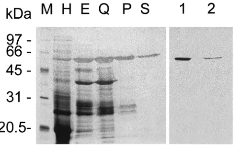
Amylase from T. molitor midguts was purified to homogeneity through preparative electrophoresis and several chromatographic steps. The yield of this prep-aration was 27.0% and the specific activity of the pur-ified enzyme was 2200 U/mg protein (Table 1). SDS– PAGE of purified amylase resulted in a single
silver-stained band with an Mrvalue of 63 000 (Fig. 1), which
has amylase activity as observed after an in-gel assay
(not shown, see Section 2 for details). This means that
the purified protein band is really an amylase. The Mr
value of our amylase is similar to that (Mr 68 000) of
the amylase purified from T. molitor by Buonocore et al. (1976), using procedures different from those described here.
The purified amylase was used to raise antibodies in a rabbit. A Western blot of midgut proteins after SDS– PAGE and incubated with the amylase antiserum showed a single band (Fig. 1, lane 1), with a migration rate identical to that of the purified amylase (Fig. 1, lane 2). Pre-immune serum was unable to react with T.
moli-tor midgut proteins (not shown). These data indicate that
the amylase antiserum recognized only amylase mol-ecules in T. molitor larval midguts.
Trypsin from T. molitor midguts was isolated pure after two chromatographic steps with a yield of 15% and a final specific activity of 0.97 U/mg protein (Table 2). SDS–PAGE of purified trypsin resulted in a single
pro-tein band with an Mrvalue of 25 000 (Fig. 2, lane Q2).
This value is identical to that of a trypsin purified from
T. molitor by Levinsky et al. (1977) using a different
procedure from that described in this paper. Gel filtration of our purified trypsin, followed by SDS–PAGE of the resulting fractions, showed that trypsin activity in the eluted fractions is proportional to the intensity of the single silver-stained bands in corresponding SDS–PAGE lanes (not shown, see Section 2).
Antibodies raised in a rabbit with purified trypsin recognized, in Western blots, a single protein in midgut homogenates (Fig. 2, lane 1) that co-migrated with pur-ified trypsin (Fig. 2, lane 2). Pre-immune serum is unable to react with T. molitor midgut proteins. These data prove that the trypsin antiserum recognizes only trypsin molecules in T. molitor larval midguts.
3.2. Trypsin precursors in midgut cells
The trypsin antiserum recognizes a protein band with
an Mrvalue larger than trypsin (Fig. 3C), if the source
tissue is boiled as soon as it is dissected. If the tissue homogenate is maintained in ice (Fig. 3B) or is
incu-bated for 1 h at 30°C (Fig. 3A), the larger trypsin
disap-pears partly or completely, respectively.
3.3. General morphology of the gut
The general morphology of the larval gut of T. molitor and the major ultrastructural features of the midgut cells have been described before (Koefoed and Zerahn, 1982; Berdan et al., 1985; Koefoed, 1987), and therefore only a brief description will be given, emphasizing only the features important for this work and those that have not been detailed before.
Fully-grown T. molitor larvae (10 determinations)
div-Table 1
Purification of T. molitor midgut amylase
Fraction Total activity (Units) Total protein (mg) Specific activity Purification factor Yield (%) (Units/mg)
Midgut homogenatea 4490 105.9 42.4 1 100.0
Preparative 5074 5.921 857 20.2 113.0
electrophoresis
Mono Q eluate 1913 2.111 906 21.4 42.6
Phenyl-Superose eluate 1230 0.918 1340 31.6 27.4
Superose eluate 1212 0.551 2200 51.9 27.0
aMidgut homogenate corresponds to the supernatant obtained after centrifuging homogenates of anterior plus middle midguts.
Fig. 1. Silver-stained (lanes M–S) SDS–PAGE (12% gel) and immu-noblotting (lanes 1 and 2) of T. molitor midgut amylase. Lane M, Mr
markers; lane H, supernatant corresponding to the homogenate of anterior plus middle midguts; lane E, preparative electrophoresis iso-late; lane Q, Mono Q eluate; lane P, Phenyl-Superose eluate; lane S, Superose eluate; lane 1 (midgut homogenate) and lane 2 (purified amylase) are Western blots stained with T. molitor amylase antiserum.
Table 2
Purification of T. molitor midgut trypsin
Fraction Total activity (Units) Total protein (mg) Specific activity Purification factor Yield (%) (Units/mg)
Midgut contentsa 3.56 185 0.019 1 100
High Q eluate 2.35 5.06 0.46 24 66
Mono Q eluate 0.523 0.54 0.97 51 15
aSupernatant corresponding to the posterior midgut contents.
ided into a foregut (0.8±0.2 mm), midgut (18±2 mm)
and hindgut (8±2 mm). The midgut has approximately
the same diameter along its length and their columnar
cells are tall (height, about 54µm; width, about 5.3µm)
with long microvilli (about 4.1µm) in the first two-thirds
of the tissue (anterior and middle midgut) and become
even taller and more slender (height, from 58 to 97µm;
width, from 4.2 to 2.8 µm) and with longer microvilli
(about 5 µm) along the posterior midgut (Fig. 4). The
midgut epithelium is more folded in the anterior and middle than in the posterior region and stands over a
very thick (about 2 µm) basal lamina.
Fig. 2. Silver-stained (lanes M–Q2) SDS–PAGE (12% gel) and immunoblotting (lanes 1 and 2) of T. molitor midgut trypsin. Lane M, Mrmarkers; lane H, supernatant corresponding to posterior midgut
contents; lane Q1, High Q eluate; lane Q2, Mono Q eluate; lane 1 (midgut homogenate) and lane 2 (purified trypsin) are Western blots stained with T. molitor trypsin antiserum.
Isolated regenerative cells and rare putative endocrine cells are seen at the base of the epithelium along the whole midgut. Endocrine cells, which occur in insects of several different orders, typically show a cytoplasm with numerous small vesicles with either clear or dark contents and lack a basal labyrinth (Nishiitsuji-Uwo and Endo, 1981; Zitnan et al., 1993; Raes and Verbeke, 1994). In T. molitor, endocrine cells show vesicles with clear contents (not shown).
Fig. 3. Putative trypsinogen in T. molitor midgut. Western blots after SDS–PAGE of T. molitor trypsin-containing samples. Gel used: 12% polyacrylamide. (A) 40µg of total proteins of posterior midgut tissue homogenate incubated for 1 h at 30°C in 100 mM Tris–HCl buffer pH 8.0; (B) same as A with tissue maintained in ice; (C) same as A with tissue boiled immediately after dissection; (D) 1.0µg of purified trypsin. A light-emitting non-radioactive method for the detection of immobilized specific antigens was used to improve sensitivity.
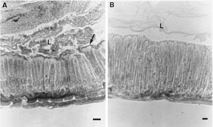
Fig. 4. Midgut cells of T. molitor larvae. (A) Cells of anterior midgut. Note apical extrusion (arrow). (B) Cells of posterior midgut. Note that these cells are taller and more slender than those in anterior midgut. L, lumen. Bar: 10µm.
although in lesser amounts. Associated with some midgut cells, mainly in anterior midgut, gregarines are observed (Fig. 5C). The gregarines do not seem to affect the cells to which they are associated, since the cells maintain their normal ultrastructure (not shown).
The basal plasma membrane and the basal part of the lateral membranes of midgut cells are highly infolded, forming long and narrow channels with openings toward the basal lamina (not shown). Some cells in anterior midgut show a less developed basal labyrinth, which suggest that they may correspond to the less frequent cells seen discharging all their apical cytoplasm into the lumen, after losing their microvilli (Fig. 6A).
3.4. Immunofluorescent visualization of amylase and trypsin in midgut cells
Fig. 5A–C shows that amylase is concentrated in a few anterior midgut cells, some of which are discharging amylase-containing material in the midgut lumen. Amyl-ase labeling in posterior midgut cells is detected only at the cell surface.
Trypsin is distributed along the posterior midgut, both at the surface and inside the cells, and is almost lacking in the anterior midgut. Some posterior midgut cells are more labeled than others. Controls made with non-immune serum were negative.
These studies agree with previous studies involving T.
molitor midgut cell fractionation that showed that
amyl-ase and trypsin are secreted by the anterior and posterior midgut, respectively (Terra et al., 1985; Ferreira et al., 1990). The results favor the view that amylase is secreted by an apocrine process (only the apical part of
the cell is discharged into the lumen), whereas trypsin may be released by an exocytic route.
It is noteworthy that the basal lamina shows fluor-escence for amylase and trypsin (Fig. 5). The simplest explanation for this phenomenon is that it is an artifact caused by the contamination of the basal lamina by digestive enzymes released into the dissection medium (the insect’s own hemolymph). T. molitor basal lamina is able to retain a large amount of fluid (Koefoed and Zerahn, 1982).
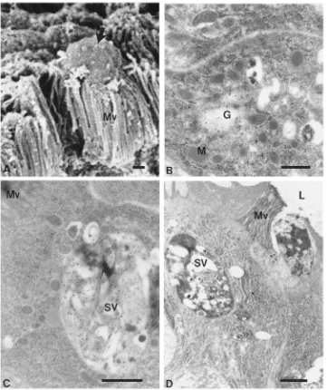
Fig. 5. Immunofluorescence visualization of amylase (A–C) and trypsin (D) in midgut cells. (A) Anterior midgut showing a single cell highly stained for amylase. (B) Anterior midgut showing cells highly stained for amylase with fluorescence concentrated in the apical cytoplasm. Note fluorescence in an apical extrusion (arrow). (C) Gregarine (arrow) associated with anterior midgut epithelium. Note that gregarine fluorescence is restricted to its surface. (D) Posterior midgut showing trypsin fluorescence along the epithelium, with some cells more labeled than others. Bars: 25µm (A, C), 10µm (B, D).
at their surface, fluorescence for trypsin is also found inside their cells (not shown).
3.5. Ultrastructural localization of amylase and trypsin in anterior midgut cells
Amylase is detected in Golgi areas (Fig. 6B), in large secretory vesicles near the cell apex (Fig. 6C), and apparently in the process of being extruded from the cell after the disappearance of microvilli and disorganization of cytoplasm (Fig. 6D). This sequence of pictures, together with those in Fig. 5, suggests that amylase is synthesized in anterior midgut cells and is packed in the Golgi area into secretory vesicles that undergo fusion, as they migrate to the cell apex. At the same time, the cell apex undergoes ultrastructural disorganization with the disappearance of cell organelles. Eventually, the api-cal cytoplasm with the large amylase-containing mem-branous structure is discharged into the midgut lumen. After extruding the apical cytoplasm, the cell apparently remains functional, since cells are found to lack the cell apex, but have all the other normal ultrastructural fea-tures (not shown).
Trypsin labeling occurs in trace amounts in anterior midgut cells in agreement with biochemical and immun-ofluorescence data (see above).
3.6. Ultrastructural localization of trypsin and amylase in posterior midgut cells
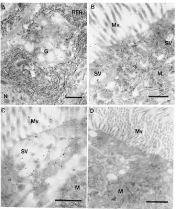
Trypsin is observed in Golgi areas (Fig. 7A) and inside small vesicles in the cell apex (Figs. 7B and 7C). Fig. 7B presents a cell (with few labeled vesicles) that corresponds to most cells displayed in Fig. 5D. In con-trast, the cell shown in Fig. 7C corresponds to the single highly labeled cell in Fig. 5D. Note that the diameter of the vesicles is close to the intermicrovillar distance (Fig. 7B) and that the vesicles do not fuse one with another (Fig. 7C), as occurs in the case of amylase-containing vesicles in the anterior midgut (Fig. 6C). Despite the weak labeling, secretory vesicles are consistently labeled above the background level, and Figs. 7B and 7C may be considered as typical. The features described for tryp-sin are those usual for proteins secreted by exocytosis (see Section 1).
sur-Fig. 6. Scanning electron microscopy (A) and immunocytochemical localization of amylase (B–D) in anterior midgut cells. (A) Luminal surface of anterior midgut. Note an apical extrusion (arrow). (B) Labeling in Golgi area. (C) Cell apex. Note heavily stained vesicle with membraneous profiles inside. (D) Cell apex with extruding secretory vesicles. Note cytoplasm and microvilli disorganization. G, Golgi; M, mitochondria; Mv, microvilli; SV, secretory vesicle. Bars: 1µm (A, C, D), 0.5µm (B).
face of midgut cells (Fig. 7D), in agreement with bio-chemical data that indicated that amylase is associated with the cell glycocalyx in T. molitor posterior midgut (Ferreira et al., 1990).
4. Discussion
4.1. Secretory mechanisms and water fluxes in T. molitor larval midguts
The larval midgut of T. molitor is about 27 mm long, with the foregut corresponding to 3%, the midgut to 67% and the hindgut to 30% of the total length. Most diges-tive enzymes are soluble and found in the midgut lumen:
the carbohydrases and a cysteine proteinase in the first two-thirds that has pH 5.6, and chymotrypsin, trypsin and a microvillar aminopeptidase in the last third that has pH 7.9 (Terra et al., 1985; Terra and Cristofoletti, 1996). Dye experiments showed that the anterior and middle midgut is water absorbing and that the posterior midgut is water secreting (Terra et al., 1985). These data, together with the finding of a low excretion rate of diges-tive enzymes, led to the proposal of an endo–ectoperi-trophic circulation of digestive enzymes and that most carbohydrate and protein digestion takes place in the anterior and posterior midgut, respectively (Terra et al., 1985 and Fig. 8).
The physiological differentiation observed along T.
Fig. 7. Immunocytochemical localization of trypsin and amylase in posterior midgut cells. (A) Trypsin labeling in Golgi area. (B) Cell apex. Note trypsin labeling in dispersed secretory vesicles. (C) Cell apex. Note trypsin labeling in secretory vesicles apparently in process of exocytosis and in microvilli. (D) Amylase labeling on the surface of microvilli. N, nucleus; RER, rough endoplasmic reticulum. Bars: 0.5µm (A, B, C), 1
µm (D).
features. The anterior and middle midgut epithelia are more folded and have more cells with apical extrusions than the posterior midgut, which possess taller and more slender cells. Anterior water absorption is assumed to be carried out by cells without apical extrusions, since these cells should correspond to those with less-developed basal labyrinths. A developed basal labyrinth is usually associated with cells undergoing water absorption (for references, see Terra et al., 1988). Posterior water secretion is thought to be carried out by all cells, since no cell differentiation is observed in the posterior midgut. Taking the activity of mitochondrial succinate dehydrogenase as an estimate of energy-consuming activities, posterior midgut cells are metabolically more active than those in other midgut regions (Ferreira et al.,
1990). This suggests that processes in the posterior region (midgut lumen alkalinization, water secretion, enzyme secretion, etc.) require more energy than those in the anterior region (water absorption, putative sugar and amino acid absorption, enzyme secretion, etc.).
As noted above, T. molitor midgut have cells showing apical extrusions, mainly at its anterior two-thirds. Some authors interpret these extrusions as holocrine (or apocrine) secretion, whereas others suggest that they are part of the cell renewal process (for review, see Terra and Ferreira, 1994). If the cells with apical extrusions were cells being replaced, one would expect that they would be devoid of digestive enzymes, the contrary being true if they were active secretory cells.
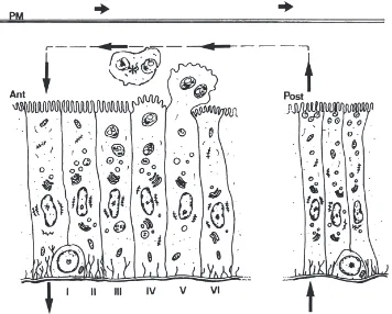
Fig. 8. Models for secretory processes and water fluxes in T. molitor. Enzymes and partially digested nutrients are suggested to be recovered from the interior of the peritrophic membrane (PM) via a countercurrent flux of the fluid (large arrows) caused by the transport of water into the posterior midgut lumen and its transfer back to the hemolymph in the anterior midgut. In the anterior midgut (Ant), absorptive columnar cells (II and III) have a more developed basal labyrinth and originate from regenerative cells at the base of the epithelium (I). As new cells arise from regenerative cells to replace lost cells, water-absorbing cells are laterally displaced and eventually lose part of the basal labyrinth and become involved in apocrine secretion. Amylase, a-glucosidase, and cellobiase are secreted by the anterior midgut cells by an apocrine mechanism. The enzymes are at first packed into secretory vesicles that fuse one with another as they migrate to the cell apex (III and IV). The cell apex undergoes ultrastructural disorganization (IV), bulges into the midgut lumen (V), and is released with large enzyme-containing structures (*). The cells recover after extrusion (VI) and initiate a new cycle of apocrine secretion. Trypsin (as trypsinogen) and carboxypeptidase occur in secretory vesicles and are secreted by exocytosis in posterior midgut (Post).
along the entire anterior midgut, certain cells are remark-ably stained with the fluorescence concentrated in the apical cytoplasm of the midgut cell. In some cells, this apical region, highly stained, bulges into the midgut lumen, suggesting an apocrine mechanism of secretion, which is corroborated by immunolocalization experi-ments performed at the ultrastructural level. At this level, it is possible to detect the presence of amylase in Golgi areas and in large secretory granules near the cell apex, where the cytoplasm shows signs of ultrastructural dis-organization.
The immunocytochemical data summarized above are in agreement with previous work by Ferreira et al. (1990). These authors introduced experimental criteria to identify microvillar enzymes, enzymes weakly and strongly associated with the cell glycocalyx, enzymes present inside and membrane-bound to vesicles of differ-ent sizes and, finally, enzymes occurring free in the cyto-sol. The criteria were based on different cell fraction-ation procedures, including differential centrifugfraction-ation. According to these criteria, Ferreira et al. (1990)
pro-posed that amylase occurs in large vesicles in the anterior midgut cells and is only associated with the cell glycocalyx in the posterior midgut. This conforms with data in the present paper (Fig. 6C and Fig. 7C). Thus the data provide strong evidence that amylase is secreted by anterior midgut cells by an apocrine process involv-ing secretory vesicles. This contrasts with apocrine mechanisms known for vertebrates. In these animals, apocrine-secreted proteins are transported inside the cells without the participation of the endomembrane
sys-tem, the Golgi apparatus and secretion granules
(Aumu¨ller et al., 1999).
activity of a marker enzyme in this preparation is as high as that after Tris treatment known to remove the cytoske-leton (Jorda˜o et al., 1995). By contrast, T. molitor pos-terior midgut microvilli prepared as before and treated with Tris result in a threefold increase in the specific activity of a marker enzyme (Jorda˜o et al., 1995). It is interesting to note that an immunocytochemistry study of apocrine-secreting vertebrate cells failed to detect the
cytoskeleton proteins villin anda-actinin in the cell apex
(Aumu¨ller et al., 1999).
In the T. molitor posterior midgut, trypsin staining is observed along the region, with fluorescence detected in small vesicles that are seen mainly around the nuclei and in the apical cytoplasm. In fact, ultrastructural data show the presence of immunolabeled trypsin in Golgi areas and inside small vesicles, which in some cells are con-centrated near the apical plasma membrane, suggesting an exocytic mechanism of secretion. The weak trypsin labeling observed in each secretory vesicle may result from a low content of the enzyme and/or may be caused by a low titre of the anti-trypsin serum. In relation to trypsin, Ferreira et al. (1990) confirmed that this enzyme occurs in small vesicles in the posterior midgut and is almost absent from the anterior midgut. This agrees with data in the present paper (Fig. 7B and C and text).
Trypsin occurs in T. molitor posterior midgut as a sol-uble precursor that converts easily into a smaller trypsin molecule (Fig. 3A). It is likely that this precursor is a trypsinogen (inactive precursor of trypsin), although, among insects, only in S. calcitrans midgut has a tryp-sinogen been identified unambiguously (Moffat and Lehane, 1990). It seems that insects may control the activity of their digestive proteinase, in the absence of inactive forms, by binding the proteinases to membrane vesicles until they are released into the midgut lumen. Thus, in larval flies, trypsin is solubilized upon exocytosis because the pH of the luminal contents causes a conformational change that hinders part of the trypsin anchor (Jorda˜o et al., 1996a). In caterpillars, after microapocrine release of membrane-bound trypsin, this enzyme is partly processed to a soluble form and partly, still bound to vesicle membranes, incorporated into the peritrophic membrane (Ferreira et al., 1994; Jorda˜o et al., 1999).
Since maltase and cellobiase have the same intracellu-lar distribution as amylase, and carboxypeptidase the same as trypsin (Ferreira et al., 1990), it is probable that all carbohydrases are released into the anterior midgut lumen by apocrine mechanisms and the peptidases into posterior midgut lumen by exocytosis.
Gregarines are found in T. molitor midgut lumen and are associated with the epithelium (Fig. 5C). Gregarines do not seem to harm the cells, since gregarine-associated cells maintain normal ultrastructure. Gregarines show amylase fluorescence only at their surface, but trypsin fluorescence is observed inside their cells. Probably T.
molitor trypsin is taken up by gregarines by endocytosis,
although the existence of a gregarine trypsin sharing epi-topes with T. molitor trypsin cannot be discounted.
The features discussed above, as well as the hypoth-eses about circulation of fluids thought to occur in T.
molitor larval midguts (Terra et al., 1985), have been
incorporated into a model of secretory processes and water fluxes in T. molitor larval midgut (Fig. 8).
4.2. A phylogenetic view of secretory mechanisms in insect midguts
Digestive enzymes are usually secreted by exocytosis in the posterior midgut, whereas alternate mechanisms may be observed in anterior midgut. Alternate mech-anisms found in anterior midgut and studied by both bio-chemical and cytological procedures are: (1) pinching-off vesicles from midgut microvilli (a microapocrine process) in Erinnyis ello (Santos et al., 1986) and in S.
frugiperda (Jorda˜o et al., 1999); and (2) apical cell
extrusions (apocrine secretion) in T. molitor (this paper). Based only on morphological evidence, we may say that, in addition to E. ello and S. frugiperda, microapocrine secretion occurs in other Lepidopteran species, such as
Manduca sexta (Cioffi, 1979), whereas apocrine secretion is observed in some Orthoptera (Heinrich and Zebe, 1973) and in many coleopteran species other than
T. molitor (Bayon, 1981; Silva and Souza, 1981; Baker
et al., 1984).
Apocrine and microapocrine secretion of digestive enzymes in insect anterior midgut cells, with the release of enzyme-containing vesicles, may be an adaptation to enhance the dispersion of secretory vesicle contents into the midgut lumen of water-absorbing tissues. The move-ment of fluid toward anterior midgut cells would prevent a uniform diffusion of material secreted by exocytosis. As posterior midgut cells secrete fluid, no problem arises in the dispersion of material released by exocytosis by these cells. Based on known examples, apocrine mech-anisms appear to be widespread among less-evolved insects, whereas more-derived insects use microapocrine mechanisms in water-absorbing tissues.
Apparent exceptions to the above rules are known. Thus, cyclorrhaphan dipteran larvae (including Musca
domestica) lack apocrine/microapocrine processes in the
Acknowledgements
This work was supported by the Brazilian research agencies FAPESP and FINEP (PRONEX program). We thank Dr C. Ferreira for helpful discussions and L.Y. Nakabayashi, W. Caldeira and M.V. Cruz for technical assistance. P.T.C. is a graduate fellow of FAPESP and A.F.R. and W.R.T. are staff members of their respective departments and research fellows of CNPq.
References
Aumu¨ller, G., Wilhelm, B., Seitz, J., 1999. Apocrine secretion—fact or artifact? Annals of Anatomy 181, 437–446.
Baker, J.E., Woo, S.M., Byrd, R.V., 1984. Ultrastructure of the gut of
Sitophilus granarius (L.) (Coleoptera: Curculionidae) with notes on
distribution of proteinases and amylases in crop and midgut. Can-adian Journal of Zoology 62, 1251–1259.
Bayon, C., 1981. Ultrastructure de l’epithelium intestinal et flore parie-tale chez la larve xylophage d’Oryctes nasicornis L. (Coleoptera: Scarabaeidae). International Journal of Insect Morphology and Embryology 10, 359–371.
Berdan, R.C., Lees-Miller, J.P., Caveney, S., 1985. Lack of cell com-munication in an epithelium: ultrastructure and electrophysiology of the midgut epithelium of the larval mealworm, Tenebrio molitor. Journal of Ultrastructural Research 90, 55–70.
Blum, H., Beier, H., Gross, H.J., 1987. Improved silver staining of plant proteins, RNA and DNA in polyacrylamide gels. Electro-phoresis 8, 93–99.
Bradford, M.M., 1976. A rapid and sensitive method for the quantit-ation of microgram quantities of protein utilizing the principle of protein–dye binding. Analytical Biochemistry 72, 248–254. Buonocore, V., Poerio, E., Silano, V., Tomasi, M., 1976. Physical and
catalytic properties ofa-amylase from Tenebrio molitor L. larvae. Biochemical Journal 153, 621–625.
Cioffi, M., 1979. The morphology and fine structure of the larval midgut of a moth (Manduca sexta) in relation to active ion trans-port. Tissue and Cell 11, 467–479.
Davis, B.J., 1964. Disc electrophoresis. II. Methods and application to human serum proteins. Annals of the New York Academy of Sciences 121, 404–427.
Erlanger, B.F., Kokowsky, N., Cohen, W., 1961. The preparation and properties of two new chromogenic substrates of trypsin. Archives of Biochemistry and Biophysics 95, 271–278.
Ferreira, C., Bellinello, G.L., Ribeiro, A.F., Terra, W.R., 1990. Diges-tive enzymes associated with the glycocalyx, microvillar mem-branes and secretory vesicles from midgut cells of Tenebrio molitor larvae. Insect Biochemistry 20, 839–847.
Ferreira, C., Capella, A.N., Sitnik, R., Terra, W.R., 1994. Digestive enzymes in midgut cells, endo- and ectoperitrophic contents, and peritrophic membranes of Spodoptera frugiperda (Lepidoptera) lar-vae. Archives of Insect Biochemistry and Physiology 26, 299–313. Graf, R., Raikhel, A.S., Brown, M.R., Lea, A.O., Briegel, H., 1986. Mosquito trypsin: immunocytochemical localization in the midgut of blood-fed Aedes algypti (L.). Cell and Tissue Research 245, 19–27.
Heinrich, D., Zebe, E., 1973. Zur Feinstruktur der Mitteldarmzellen von Locusta migratoria in verschiedenen Phasen der Verdauung. Cytobiologie 7, 315–326.
Jorda˜o, B.P., Terra, W.R., Ferreira, C., 1995. Chemistry of microvillar membranes purified from brush borders isolated from the larval midgut from one Coleoptera and two Diptera species. Insect Bio-chemistry and Molecular Biology 25, 417–426.
Jorda˜o, B.P., Terra, W.R., Ribeiro, A.F., Lehane, M.J., Ferreira, C., 1996a. Trypsin secretion in Musca domestica larval midguts: a bio-chemical and immunocytobio-chemical study. Insect Biochemistry and Molecular Biology 26, 337–346.
Jorda˜o, B.P., Lehane, M.J., Terra, W.R., Ribeiro, A.F., Ferreira, C.F., 1996b. An immunocytochemical investigation of trypsin secretion in the midgut of the stablefly Stomoxys calcitrans. Insect Biochem-istry and Molecular Biology 26, 445–453.
Jorda˜o, B.P., Capella, A.N., Terra, W.R., Ribeiro, A.F., Ferreira, C., 1999. Nature of the anchors of membrane-bound aminopeptidase, amylase, and trypsin and secretory mechanisms in Spodoptera
frug-iperda (Lepidoptera) midgut cells. Journal of Insect Physiology 45,
29–37.
Koefoed, B.M., 1987. The ability of an epithelium to survive removal of the basal lamina by enzymes: fine structure and content of sodium and potassium of the midgut epithelium of the larva of
Tenebrio molitor after withdrawal of the basal lamina by elastase—
a short note. Tissue and Cell 19, 65–70.
Koefoed, B.M., Zerahn, K., 1982. Transport of sodium and potassium across the isolated midgut of the larvae of Tenebrio molitor related to the fine structure of the epithelium. Journal of Experimental Biology 98, 459–463.
Laemmli, U.K., 1970. Cleavage of structural proteins during the assembly of the head of bacteriophage T4. Nature 227, 680–685.
Levinsky, H., Birk, Y., Applebaum, S.W., 1977. Isolation and charac-terization of a new trypsin-like enzyme from Tenebrio molitor L. larvae. International Journal of Peptide and Protein Research 10, 252–264.
Marana, S., Ribeiro, A.F., Terra, W.R., Ferreira, C., 1997. Ultrastruc-ture and secretory activity of Abracris flavolineata (Orthoptera: Acrididae) midguts. Journal of Insect Physiology 43, 465–473. Moffat, M.R., Lehane, M.J., 1990. Trypsin is stored as an inactive
zymogen in the midgut of Stomoxys calcitrans. Insect Biochemistry 20, 719–723.
Nishiitsuji-Uwo, J., Endo, Y., 1981. Gut endocrine cells in insects: the ultrastructure of the endocrine cells in the cockroach midgut. Biomedical Research 2, 30–44.
Noelting, G., Bernfeld, P., 1948. Sur les enzymes amylolytiques. III. Lab-amylase: dosage d’activite´ et controˆle de l’absence d’a -amyl-ase. Helvetica Chimica Acta 31, 286–290.
Raes, H., Verbeke, M., 1994. Light and electron microscopical study of two types of endocrine cell in the midgut of the adult worker honeybee (Apis mellifera). Tissue and Cell 26, 223–230. Santos, C.D., Ribeiro, A.F., Terra, W.R., 1986. Differential
centrifug-ation, calcium precipitation and ultrasonic disruption of midgut cells of Erinnyis ello caterpillars. Purification of cell microvilli and inferences concerning secretory mechanisms. Canadian Journal of Zoology 64, 490–500.
Silva, I., Souza, V.B.V., 1981. Ultrastructural aspects of the anterior midgut of Oncideres s. saga (Dalman, 1823) larva (Coleoptera: Cerambycidae). Revista Brasileira de Entomologia 25, 103–112. Silva, C.P., Ribeiro, A.F., Gulbenkian, S., Terra, W.R., 1995.
Organi-zation, origin and function of the outer microvillar (perimicrovillar) membranes of Dysdercus peruvianus (Hemiptera) midgut cells. Journal of Insect Physiology 41, 1093–1103.
Stefanini, M., De Martino, C., Zamboni, L., 1967. Fixation of ejacu-lated spermatozoa for electron microscopy. Nature 216, 173–174. Terra, W.R., Cristofoletti, P.T., 1996. Midgut proteinases in three divergent species of Coleoptera. Comparative Biochemistry and Physiology 113B, 725–730.
Terra, W.R., Ferreira, C., 1994. Insect digestive enzymes: properties, compartmentalization and function. Comparative Biochemistry and Physiology 109B, 1–62.
Terra, W.R., Espinoza-Fuentes, F.P., Ribeiro, A.F., Ferreira, C., 1988. The larval midgut of the housefly (Musca domestica): ultrastruc-ture, fluid fluxes and ion secretion in relation to the organization of digestion. Journal of Insect Physiology 34, 463–472.
Towbin, H., Staehelin, T., Gordon, J., 1979. Electrophoretic transfer of proteins from polyacrylamide gels to nitrocellulose sheets: pro-cedure and some applications. Proceedings of the National Acad-emy of Sciences (Washington) 76, 4350–4354.
Wigglesworth, V.B., 1972. The Principles of Insect Physiology, 7th edn. Methuen, London.