* corresponding author: riswanhk @yahoo.com
Serum calcium ions, ratio of calcium/
creatinine urine and bone mass density
in perimenopausal and postmenopausal
women
Riswan Hadi Kusuma1*, Windarwati2, Harjo Mulyono2
1Academic Hospital, UGM,2Department of Patology Clinic, Faculty of Medicine,
Universitas Gadjah Mada, Yogyakarta
ABSTRACT
Early diagnosis of osteoporosis is important to take early prevention and treatment. Dual energy X-ray absorbtiometry (DEXA) scan is gold standard diagnosis of osteoporosis. However, it is high technology and high cost. Biochemical marker using calcium could be developed for diagnosis of osteoporosis. The study was conducted to evaluate the correlation between serum calcium ions level as well as urine calcium and urine creatinine (UCa/UCr) ratio and Bone Mineral Density (BMD) in perimenopausal and postmenopausal women. This was an observational study with cross-sectional design involving women aged 45-75 years who fulfilled the inclusion and exclusion criteria. Bone Mineral Density was measured using DEXA method. Serum and urine calcium ions were measured using ISE method, while serum and urine creatinine were determined using Jaffe method. Ratio of UCa/UCr were then calculated. The correlation between serum calcium ions as well as the ratio UCa/UCr and BMD of subjects were then analyzed. A total 63 subjects were involved in this study consists of 21 normal subjects, 22 subjects with osteopenia and 20 subjects with osteoporosis. Negative correlation between BMD and age (r=-0.591; p=0.001) and positive correlation between BMD and body mass index (BMI) (r=0.432; p=0.001) were observed. No correlation between serum calcium ions and BMD (r= -0.145; p=0.258), however negative correlation between UCa/UCr ratio and BMD (r=-0.310; p=0.013) were reported. Furthermore, no correlation was found between serum calcium ions and lumbar BMD (r=0.036; p=0.778), while negative correlation was found between UCa/UCr ratio and lumbar BMD (r= -0.414; p=0.001). In conclusion, there is no correlation between serum calcium ions levels and BMD, however there is a weak negative correlation between UCa/UCr ratio and BMD.
ABSTRAK
p=0,001) dan hubungan positif antara BMD dan indeks masa tubuh (IMT) ((r=0,432; p=0,001). Tidak terdapat hubungan antara ion kalsium serum dan BMD (r=-0,145; p=0,258), namun terdapat hubungan negatif antara rasio UCa/UCr dan BMD (r=0,310; p=0,013). Selanjutnya terbukti tidak ada hubungan antara ion kalsium serum dan BMD lumbal (r= 0.036; p=0,778), namun demikian terdapat hubungan negatif antara rasio UCa/UCr dan BMD lumbal (r= -0,414; p=0,001). Dapat disimpulkan, tidak terdapat hubungan antara kadar ion kalsium serum dengan BMD, namun terdapat hubungan negatif lemah antara rasio UCa/UCr dengan BMD.
Keywords:osteoporosis - calcium ions - serum - urine - DEXA
INTRODUCTION
The increase of life expectancy and the lack of knowledge concerning osteoporosi prevention leads to the high risk of osteroporosis
of women population in Indonesia.1
Osteoporosis is defined as a systemic bone disease characterized by low bone mass density (BMD) and microarchitectural deterioration of bone tissue, leading to enhanced bone fragility and a consequent increase in fracture risk.2,3The
prevalence of osteoporosis is growing, especially as the number of postmenopausal women in the population continues to rise. An estimated 52 million women aged fifty years plus are expected to be affected by osteoporosis and osteopenia by 2010 and 61 million are expected to be affected by 2020.4
Concern has been addressed recently about the early screening of osteoporosis in perimeno-pausal and postmenoperimeno-pausal women. This early screening is important to take action about early prevention and treatment of the osteoporosis. Several osteoporosis examinations including radiological, biopsy and biochemical examinat-ions have been developed base on reduction of
BMD in the osteoporosis.3,5 Radiological
examination to assess BMD with dual X-ray method absorbtiometry (DEXA) is the gold
standard for diagnosing osteoporosis.6
However, the radiological examination needs high technology and cost. Therefore, it can not be applied in all health care centers. Whereas, the biopsy examination is too invasive and the
results on one part of skeleton cannot be applied to the other skeleton.7
In order to obtain an effective and efficient method, several biochemical markers reflecting a bone state or bone remodeling process have being developed for diagnosing osteoporosis. Calcium, well known as a bone resorption marker, is recommended as biochemical marker for osteoporosis screening. Three forms of calcium exist in the blood i.e. calcium ions, protein-bound calcium and complex-bound calcium. The serum calcium ions is the active form reflecting physiological and pathological conditions of the body.8,9
This study was conducted to evaluate the correlation between serum calcium ions level and BMD in perimenopausal and post-menopausal women. The correlation between ratio of urine calcium and urine creatinine (UCa/UCr) for 24-hour urinary excretion was also evaluated. This UCa/UCr ratio can be used to estimate the mean calcium excretion in the urine.10,11
MATERIALS AND METHODS
Subjects
This was an observational study with cross-sectional design conducted in the Integrated
Services Post of Elderly (Pos Pelayanan
Terpadu Lanjut Usia/Posyandu Lansia) in
fulfilled the inclusion and exclusion criteria. The inclusion criteria were perimenopausal and postmenopausal women, while the exclusion criteria were patients with a history of malignancy, underwent chemotherapy or radiation therapy, had renal failure or renal dysfunction (serum creatinine > 1.3 mg/dL or creatinine clearance <88 mL/minute/1.73m2 or urine creatinine levels low/high), had metabolic bone and joint diseases, history of surgical removal of the ovaries, underwent a hormone replacement therapy (HRT) and unable to collect samples of serum and urine appropriatly. The study would be conducted after obtained approval from the Medical and Health Research Ethics Committee, Faculty of Medicine, Universitas Gadjah Mada, Yogyakarta.
Procedure of study
On the day when study started, subjects were consecutively selected. An explanation concerning the background, objectives and benefits of the study was given. Subjects who
fulfilled the inclusion and exclusion criteria were given an informed consent to be signed. Anamnesis was then performed to gather informations of subject characteristics. Physical examination including body weight and body height measurement, Body Mass Index (BMI) calculation was also conducted. Bone Mineral Density (BMD) examination was conducted followed by serum and urine ionized calcium examination as well as serum and urine creatinine examination. Ratio of urine calcium and urine creatinine (UCa/UCr) were then calculated. The correlation between serum calcium ions and the ratio UCa/UCr with BMD of subjects was analyzed.
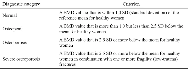
The BMD measurement to diagnose the osteoporosis of women subjects was conducted in Department of Radiology, Dr. Sardjito General Hospital using a dual-energy X-ray absorptiometry (DEXA) scan. The BMD value obtained was used to calculate T scores which used to evaluate degree of osteoporosis of the subjects according to World Health Organization criteria (TABLE 1).
TABLE 1. Diagnosis of osteroporosis based on BMD value
Serum and urine calcium ions were measured using ISE method in the Clinical Laboratory Installation, Dr. Sardjito General Hospital using a Beckman Coulter Synchron
chemistry Analyzer CX Pro. Subject fasted for 10 hours before serum and urine sample were taken. No vigorous physical activities were
libitum was provided to subjects. Serum and urine creatinine were measured using Jaffe method in the same laboratory. Assessment of renal function was performed using serum creatinine levels or creatinine clearance or urine creatinine using the Cockcroft-Gault formula.
Statistical analysis
Diagnostic performance tests including calibration, precision and accuracy was conducted for each measurement method. The precision of measurement method was expressed by mean, standard deviation (SD) and calculated the coefficient of variation (CV) value, whereas the accuracy was expressed by the closeness of a measured value to a standard or true value. Statistical analysis used were the Kolmogorov-Smirnov, Chi-square, one-way Anova and Pearson correlation. Value of p < 0.05 with 95% Confidence Interval (95% CI) was considered as statistically significant.
RESULS
Characteristics of subjects
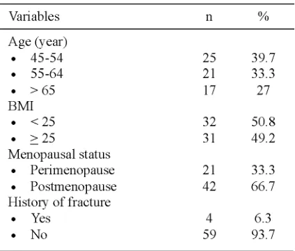
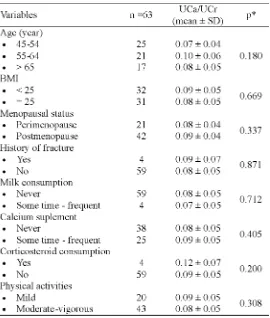
Sixty-three eligible subjects were involved in this study. The characteristics of subjects are presented in TABLE 2.
TABLE 2. Characteristics of subjects
TABLE 2. Characteristics of subjects (lanjutan
The status of BMD of subjects
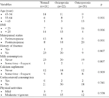
The BMD status of subjects risk factors of osteoporosis is presented in TABLE 3. BMD examination was performed on hip and lumbar regions. The BMD value was then used to determine T-score which would be used to determine osteoporosis status of subjects based on WHO criteria. Among 63 subjects who examined, 21 (33.3%) subjects had normal BMD, 22 (34.9%) had osteopenia and 20 subjects (31.7%) subjects had osteoporosis.
TABLE 3. Status of BMD of subjects based on risk factors of osteoporosis
* One way Anova (significant different if p<0.05)
Further analysis showed that there was a negative correlation between BMD and age (r= -0.591; p=0.001) and positive correlation
between BMD and BMI (r=0.432; p=0.001) as presented in TABLE 4.
TABLE 4. Correlation between BMD status and age or BMI
*Pearson correlation test (significant different if p<0.05)
Serum calcium ions of subjects
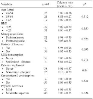
The mean of serum calcium ions levels and the ratio of UCa/UCr based on the risk factors of osteoporosis is presented in TABLE 5 and 6. No significantly difference was observed in serum calcium ions levels and in the ratio of
TABLE 5. Mean of serum calcium ions (mg/dL) based on the risk factors of osteoporosis
TABLE 6. Mean of UCa/UCr ratio based on the characteristics of subjects
Moreover, no significant difference was also observed in the mean serum calcium ions levels and the ratio of UCa/UCr based on the
osteoporosis diagnostic category (p>0.05) as presented in TABLE 7.
*One way Anova (significant different if p<0.05)
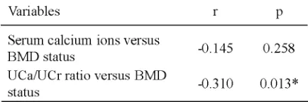
Correlation between serum calcium ions or UCa/UCr ratio and BMD
The correlation between serum calcium ions levels and BMD and between UCa/UCr ratio and BMD are presented in TABLE 8. No significant correlation was observed between serum calcium ions level and BMB (r= -0.145, p=0.258), however weak significant negative correlation was observed between UCa/UCr ratio and BMD (r= -0.310, p=0.013).
TABLE 8. Correlation between serum calcium ions levels or UCa/UCr ratio and BMD
*Pearson correlation test (significant different if p<0.05)
Further analysis showed that no correlation was observed between serum calcium ions levels and lumbar BMD (r=0.036, p=0.778), whereas the ratio of UCa/UCr showed a moderate negative correlation with lumbar BMD (r=-0.414, p=0.001). The correlation between the ratio of UCa/UCr and lumbar BMD was stronger than that between the ratio of UCa/ UCr and BMD (r=-0.414 vs. r=-0.310).
TABLE 9. Correlation between serum calcium ions levels or UCa/UCr ratio and lumbar BMD
*Pearson correlation test (significant different if p<0.05)
DISCUSSION
This study showed that age, BMI and menopausal status were significantly associated with osteroporosis status subjects, whereas history of fracture, milk consumption, calcium suplement, corticosteroid consumption and physical activities were not associated with osteoporosis status in perimenopause and postmenopause women. The results are consistent with previous studies.2,12Bone mass
density has changed with age both in men and women. In the beginning, the process of bone remodeling is in balance or showed a surplus and no reduction in bone mass. However, as humans get older, the bone formation process becomes inadequate, resulting in decreased bone mass. In women, this process occurs from the third decade of human life or several years before menopause.13,14
This study also showed that no significantly difference was observed in serum calcium ions levels and in the UCa/UCr ratio based on the risk factors osteoporosis namely age, BMI, menopausal status, history of fracture, milk consumption, calcium supplements, and corticosteroid consumption as well as physical activities. These results are consistent with previous studies conducted by Georgeet al.that reported no significant difference in serum calcium levels between subjects with low BMD
and normal BMD.15 The concentration of
calcium ions in extracellular fluid is always maintained in contant state through calcium homeostatis mechanism. The most important hormones for maintaining calcium levels in the body are parathyroid hormone (PTH) and 1,25-dihydroxyvitamin D (the active form of vitamin D) and calcitonin.9 The major regulator is PTH,
high.9,16 This may underlie the absence of
difference of serum calcium ions levels in the normal bone density, osteopenia and osteoporosis.
The mean UCa/UCr ratio increases along with the decrease of BMD. The mean UCa/UCr ratio was higher in both osteopenia dan osteoporosis groups compare to normal group, however it was not significantly different. Previous studies showed that the increase of UCa/UCr ratio was paralel with the increase of the severity of osteoporosis. Urinary calcium levels of postmenopausal women with osteoporosis was hingher than those without osteoporosis.16Another study also showed that
the mean UCa/UCr ratio of postmenopausal women with osteoporosis was higher than those without osteoporosis. Moreover, the mean UCa/ UCr ratio of postmenopausal women was higher
than pramenopausal women.17Perimenopause
and postmenopause are associated with the decrease in estrogen levels in the body that leads to the decrease of urinary calcium resoption in the kidney.18
In this study, no correlation was observed between serum calcium ions levels and BMD or lumbar BMD, whereas the ratio of UCa/UCr showed a weak negative correlation with BMD and a moderate negative correlation with lumbar BMD. This results are consistent with those previous studies.19-22Ratio of UCa/UCr is a
biochemical marker that is used at an early stage to assess bone metabolism.23 The increase of
urinary calcium excretion and calcium malabsorption may be a predictor of low BMD in postmenopausal women,24and may indicate
an increase in bone resorption by osteoclasts.11
CONCLUSION
In conclusion, there is no correlation between serum calcium ions level and BMD, however there is a weak negative correlation
between the UCa/UCr ratio and BMD in perimenopause and postmenopause women. Ratio of UCa/UCr can be considered as biochemical markers in osteoporosis diagnosing in subject with normal renal function. Further study should be conducted to evaluate changes in other biochemical markers with the decrease of BMD.
ACKNOWLEDGEMENTS
Authors would like to thank Head of Department of Clinical Pathology as well as Head of Department of Radiology, Universitas Gadjah Mada/Dr. Sardjito General Hospital, Yogyakarta for their support in this study. We would also like to thank all subjects who willing to participate in this study.
REFERENCES
1. Menteri Kesehatan Republik Indonesia. Keputusan Menteri Kesehatan Republik Indonesia Nomor 1142/MENKES/SK/XII/2008 Tentang pedoman pengendalian osteoporosis. Jakarta: Kepmenkes RI, 2008.
2. Stabler CAH. Osteoporosis from pathophysiology to treatment. Washington: AACC Press. 2004. 3. Nelson HD, Helfand M, Woolf SH, Allan J.
Screening for postmenopausal osteoporosis: a review of the evidence for the US preventive services task force. Ann Intern Med 2002;137(6):529-41.
4. Chesnut III CH, Skag A, Christiansen C, Recker R, Stakkestad JA, Hoiseth A,et al. Effects of oral ibandronate administered daily or intermittently on fracture risk in postmenopausal osteoporosis. J Bone Miner Res 2004;19(8):1241-9.
5. Seibel JM. Biochemical markers of bone turnover part I: biochemistry and variability. Clin Biochem Rev 2005; 26(4):97-122.
6. Thomakos N, Liakakos T. Diagnostic methods in osteoporosis. Arch Hellenic Med 2000; 17(2):146-51.
8. Lewandrowski K. Clinical chemistry. Philadelphia: Lippincott Williams and Wilkins, 2002.
9. Guyton AC, Hall JE. Buku ajar fisiologi kedokteran, edisi 11. Jakarta: Penerbit Buku Kedokteran EGC, 2008.
10. Nicoll D, McPhee SJ, Pignone M, Lu CM. Pocket guide to diagnostic Test, 5th
edition, New York: McGraw-Hill, 2008.
11. Burtis CA, Ashwood ER, Bruns DE. Tietz textbook of clinical chemistry and molecular diagnostics, 4th
Edition, Philadelphia: Saunders Elsevier, 2006. 12. Rudiansyah M, Kertia N, Achadiyono DN, Probosuseno, Purnomo S. Hubungan roboh dengan bone mass densitydi. Klinik Lansia RSUP Dr. Sardjito Yogyakarta. Berkala Kesehatan Klinik 2007; 8(2):82-88.
13. Seeman E. Invite review: pathogenesis of osteoporosis. J Appl Physiol. 2003; 95(5):2142-51.
14. Warming L, Hassager C, Christiansen C. Changes in bone mineral density with age in men and women: a longitudinal study. Osteoporosis Int 2002; 13(2):105-12.
15. George J, Ganesh HK, Acharya S, Bandgar TR, Shivane V, Karvat A,et al.Bone mineral density and disorders of mineral metabolism in chronic liver disease. World J Gastroenterol 2009; 15(28):3516-22.
16. Cohen AJ, Roe FJC. Review of risk factors for osteoporosis with particular reference to a possible aetiological role of dietary salt. Food Chem Toxicol 2000; 38: 237-53.
17. Nordin BE, Need AG, Morris HA, Horowitz M. The nature and significance of the relationship between urinary sodium and urinary calcium in women. J Nutr 1993; 123(9):1615-22.
18. Väänänen HK, Härkönen PL. Estrogen and bone metabolism. Maturitas 1996; 23(Suppl):S65-9. 19. Tanzil Y. Hubungan kadar kalsium urin dengan
osteoporosis pada wanita postmenopause. [Tesis]. Yogyakarta: Program Studi Patologi Klinik, Sekolah Pasca Sarjana Universitas Gadjah Mada; 2007.
20. Lee YH, Rho YH, Choi SJ, Ji JD, Song GG. Predictors of bone mineral density and osteoporosis in patients attending a rheumatology outpatient clinic. Rheumatol Int 2003; 23(2):67-9.
21. Sakuma M, Endo N, Oinuma T, Hayami T, Endo E, Yazawa T,et al.Vitamin D and intact PTH status in patients with hip fracture. Osteoporos Int 2006; 17(11):1608-14.
22. Freundlich M, Alonzo E, Bellorin-Font E, Weisinger JR. Reduced bone mass in children with idiopathic hypercalciuria and in their asymptomatic mothers. Nephrol Dial Transplant 2002; 17(8):1396-401.
23. Lakatos P, Takacs I, Buki B, Nemeth J, Horvath C. Urinary calcium excretion: normal values for urinary calcium/creatinine ratio in Hungary. Orv Hetil 1997; 138(22):1405-9.