Tatas H.P. Brotosudarmo, and Richard J. Cogdell
* Corresponding author. Tel/Fax : 0044-141-330 7264/3779 Email address : [email protected]
STUDY ON THE STRUCTURAL BASIS OF PERIPHERAL LIGHT HARVESTING COMPLEXES
(LH2) IN PURPLE NON-SULPHUR PHOTOSYNTHETIC BACTERIA
REVIEW
Tatas H.P. Brotosudarmo1,2,*, and Richard J. Cogdell1
1
Division of Biochemistry & Cell Biology, University of Glasgow Biomedical Research Building, 126 University Place, G12 8TA, United Kingdom b
Ma Chung Research Center for Photosynthetic Pigments, Ma Chung University Jl. Villa Puncak Tidar N/01, Malang 65151, Indonesia
Received May 20, 2010; Accepted August 26, 2010
ABSTRACT
Photosynthesis provides an example of a natural process that has been optimized during evolution to harness solar energy efficiently and safely, and finally to use it to produce a carbon-based fuel. Initially, solar energy is captured by the light harvesting pigment-protein complexes. In purple bacteria these antenna complexes are constructed on a rather simple modular basis. Light absorbed by these antenna complexes is funnelled downhill to reaction centres, where light drives a trans-membrane redox reaction. The light harvesting proteins not only provide
the scaffolding that correctly positions the bacteriochlorophyll a and carotenoid pigments for optimal energy transfer
but also creates an environment that can modulate the wavelength at which different bacteriochlorophyll molecules absorb light thereby creating the energy funnel. How these proteins can modulate the absorption spectra of the bacteriochlorophylls will be discussed in this review.
Keywords: photosynthesis, peripheral light harvesting complex, H-bonding, energy transfer
INTRODUCTION
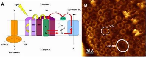
In the early processes of photosynthesis, sunlight is absorbed and this excitation energy is then funnelled to the reaction centre (RC), where the primary charge separation takes place (Fig. 1A) [1-2]. The initial absorption of solar energy occurs in light harvesting pigment-protein complexes that surround the reaction centres (Fig. 1) [3-5]. In principle, photosynthesis could have evolved just with RCs. However, this would have meant that except under very high intensity light there would be a relatively long time-gap between two photons reaching the same RC. This would have cause a major problem because several of the redox reactions that take place within the RCs require multiple one-electron turnovers. If the RCs had to wait too long between the arrivals of the consecutive photons then back reactions would become favourable. In this case the whole charge-separation process would become inefficient. One of the important function of a LH system is to increase cross-sectional area for photon capture in order to supply the RCs with sufficient numbers of photons, so that the forward electron-transfer reactions take place frequently enough and the back reactions are reduced to a minimum [2].
In the purple bacteria there are two types of light harvesting (LH) complexes, the peripheral (LH2) and the core (LH1). Both LH complexes contain a pair of small
(5-7 kDa) transmembrane polypeptides, called α and β, that oligomerise to form the intact native complexes. The N-termini of these apoproteins are located at the cytoplasmic surface of the photosynthetic membrane and
Fig 1. A schematic diagram of the photosynthetic
membrane of a typical purple bacterium (A). The major integral membrane protein involved in the light reaction of photosynthesis are displayed. The yellow arrows show energy transfer and the red arrows the redox reactions involved their simple cyclic electron transport pathway. The reaction centre (RC) reduces the secondary electron acceptor (ubiquinone, QB) which
has to pass through the LH1 complex in order to deliver its reducing equivalents to cyclic electron pathway. (B) An image of the photosynthetic membrane from Phs.
molischianum taken by atomic force microscopy (AFM)
Tatas H.P. Brotosudarmo, and Richard J. Cogdell
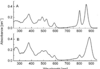
Fig 2. Room temperature absorption spectra of LH2 and LH1 complexes from Rhodopseudomonas acidophila
10050.
Fig 3. First step of isolation and purification of LH2 and LH1-RC complexes from Rps. palustris. The photosynthetic membrane was solubilized by 1% (v/v) LDAO (Fluka Biochemicals). Then the solubilized materials were gently layered onto the top of the sucrose gradients. After high-speed centrifugation (149,000 g at 4 °C) for 16 h the two major LH complexes were separated. The two complexes have different colour, the LH2 is red (middle) and the LH1-RC (core) is pink (bottom). The low-density cellular materials and denatured complex are yellow (top). The absorption spectra of LH2 and LH1-RC (core) complexes were controlled and the complexes were collected from the tubes.
the C-termini at the periplasmic side. These polypeptides non-covalently bind bacteriochlorophyll a (Bchl a) and carotenoid (Car) pigments, which function to absorb the solar energy.
Fig. 2 shows the absorption spectrum of LH2 and LH1 complexes from Rhodopseudomonas acidophila
10050 at room temperature. The Bchl a molecules in the LH1 complex have a strong near infrared (NIR) Qy
absorption band at ~880 nm, while the LH2 has two strong Qy transition band at ~800 and ~850 nm. The
position of Qy transition band of Bchl a molecules is
very sensitive to its environment [6], such as aggregation, local pigment-protein interaction, central metal ions and axial binding, macrocycle deformation as well as hydrogen binding. Different binding environment of Bchl a molecules in these two complexes is functionally important as it creates energy gradient and facilitate the energy transfer from LH2 to LH1 and down to RC. The question of how the binding pigment-protein interaction modulate the absorption spectra of Bchl a molecule for optimal energy transfer will be addressed in this review.
GETTING THE STRUCTURE OF LH2
Isolation and purification
A detailed understanding of the function of proteins requires knowledge of their three-dimensional structure. X-ray crystallography has been recognized to be an important tool in resolving detailed structural information of proteins. Working with membrane proteins such as LH2 complexes requires an appropriate detergent in order to maintain the membrane protein in solution. In this case, the photosynthetic membrane has to be solubilized by addition of the detergent (e.g. lauryldimethylamine N-oxide, LDAO) prior to isolation and purification. One of the most joyful things about purifying LH2 antenna complexes is that they have such beautiful array of colours, ranging from brown and pinks to deep reds and purple. Also their spectral integrity can be monitored easily by eye, making the purification procedure much easier that for colourless protein. Ones the cells are broken by mechanical disruption in a French pressure cell, the membrane pellets are re-suspended in 20 mM Tris-HCl, pH 8.0, their concentration is adjusted to an OD850 of 50 cm-1 and
they are solubilized in 1% (v/v) LDAO at room temperature. Any un-solubilized materials can be removed by low-speed centrifugation. The first step of the isolation and purification of LH2 and LH1-RC is described in Fig. 3. To ensure the high purity, the LH2 complexes were loaded onto a DE52 cellulose anion exchanger column. The purity of the LH2 complex can be monitored by measuring the ratio of the absorption maximum at ~850 nm band to that at~280 nm band [7]. The Qy transition bands of Bchl a are at ~800-900 nm,
Tatas H.P. Brotosudarmo, and Richard J. Cogdell
Fig 4. Typical apparatus used in vapour diffusion
method for the crystallisation of LH2 complexes. The vapour diffusion method is the most widely used and it is proven to be very successful in crystallizing proteins
(see statistics presented by http://www.mpdb.ul.ie/index.asp). There are two common methods in vapour diffusion, hanging drops (A) and sitting drops (B). For hanging drops (A), the protein droplet hangs from the lid (D), whereas for the case of sitting drops (B), the protein droplet sits in the bridge well (E). Both entail a droplet containing purified protein, buffer, and precipitant being allowed to equilibrate with a larger reservoir containing similar buffers and precipitants in higher concentrations. Initially, the droplet of protein solution contains a lower concentration of precipitant than required to induce precipitation. As water vaporises from the drop to the reservoir, the precipitant concentration increases to a level optimal for crystallisation. The vapour diffusion plate from EasyXtal (QIAGEN) (C) offers 24-well plates easier setup for hanging or sitting drop. (E) Bridges are needed for sitting drop. (F) The first crystal of LH2 from Rps. acidophila
10050 was successfully grew in 1983. (G) The optimized crystal of LH2 complex of Rps. acidophila strain 10050. The crystal was grown from phosphate in the presence of benzamidine hydrochloride and β-octyl-glucoside. It is ~1 mm across in its longest dimension. (Crystal images are courtesy of Prof. R.J. Cogdell FRS).
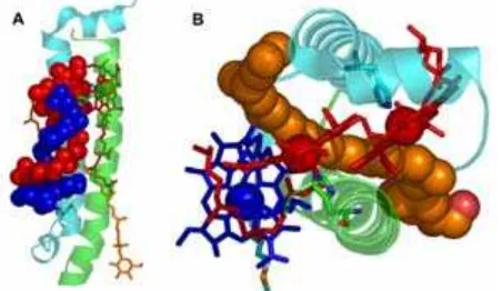
Fig 5. The high resolution (2.0 Å) structure of LH2
complex from Rps. acidophila 10050 (PDB: 1NKZ) [16]. Front (A) and side (B) views of the LH2 complex showing nine-membered circular module of α- (cyan) and β- (green) polypeptides with B800 Bchl (blue), B850 Bchl (red) and the carotenoid (orange)
Crystallisation
Generally proteins crystallize when they are gradually induced to precipitate. This is usually achieved by equilibration with precipitant, e.g. ammonium sulphate or polyethylene glycol, which influence the solubility of the protein yet do not denature them. During the process of crystallization of membrane proteins, very often the precipitant reacts with the detergent and causes the detergent to phase separate. When phase separation happens the membrane protein usually denatures in the oily detergent phase and crystals will never be formed. The problem was then overcome when Michel [8-9] and Garavito [10] independently discovered that addition of specific small molecules, e.g. heptane-1,2,3-triol, benzamidine hydrochloride, etc. [11] could alter the phase boundaries. This small amphiphile shifts the phase separation point to be above the critical precipitation point, so that the crystallization becomes possible.
The first success of crystallizing LH2 was achieved by Richard J. Cogdell group in early 1980s [12]. Unfortunately, these crystals looked very beautiful under microscope but diffracted X-rays only to about 12 Å (Fig. 4F). The reason of this poor disorder was likely to be the variation in lipid content [13]. An optimisation on the purification protocols was then improved by washing the LH2 complex in β-octylglucoside, and set up the crystallisation in this detergent. Large crystals grew and diffraction to 3.2 Å can be achieved in the mid-1990s (Fig. 4G).
STRUCTURE OF LH2 AND THE ENERGY TRANSFER
The high-resolution X-ray structure of the LH2 complexes [14-16] from Rhodopseudomonas (Rps.)
acidophila strain 10050 and low-light adapted strain
7050 show a remarkable symmetry in the arrangement of the pigments embedded in the protein matrix (Fig. 5). These complexes are modular. Each module consists of a protein heterodimer (αβ), which binds three Bchl a pigments and one carotenoid molecule. Nine such modules, αβ-polypeptides, are arranged circularly to form the single LH2 complex. The α -polypeptide is located inside the ring and the β -polypeptide is on the outside (Fig. 5). Inside the protein matrix the bacteriochlorin rings of the Bchl a molecule are organised in two ways (Fig. 5). Nine monomeric Bchl a molecules have their bacteriochlorin rings oriented parallel to the plane of the membrane (blue in Fig. 5), and absorb the light with absorption maximum (λmax) at ~800 nm. They are called B800 Bchl as. These
Tatas H.P. Brotosudarmo, and Richard J. Cogdell
Fig 6. The binding pocket of the B850 Bchl as (A and B) and the B800 Bchl a (C). The coordinates for this figure were taken from the high resolution (2.0 Å) structure of the LH2 complex from Rps. acidophila 10050 (PDB: 1NKZ) [16]. (B) The electron density of a B850 Bchl a
molecule shows that the bacteriochlorin ring is rather bent and the histidine residue, which is the fifth coordinated to the central Mg2+atom, is clearly seen. (D) The spatial organisation and distances between the Bchl
a pigments in LH2. The numbers indicate the size of LH2 and centre-to-centre distances between the macrocyles of the Bchl a molecules in Å. The arrows indicate the direction of the Qy transition moments. The phytol chain
is cropped off for clarity.
Fig 7. Diagram showing (A) the interaction of the phytol chain of the B800 Bchl a (dark spheres) and β-bound Bchl a (grey spheres) and (B) the position of all-trans
carotenoid rhodopin-glucoside in the heterodimeric pair of αβ-polypeptides. The coordinate is taken from high resolution (2.0 Å) structure of LH2 complex from Rps.
acidophila 10050 [16].
each other (Fig. 6D). A further eighteen Bchl a
molecules have their bacteriochlorin rings oriented perpendicular to the membrane plane. They are responsible for the absorption band at about 850 nm. The B850 Bchl as (red in Fig. 5 and 6D) sit very close to each other and, when viewed from above, superficially
resembles the blades of a turbine. The central Mg2+ atoms of these B850 Bchl as are separated 0.95 nm within one αβ pair, and by 0.88 nm from one αβ pair to the next (Fig. 6D). The 2.0 Å resolution structure suggests that there is a single well-defined carotenoid rhodophin–glucoside per αβ pair. In case of
Phaeospirillum (Phs. previously Rhodospirillum)
molischianum it has been found that the LH2
complexes are α8β8 octamers [17]. Until now, it is not
clear what structural features cause one to be nonamer and the other octamer, or indeed whether this difference has any significant functional consequences.
Looking at the binding site, the B850 Bchl as have their central Mg2+ liganded to the imidazole ring sidechain of histidine residues, α-His31 and β-His30 (Fig. 6A and B) [15-16]. A hydrogen bond is formed between the acetyl group in ring A and the phenol sidechain of α-tyrosine44 for α-B850 Bchl as and the indole sidechain of α-tryptophan45 for β-B850 Bchl a
molecules (Fig. 6A) [16]. The B800 Bchl as are stabilised by coordination between the Mg+2 of the Bchl
a and COO-α-Methionine1, and a H-bond between the acetyl group of Bchl a and the guanidinium group of β -Arginine20 (Fig. 6C) [16]. On a closer inspection, there is an interesting interaction between Bchl a molecules of B850 and B800, within an αβ-apoprotein pair, conducted by their hydrophobic phytyl chains. The B800 phytyl chain from B800 folds around the phytyl chain of β-B850 Bchl, crossing it and passes across the macrocycle ring of the β-B850 (Fig. 7A). The B800 phytyl chain interacts via van der Waals contact with rings A and D of the β-B850 macrocycle.
The Car found in the Rps. acidophila strain 10050
is rhodopin-glucoside. The X-ray structure of LH2 from
Rps. acidophila also reveals interactions between the
Car and the polypeptide as well as between Car with the Bchl a molecules (Fig. 7B). In the LH2 complex from Rps. acidophila 10050, the rhodopin-glucoside passes in close contact (3.4 Å) to the edge of the bacteriochlorin ring of the B800 Bchls. The polyene chain then runs perpendicular to the macrocycle of the
α-B850 Bchl a (Fig. 7B). The glucosyl group of the rhodopin-glucoside molecule is located in a hydrophilic binding pocket on the cytosolic side of the transmembrane-spanning protein. This Car has an extremely important structural role in LH2. It holds the
αβ-polypeptide pairs together (Fig. 7B). In a mutant that lacks Car the LH2 complexes fail to assemble [18].
Tatas H.P. Brotosudarmo, and Richard J. Cogdell
Fig 8. Room temperature (RT) steady state absorption spectra of the B800-850 (black line), the B800-820 (dashed line) LH2 complexes from Rps. acidophila 7050 and B800-low-850 (dotted line) LH2 complexes from
Rps. palustris 2.1.6.
Fig 9. Comparisons of the α- and β-bound Bchl a in B850 (green) and B820 (cyan) LH2. A. Diagram showing the interaction of the C3-acetyl group of Bchl a with the
key potential H-bonding residues. B. Highlighting the twisting of the C3-acetyl group of Bchl a with respect to
the bacteriochlorin plane. The coordinates used to produce this figure were taken from the high resolution (2.0 Å) structure of B800-850 LH2 complex from Rps.
acidophila 10050 [16] and the 3.0 Å resolution structure
of the B800-820 LH2 from Rps. acidophila 7050 (PDB: 1IJD) [14].
Rps. acidophila are excited, the energy is transferred to
the Bchl as within 61 fs [21]. About two-thirds of this energy is transferred to the B850 molecules and one-third to the B800 molecules [22]. Upon the excitation of B800 Bchl as, the hopping of excitation between B800 monomers occurs in 1.5 ps in the case of Rps.
acidophila 10050 [23] and 1-3 ps for Phs. molischianum
[24]. The B800→B850 energy transfer in Rps. acidophila
takes place with a time constant of 0.7-0.9 ps at room temperature [23] and only slows down to 1.8-2.4 ps at 1.4K [25]. Exciton relaxation in the B850 ring of the LH2 from Rba. sphaeroides has been measured to take place on the 100-200 fs timescale [26]. In Rps. acidophila
10050 the exciton relaxation in B850 ring has been recorded to be 160 fs [27]. The LH2→LH1 energy
transfer has been measured to be 3 ps at 296 K in
Rba. sphaeroides [28] and LH1→RC transfer has been
recorded to be about 20-50 ps in Rba. sphaeroides
[29].
VARIANTS OF LH2 COMPLEXES
In some species of purple bacteria, such as Rps.
acidophila 7050/7750 or Rps. palustris 216, the Qy
absorption band of the Bchl in the LH2 complexes can vary depending on the growth conditions. When Rps.
acidophila strain 7050 and 7750 are grown under
low-light conditions, the B800-850 LH2 is replaced by a different LH2 complex with the Qy absorption bands at
800 and 820 nm (Fig. 8) [7,14,30]. The ability to change the type of LH2 in response to growth at different light intensities is related to the presence of multiple αβ-polypeptides, which are (in the case of Rps.
acidophila) encoded by at least four different
αβ-apoprotein gene pairs [30-31]. In order to distinguish these two types of peripheral LH2 complexes they are often referred to as B800-850 and B800-820 complexes, or LH2 and LH3, respectively.
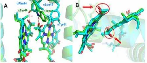
The origin of this spectral variation comes from the fine-tuning of the electronic energy levels of the “B850” molecules, by altering the binding site of the Bchl a in the protein matrix. In the B800-850 complex, the C3-acetyl group of the α-bound B850 Bchl a
molecule is H-bonded to the αTrp45 residue and the C3-acetyl group of the β-bound B850 Bchl a molecule is
H-bonded to the αTyr44 residue (Fig. 9A) [14]. In contrast, in the B800-820 complex, the respective residues in these positions, i.e. αPhe44 and αLeu45, are unable to form hydrogen bonds (Fig. 9A). Instead the C3-acetyl group of the αB820 Bchl a molecules is
Tatas H.P. Brotosudarmo, and Richard J. Cogdell
Raman studies of these mutants identified the breakage of one or two H-bonds, respectively, between the protein residues and the respective C2-acetyl carbonyl group of
the B850 Bchl a molecules [33]. The removal of a H-bond to the acetyl carbonyl group was signalled by a shift of the Raman peak expected for interaction-free acetyl carbonyl, i.e. 1635 cm -1 in the wild-type LH2 to 1659 cm-1. Similarly it is also observed in the Phs.
molischianum mutant, when αTyr43 in the B800-850
LH2 is replaced a Phe in the B800-820 LH2 [36]. The B800-820 LH2 mutant from Rs. molischianum shows loss of H-bound C2-acetyl RR-stretching mode of B850
Bchl a molecule at 1642 cm-1 and a dramatic increased of free-from-interaction acetyl carbonyls RR-stretching mode at 1663 cm-1. The importance of the H-bonding residues has also been shown in controlling the site-energy of Bchl a molecules [6,37].
This phenomenon of chromatic adaptation is also observed in Rps. palustris. When Rps. palustris 216 is grown under low light intensity, it replaces the B800-850 LH2 complexes with the B800-low-850 LH2 complexes (Fig. 8 dotted line). The complete genome of Rps.
palustris has been sequenced [38]. This ability to adapt
is related to the five different pucBA genes present in the
Rps. palustris genome (pucBA-a, b, c, d, and e), which
their expression is regulated by light intensity [39-40]. The regulation of the LH2 complex from Rps. palustris is even more complicated as 6 bacteriophytochrome (Bph)-like genes have also now been identified in the genome [38]. Four of these genes are located near to genes coding for photosynthetic LH-apoproteins or pigment biosynthetic genes. This suggests that these Bphs are also involved in the regulation of the PSU. The regulation of the LH2 complex in Rps. palustris is, therefore, influenced by the light intensity as well as the light quality.
In Rps. palustris 261, the structural explanation of
the spectral changes going from B800-850 to B800-low-850 LH2 is still a matter of debate [41-44]. The ability to shift the type of LH2 allows these bacteria to be able to grow at ten times lower light intensity than most species that cannot do this. It is not currently understood how changing the types of LH2 allows the bacteria to grow photosynthetically at these lower light intensities.
It was reported in 2002 that Hartigan et al. managed to isolate the B800-only LH2 from the low-light B800-low-850 LH2 by employing a strong anion exchanger as additional purification protocol [45]. The B800-LH2 has been crystallized and a structural model based on the low-resolution crystals (7.5 Å) has been proposed [45]. This model suggests that this complex is an octamer and that each of its αβ-subunit binds an extra B800 Bchl a relative to LH2 from Phs.
molischianum. An AFM study of LL membranes from
Rps. palustris also supports that the low light grown LH2
is predominantly octameric [46]. The unusual B800-LH2 (LH4) complex, which has Qy absorption band at
800 nm only, is encoded by pucBd and pucAd, producing LH4 PucBdAd peptides, and is regulated by
two Bph genes, Rpa3015 (Bph4) and Rpa3016 (Bph5) [47]. The PucAd apoprotein does not contain Tyr and
Trp, but rather Phe and Met at the position 44 and 45, thus unable to H-bonding with the C3-acetyl group of
both Bchl a pairs.
A trial expressing two LH2 gene pairs, which encode the high-light (pucBAa) and the low-light
(pucBAd) proteins, from Rps. palustris in Rba.
sphaeroides has been carried out and the synthesis of
the high-light B800-850 LH2 complex (pucBAa) has
been a great success [43]. However expressing pucBd
and pucAd geen pairs in Rba. sphaeroides resulted
unstable low-light B800-830 LH2 complexes.
Studies on the native B800-low-850 LH2 complexes (Fig. 8 dotted line) have been carried out [42,48]. The polypeptide composition of the B800-low-850 LH2 complexes has been characterised by mass spectroscopy. It was found that B800-low-850 LH2 complexes contain mixtures of pucBAa, pucBAd and
pucBe polypeptides [49]. Previously, it was
hypothesised that the LH2 fro Rps. palustris may have a heterogeneous polypeptides composition [42]. This hypothesis suggests that the LL LH2 from Rps.
palustris is composed of a rings that consists of a
Tatas H.P. Brotosudarmo, and Richard J. Cogdell
crystal structure are still rather at low-resolution diffraction (6.7 Å) [49]. From these low-resolution data, the best molecular replacement solutions suggest that these LL LH2 complexes are nonameric. However, definite structural conclusion must await better quality crystals.
SUMMARY
In this review it is shown that the polypeptides in LH2 do not just provide inert scaffolding for the pigments – their role is far subtler. They are able to control the electronic properties of the pigments, for example, the absorption of Bchl, which can be altered by changing the number of hydrogen bonds between Bchl and the protein backbone. Understanding the mechanism of this optical tuning is particularly important, as in the purple bacteria the funnelling of the excitation energy (LH2, B800→B850, LH2→LH1, B850→B875) towards the reaction centre is strongly controlled by the gradient energy of the electronic transition of the Bchl Qy
transition band of the different light harvesting complexes.
ACKNOWLEDGEMENT
RJC acknowledges Biotechnology & Biological Science Research (BBSRC), UK. THPB is grateful for the fellowship from the European Commission through the Human Potential Program (Marie-Curie RTN BIMORE, Grant No. MRTN-CT-2006-035859) and from the United States Department of Energy, the Photosynthetic Antenna Research (DE-SC0001035).
REFERENCES
1. Blankenship, R.E. 2002, Molecular mechanisms of
photosynthesis, Blackwell Science, Oxford.
2. Cogdell, R.J., Gardiner, A.T., Hashimoto, H., Brotosudarmo, T.H., 2008, Photochem. Photobiol.
Sci., 7, 1150–1158.
3. Scheuring, S., and Sturgis, J.N., 2009, Photosynth.
Res., 102, 197–211.
4. Bahatyrova, S., Frese, R.N., Siebert, C.A., Olsen, J.D., Van Der Werf, K.O., Van Grondelle, R., Niederman, R.A., Bullough, P.A., Otto, C., and Hunter, C.N., 2004, Nature, 430, 1058–1062.
5. Hu, X., Ritz, T., Damjanovic, A., Autenrieth, F., and Schulten, K., 2002, Q. Rev. Biophys., 35, 1, 1–62. 6. Cogdell, R.J., Howard, T.D., Isaacs, N.W.,
McLuskey, K., and Gardiner, A.T., 2002,
Photosynth. Res., 74, 135–141.
7. Cogdell, R.J., and Scheer, H., 1985, Photochem.
Photobiol., 42, 6, 669–678.
8. Michel, H., and Oesterhelt, D., 1980, Proc. Natl.
Acad. Sci. U.S.A., 77, 3, 1283–1285.
9. Michel, H., 1982, EMBO J., 1, 10, 1267–1271. 10. Garavito, R.M., and Rosenbusch, J.P., 1980, J.
Cell. Biol., 86, 327–329.
11. Michel, H., 1982, J. Mol. Biol., 158, 567–572. 12. Papiz, M.Z., Hawthornthwaite, A.M., Cogdell, R.J.,
Woolley, K.J., Wightman, P.A., Ferguson, L.A., and Lindsay, J.G., 1989, J. Mol. Biol., 209, 833–835. 13. Cogdell, R.J., and Hawthornthwaite, A.M., In The
photosynthetic reaction center; Eds. Deisenhofer,
J., Norris, J. R., Academic Press, INC., 1993, Vol. 1.
14. McLuskey, K., Prince, S.M., Cogdell, R.J., and Isaacs, N.W., 2001, Biochemistry, 40, 30, 8783– 8789.
15. McDermott, G., Prince, S.M., Freer, A.A., Hawthornthwaitelawless, A.M., Papiz, M.Z., Cogdell, R.J., and Isaacs, N.W., 1995, Nature,
374, 517–521.
16. Papiz, M.Z., Prince, S.M., Howard, T., Cogdell, R.J., and Isaacs, N.W., 2003, J. Mol. Biol., 326, 1523–1538.
17. Koepke, J., Hu, X., Muenke, C., Schulten, K., and Michel, H., 1996, Structure, 4, 581–597.
18. Hunter, C.N., Hundle, B.S., Hearst, J.E., Lang, H.P., Gardiner, A.T., Takaichi, S., and Cogdell, R.J., 1994, J. Bacteriol., 176, 12, 3692–3697.
19. Sundström, V., and van Grondelle, R., In
Anoxygenic Photosynthesis Bacteria, vol 2.
Advances in Photosynthesis; Ed. Blankenship, R.
E., Madigan, M.T., and Bauer, C.E., Kluwer Academic Publisher, Dordrecht, 1995.
20. Sundström, V., Pullerits, T., and van Grondelle, R., 1999, J. Phys. Chem. B, 103, 2327–2346.
21. Polli, D., Cerullo, G., Lanzani, G., De Silvestri, S., Hashimoto, H., and Cogdell, R.J., 2006, Biophys. J., 90, 7, 2486–2497.
22. MacPherson, A.N., Arellano, J.B., Fraser, N.J., Cogdell, R.J., and Gillbro, T., 2001, Biophys. J.,
80, 2, 923–930.
23. Kennis, J.T.M., Streltsov, A.M., Vulto, S.I.E., Aartsma, T.J., Nozawa, T., and Amesz, J., 1997, J.
Phys. Chem. B, 101, 7827–7834.
24. Novoderezhkin, V., Wendling, M., and van Grondelle, R., 2003, J. Phys. Chem. B, 107, 11534–11548.
25. Kennis, J.T.M., Streltsov, A.M., Permentier, H., Aartsma, T.J., and Amesz, J., 1997, J. Phys.
Chem. B, 101, 8369–8374.
26. Agarwal, R., Rizvi, A.H., Prall, B.S., Olsen, J.D., Hunter, C.N., and Fleming, G.R. 2002, J. Phys.
Chem. A, 106, 7573–7578.
Tatas H.P. Brotosudarmo, and Richard J. Cogdell
Cogdell, R.J., Springate, E., and Turcu, E., 2009,
Phys. Rev. Lett., 102, 5, 057402.
28. Hess, S., Chachisvilis, M., Timpmann, K., Jones, M.R., Fowler, G.J.S., Hunter, C.N., and Sundstrom, V., 1995, Proc. Natl. Acad. Sci. U.S.A., 92, 12333– 12337.
29. Visscher, K.J., Bergstrom, H., Sundström, V., Hunter, C.N., and Vangrondelle, R., 1989,
Photosynth. Res., 22, 211–217.
30. Gardiner, A.T., Takaichi, S., and Cogdell, R.J., 1993, Biochem. Soc. Trans., 21, 6S.
31. Bissig, I., Brunisholz, R.A., Suter, F., Cogdell, R.J., and Zuber, H., 1988, Z. Naturforsch., C: Biosci., 43, 77–83.
32. Sturgis, J.N., Jirsakova, V., Reiss-Husson, F., Cogdell, R.J., and Robert, B., 1995, Biochemistry,
34, 33, 517–523.
33. Fowler, G.J., Sockalingum, G.D., Robert, B., and Hunter, C.N., 1994, Biochem. J., 299, 3, 695–700. 34. Olsen, J.D., Sturgis, J.N., Westerhuis, W.H., Fowler,
G.J., Hunter, C.N., and Robert, B., 1997,
Biochemistry, 36, 12625–12632.
35. Fowler, G.J.S., Visschers, R.W., Grief, G.G., Vangrondelle, R., and Hunter, C.N., 1992, Nature,
355, 848–850.
36. Sauer, P.R.R., Lottspeich, F., Unger, E., Mentele, R., and Michel, H., 1996, Biochemistry, 35, 6500– 6507.
37. Cogdell, R.J., Gall, A., and Köhler, J., 2006, Q. Rev.
Biophys., 39, 227–324.
38. Larimer, F.W., Chain, P., Hauser, L., Lamerdin, J., Malfatti, S., Do, L., Land, M.L., Pelletier, D.A., Beatty, J.T., Lang, A.S., Tabita, F.R., Gibson, J.L., Hanson, T.E., Bobst, C., Torres, J.L., Peres, C., Harrison, F.H., Gibson, J., and Harwood, C.S., 2004, Nat. Biotechnol., 22, 55–61.
39. Tadros, M.H., and Waterkamp, K., 1989, EMBO J.,
8, 1303–1308.
40. Tadros, M.H., Katsiou, E., Hoon, M.A., Yurkova, N., and Ramji, D.P., 1993, Eur. J. Biochem., 217, 3, 867–875.
41. Evans, M.B., Hawthornthwaite, A.M., and Cogdell, R.J. 1990, Biochim. Biophys. Acta, 1016, 1, 71–76. 42. Gall, A., and Robert, B. 1999, Biochemistry, 38, 16,
5185–5190.
43. Fowler, G.J.S., and Hunter, C.N., 1996, J. Biol.
Chem., 271, 23, 13356–13361.
44. Nishimura, Y., Shimada, K., Yamazaki, I., and Mimuro, M., 1993, FEBS Lett., 329, 3, 319–323. 45. Hartigan, N., Tharia, H.A., Sweeney, F., Lawless,
A.M., and Papiz, M.Z., 2002, Biophys. J., 82, 2, 963–977.
46. Scheuring, S., Goncalves, R.P., Prima, V., and Sturgis, J.N., 2006, J. Mol. Biol., 358, 83–96.
47. Evans, K., Fordham-Skelton, A.P., Mistry, H., Reynolds, C.D., Lawless, A.M., and Papiz, M.Z., 2005, Photosynth. Res., 85, 169–180.
48. van Mourik, F., Hawthornthwaite, A.M., Vonk, C., Evans, M.B., Cogdell, R.J., Sundström, V., and Vangrondelle, R., 1992, Biochim. Biophys. Acta,
1140, 85–93.
49. Brotosudarmo, T.H.P., 2009, PhD Thesis, University of Glasgow.
50. Brotosudarmo, T.H., Kunz, R., Bohm, P., Gardiner, A.T., Moulisova, V., Cogdell, R.J., and Kohler, J., 2009, Biophys. J., 97, 1491–1500.
51. Moulisova, V., Luer, L., Hoseinkhani, S., Brotosudarmo, T.H., Collins, A.M., Lanzani, G., Blankenship, R.E., and Cogdell, R.J., 2009,
Biophys. J., 97, 3019–3028.
![Fig 5. The high resolution (2.0 Å) structure of LH2 complex from Rps. acidophila 10050 (PDB: 1NKZ) [16]](https://thumb-ap.123doks.com/thumbv2/123dok/871857.820775/3.595.48.299.548.671/fig-high-resolution-structure-complex-rps-acidophila-pdb.webp)