13. Poupard, M., Ngom Gueye, N.F., Thiam, D., Ndiaye, B., Girard, P.M., Delaporte, E., et al.. Quality of life and depression among HIV-infected patients receiving efavirenz- or protease inhibitor-based therapy in Senegal, HIV Med. , 2007 Mar;8(2):92-5.
14. Ribs, T.A., Begley, K., Smith, D.E., Sarangapany, J., Callaghan, A., Kelly, M., et al. Efavirenz and Chronic Neuropsychiatric Symptoms: Results, HIV Medicine, 2006 ; 7(8):544-548.
15. Clifford, B.D., Evans, S., Yang, Y., Acosta, E.P., Pham, D., Goodkin, K., et al. Impact of Efavirenz on Neuropsychological Performance and Symptoms in HIV-Infected Individuals, Ann Intern Med., 2005; 143:714-721.
30
M. Yusuf Hamra, et al Acta Interna - The Journal of Internal Medicine
16. Seidman, D.Depression, Smoking, and Quitting, www.danielfseidman.com.2010 17. Oman, D., Shapiro, S.L., Thoresen, C.E., Plante,
T.G., Flinders, T. Meditation Lowers Stress and Supports Forgiveness Among College Student: A Randomized Controlled Trial, Journal of American College Health, 2008; Vol. 56, No.5. 18. Nidich, S., and Myers, H. New studies show
reduced depression with Transcendental Meditation, www.eurekalert.org. 2010
19. Woolery, A., Myers, H., Sternlieb, B., Zeltzer, L. A Yoga Intervention for Young Adults with Elevated Symptoms of Depression, Alternative Therapies, Vol.10, No.2. 2004.
R E V I E W A R T I C L E
ACUTE PULMONARY EMBOLISMS
Diagnosis and Management
Kartika Widayati Taroeno-Hariadi
* Hematology and Medical Oncology, Department of Internal Medicine, Dr. Sardjito General Hospital/Faculty of Medicine, Universitas Gadjah Mada, Yogyakarta , Indonesia
ABSTRACT
Acute pulmonary embolisms is a major cause of complications and death associated in surgery, medical illnesses, injury, and also may occurs after a long-distance air travel. It is often originating from deep-vein thrombosis and has a wide spectrum of clinical manifestation ranging from asymptomatic, incidentally discovered emboli, to massive embolism causing immediate death. Incidence of pulmonary embolism ranges from 23-69 cases per 100,000 populations. Case fatality rates vary widely depending on the severity of the cases; at an average case fatality rate within 2 week of diagnosis of approximately 11%. It may have chronic sequele as post thrombotic syndrome and chronic thromboembolism pulmonary hypertension.
Acute pulmonary embolism is often difficult to diagnose. The predisposing factors for pulmonary embolisms consist of hereditary factors, acquired factors, and probable factors. Patients with symptoms of dyspnea, chest apnea, tachypnea or tachycardia arise suspiciousness of pulmonary embolisms therefore should be screened their probability for developing the disease. Low risk patients will then be evaluated for d-dimer test. Treatment should be initiated promptly in high risk patients, followed by imaging procedure evaluation. Chest radiographs, CT scan arteriography, VQ scan are performed to either include or exclude diagnosis of pulmonary embolisms.
Treatments consist of thrombolysis for acute and unstable massive pulmonary embolisms, and anticoagulation with heparin for stable acute pulmonary embolism. A meta-analysis of several major trials showed that low molecular weight heparin is at least as effective as unfractionated heparin in preventing the recurrence of venous thromboembolism events and at least as safe with respect to the rate of major bleeding.
This review will further describe in detail the pathomechanisms, diagnosis, and management of acute pulmonary embolisms.
INTRODUCTION
P u l m o n a r y e m b o l i s m s ( P E ) i s a cardiovascular and cardiopulmonary symptoms caused by oclusion of the main pulmonary vessels by embolic process. Embolism is originated from detachment of thrombus – ussually from deep vein
1,2
thrombosis- and then propagated . Pulmonary embolisms (PE) and deep vein thrombosis (DVT) is one spectrum of disease. About 79% of patients with PE have DVT in their legs and 50 % of patients with
3 DVT will progress to PE .
In US incidence of PE reaches 1 per 1000 population; with 15 % mortality within 3 month of diagnosis. Mortality due to PE reaches 300,000 cases per year that make it as deadly as acute
3 coronory syndrome .
Eventhough in some certain countries incidence of venous thromboembolisms is not frequent, awareness of it should be routinely performed especially for highrisk groups of patients 4
.
Diagnosing PE is not easy. Lack of awareness and non specific symptoms and signs of PE make it difficult to diagnose at early course of disease and many of PE are diagnosed post mortemly.
This review is aimed at describing clinical manifestations of pulmonary embolisms, its pathomechanisms, diagnostic procedures, and management based on the most recent literatures and publications.
Pathophysiology
Thrombi arises from deep veins in the lower extremity, propagate in the proximal veins include popliteal veins and their upper veins, in which is
3
easier to form embolization . Emboli migrates from right ventricle and plugs pulmonary arterial.
In acute PE, anatomical obstruction will decrease pulmonary function, followed by secretion of vasoactive and bronchoactive mediators that lead
Kartika Widayati Taroeno-Hariadi Acta Interna - The Journal of Internal Medicine
to impairment of ventilation-perfusion. Increasing of right ventricle wall pressure cause ventricle dilatation, dysfunction, and ischemic of right ventricle. Death may caused by right ventricular
3 failure .
Risk Factors
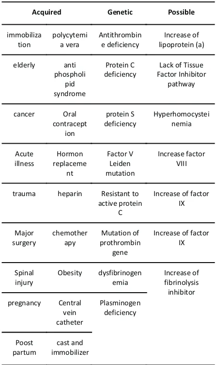
Many health's condition and genetic factors increase the risk of PE. All risk factors of DVT become risk factors of PE. Table 1 gives summary of the risk factors of PE including hereditary and acquired risk factors.
Acquired Genetic Possible
immobiliza
Lack of Tissue Factor Inhibitor
trauma heparin Resistant to
active protein C
Increase of factor IX
Increase of factor IX
Table 1. Risk Factors of Venous
Thromboembolisms (Deep Vein Thrombosis
3,5
and Pulmonary Embolisms)
Diagnosis approach
Symptoms of PE are not specific. Dyspneu, tachycardia, pleuritic pain, and hemoptysis are
sometimes found in patients with PE as well as other diseases. Cough, palpitation, light headedness, fever, and rales are common symptoms and signs which may be obscured by presenting comorbid illness. Symptoms and signs of pulmonary hypertension like elevation of jugular vein pressure, loud P2, gallops on the right ventricle, and elevation of right heart configuration may rise suspicion to PE b u t n e i t h e r s e n s i t i v e n o r s p e c i f i c . Electrocardiography showed unexplained sinus tachycardia, and S1Q3T3 pattern in massive pulmonary embolisms, right bundle branch block, P pulmonale, and right axis deviation.
Patient with PE usually present with hypoxemia, but somehow blood gas analysis showed normal results in large number of patients. Cyanosis, hypotension, and syncope may preceed a
2,6,7
fatal and massive PE that lead to death . Troponin may increase in PE but should not be used as diagnosis marker; its indication is recommended for
8 prognostic stratification .
Upon suspicion of PE, physician should perform a comprehensive and detailed anamnesis, assessment of prediction probability of PE and its risk factors, biomarker test and diagnostic imaging.
A s i m p l e m e t h o d t o p r e d i c t thromboembolism is developed by Wells et al using Clinical Prediction Rules for Pulmonary
9,10,11
Embolism . Beside Wells criteria, it has been developed other prediction criteria using more complicated items, radiographic imaging, and electrocardiography that should be interpreted by an
1,5,12 expert .
Tabel 2. Wells Clinical Prediction Rule 9 for Pulmonary Embolism (PE)
Clinical feature Points
Clinical symptoms of DVT 3
Other diagnosis less likely than PE 3
Heart rate greater than 100 beats per minute 1.5
Immobilization or surgery within past 4 weeks 1.5
Previous DVT or PE 1.5
Hemoptysis 1
Malignancy 1
Total points 12.5
Risk score interpretation (probability of PE): >6 points: high risk (78.4%);
2 to 6 points: moderate risk (27.8%); <2 points: low risk (3.4%).
32
Acute Pulmonary Embolism
Diagnostic Imaging
Patients with low probability of PE at outpatient setting should undergo Dimer test. D-dimer is a method to measure degradation product of cross linked-fibrin in the blood circulation. This method has a high sensitivity value but low specificity. Increasing of D-dimer is almost always happened in patients with emboli, as well as in elderly, trauma, post surgery, pregnancy, inflammation, and cancer. In patients with low risk probability and high D-Dimer results using highly sensitive D-Dimer assay should be followed by
12
imaging diagnostic procedures . In publication by Gibson et al D-Dimer test should be performed after clinical probability assessment or clinical prediction rule has done. High clinical probability patients should undergo other testing, regardless the
D-12,20 Dimer outcome .
Treatment should be initiated promptly for high risk probability patients, while concommitantly
carrying out diagnostic procedures such as helical CT scanning, ventilation-perfusion scanning, pulmonary arteriography until diagnosis is
12
established . Ventilation-perfusion scanning has the important role in diagnosing PE for almost 3 decades. Positive results from ventilation-perfusion scanning (V/Q scanning) gives high sensitivity for thromboembolism. However many large trial showed patients with emboli showed normal
12 ventilation-perfusion scanning .
Computed Tomography scanning has superiority compared with ventilation-perfusion scanning in term of either sensitivity or specificity. Helical CT scan has 57-100% sensitivity and 78-100% specificity. If the helical CT showed negative for PE, the likelihood of PE diagnosis is low, but can not directly exclude PE diagnosis as well as
12
ventilation perfusion scanning . A negative result of multidetector-row CT can safely rule out PE
12,27 diagnosis .
12 Figure.1 Diagnostic approachs of pulmonary embolism
33 Volume 1, Number 1, Juli 2011
Acta Interna - The Journal of Internal Medicine
Evaluation of veins in lower extremities should always be performed as PE arise from DVT. Duplex USG will be positive in 10-20% patients without symptoms in their leg, and USG of lower extremities' veins will positive in 50% patients with emboli. Normal USG duplex does not exclude
12 diagnosis of emboli .
Gold standard in PE diagnosis is pulmonary angiography. This invasive procedure needs specialistic technique and expert interpreter. Pulmonary angiography should be performed in condition where other non invasive diagnostic
12 procedures are not conclusive .
Treatment
Upon established diagnosis, patient with PE should be hospitalized and resting for the first 48
3
hours . If there were no contraindications, anticoagulation with heparin or low molecular weight heparin should be instituted. Survival will be improved with anticoagulation but recurrence rate is
3
still 5-10% within 1 year post diagnosis . Warfarin should be given concurrently with heparin at the first d a y o f t r e a t m e n t . M i n i m a l d u r a t i o n o f
3
anticoagulation is 5-days .Low molecular weight heparins have the similar efficacy as unfractionated heparin and have some advantages over it in term of:
higher bioavailability, more predictable dose, heparin and have some advantages over it in term of: higher bioavailability, more predictable dose, convenient in usage, no need for monitoring, and f e w e r i n c i d e n c e o f h e m o r r h a g e a n d
3,18,23 thrombocytopenia .
Direct thrombin inhibitor is indicated for t h r o m b o o s i s a n d h e p a r i n - i n d u c e d t h r o m b o c y t o p e n i a . I n h i b i t o r f a c t o r X a (rivuroxaban, apixaban, idraparinux and aptamers)
3,15 are still in phase 3 trial for PE .
Massive PE needs more aggresive treatment. Sodium chloride infusion, vasopressor, oxygenation, and intubation should be initiated in
16 cardiorespiratory failure .
Thrombolytic is indicated in PE presenting with cardiogenic shock, or systemic hypotension. In sub massive PE without shock thrombolytic treatment is still debatable. No significant difference between thrombolytic therapy and heparinization in
17 PE, unless in severe and acute PE .
In massive, acute PE in which thrombolytic therapy is contraindicated, mechanical pulmonary
3 embolectomy should be performed .
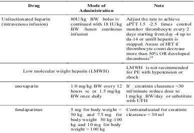
1,23 Table 3. Initial treatment for PE management
Drug Mode of continued with 18 IU/kg BW /hours continous infusion
Adjust the rate to achieve aPTT 1.5 -2.5 times control monitor thrombocyte every 2 days starting from day -4 up to da-14 or untill heparin is stopped. Aware of HIT if thrombocyte count decrease more than 50% OR developed thrombosis18
Low molecular weight heparin(LMWH) LMWH is not recommended for PE with hypotension or shock
enoxaparin 1.0 mg/kgBW every 12 hours sc or 1.5 mg/kg BW once daily
If creatinin clearance <30 ml/minute reduce dose to 1 mg/kg BW/day or substitute
with UFH
fondaparinux 5 mg for body weight < 50 kg and 7.5 mg for body weight 50 kg-100 kg and 10 mg for body weight > 100 kg
Contraindicated for creatinin clearance < 30 ml
34
Kartika Widayati Taroeno-Hariadi
Prognosis
Almost all patient with PE will survive after received adequate anticoagulation. Overall mortality rate within 3 months after diagnosis is 15-18%. Survival is determined by present or abscence of syncope or shock in the first hours of PE onset. P o s t t h r o m b o t i c s y n d r o m e a n d c h r o n i c thromboembolic pulmonary hypertension is a
19,25 common sequalae of PE .
Recurrency is still a major problem in acute PE. Recurrent VTE is found among 17.6% patients with symptomatic PE and among 9.5% patients DVT without symptomatic PE. Relative risk for recurrence VTE in PE is 2.2 (95% confident interval
21
1.3-3.7,p=0.005) . Predictors of recurrence in thrombosis are unprovoked VTE, thrombophillia, primary DVT, shorter episode of anticoagulation
22,24
(up to 6 months), and elderly . Persistent right ventricular dysfunction at hospital discharge from first episode of PE is also associated with right
26
ventricular dysfunction . Nomogram for predicting recurrency in unprovoked VTE has been developed by scoring sex, location of VTE, and d-Dimer
28 results .
SUMMARY
Physicians should aware of pulmonary embolism. Diagnosing PE is not always easy. Patients with unexplained dyspneu, tachycardia, hemoptisis, with or without deep vein thrombosis should be assessed probability of PE with clinical probability test developed by Wells. Those with high and moderate probability should be initiated treatment while waiting further test. Higher d-dimer value directs to chest x ray and multidetector chest CT. If PE was detected by imaging, anticoagulation is continued for up to 6 month. Unprovoked VTE needs lifelong anticoagulation.
REFERENCES
1. Konstantinides S. Acute Pulmonary Embolism. N Engl J Med 2008;359:2804-13.
2. G o l d h a b e r S . Z . P u l m o n a r y thromboembolism in Kasper D.L., Braunwald E., Fauci A.S.,Harrison's Principles of Internal Medicine (16th ed.). New York, NY: McGraw-Hill. 2005 pp. 1561–65.
3. Tapson V.F. Acute Pulmonary Embolism. N Engl J Med 2008;358:1037-52.
4. Leizorovicz A, Turpie A.G.G., Cohen A.T., Wong L., Yoo M.C., Dans A. Epidemiology
of venous thromboembolism in Asian patients undergoing major orthopedic surgery without thromboprophylaxis: the SMART Study. J Thromb Haemost 2005; 3:28-34.
5. Torbicki A, Perrier A, Konstantinides S. Guidelines on the diagnosis and management of acute pulmonary embolism: the Task Force for the Diagnosis and Management of Acute Pulmonary Embolism of the European Society
of Cardiology (ESC). Eur. Heart J.
2008;29:(18): 2276–315.
6. Goldhaber S.Z., Elliott C.G. Acute Pulmonary E m b o l i s m : P a r t I E p i d e m i o l o g y, Pathophysiology, and Diagnosis. Circulation. 2003;108:2726-2729
7. Goldhaber S.Z. and Elliott C.G. Acute Pulmonary Embolism: Part II: Risk Stratification, Treatment, and Prevention.
Circulation 2003;108;2834-2838
8. Becattini C., Vedovati M.C., Agnelli G. Prognostic Value of Troponins in Acute Pulmonary Embolism A Meta-Analysis.
Circulation. 2007;116:427-433
9. Wells P.S.., Ginsberg J.S., Anderson D.R. Use of a clinical model for safe management of patients with suspected pulmonary embolism.
Ann Intern Med 1998;129:997- 1005.
10. Wells P.S., Anderson D.R., Rodger M. Excluding pulmonary embolism at the bedside without diagnostic imaging: management of patients with suspected pulmonary embolism presenting to the emergency department by using a simple clinical model and D-dimer. Ann Intern Med: 2001;1:35:98-107.
D-Dimer Testing to Determine the Duration of Anticoagulation Therapy. N Engl J Med 2006;355:1780-9.
Acta Interna - The Journal of Internal Medicine
thromboembolism in patients with cancer. N Engl J Med 2003; 349 (2): 146–53.
Dabigatran versus Warfarin in the Treatment of Acute Venous T h r o m b o e m b o l i s m .
15. Schulman S, Kearon C, Kakkar A.K., Mismetti, Schellong S, Eriksson H for the RE-COVER Study Group.
N E n g l J M e d
2009;361:2342-52.
16. Kucher N, Rossi E,De Rosa M, Goldhaber S . Z . M a s s i v e P u l m o n a r y E m b o l i s m
Circulation 2006;113:577-582
17. Wan S., Quinlan D.J., Agnelli G., Eikelboom J.W. Thrombolysis Compared With Heparin for the Initial Treatment of Pulmonary E m b o l i s m A M e t a - A n a l y s i s o f t h e Randomized Controlled Trials. Circulation. 2004;110:744-749.
18. Warkentin T.E., Greinacher A., Koster A., Lincoff A.M. Treatment and prevention of heparin-induced thrombocytopenia: American College of Chest Physicians Evidence-Based Clinical Practice Guidelines (8th edition).
Chest 2008;133:Suppl:340S-380S.
19. Fedullo P.F. Auger W.R, Kerr K.M., Rubin L.J. Chronic Thromboembolic Pulmonary Hypertension. N Engl J Med 2001;345:1465-1472.
20. G i b s o n N . S . , S o h n e M . , G e r d e s , V.E.A.,Nijkeuter M.,Buller H.R. The I m p o r t a n c e o f C l i n i c a l P r o b a b i l i t y Assessment in Interpreting a Normal d-Dimer in Patients with Suspected Pulmonary Embolism. Chest 2008:134:789-793.
21. Eichinger S., Weltermann A., Minar E., Stain M., Scho'nauer V.,Schneider B. Symptomatic Pulmonary Embolism and the Risk of Recurrent Venous Thromboembolism. Arch Intern Med 2004:164:92-96
22. Prandoni P., Noventa F., Ghirarduzz A., Pengo V., Bernard E. The Risk of Recurrent Venous Thromboembolism after Discontinuing Anticoagulation in Patients with Acute Proximal Deep Vein Thrombosis or Pulmonary Embolism. A Prospective Cohort Study in 1,626 Patients.Haematologica 2007; 92:199-205.
23. Mismetti P.,Quenet S., Levine M. Enoxaparin in the Treatment of Deep Vein Thrombosis With or Without Pulmonary Embolism An Individual Patient Data Meta-analysis Chest 2005; 128:2203–2210.
24. Nijkeuter M., So¨hne M, Tick L.W. The Natural Course of Hemodynamically Stable Pulmonary Embolism: Clinical Outcome and Risk Factors in a Large Prospective Cohort Study. Chest 2007; 131:517–523.
25. Becattini C.,Agnelli G.,Pesavento R. Incidence of Chronic Thromboembolic Pulmonary Hypertension after a First Episode o f P u l m o n a r y E m b o l i s m . C h e s t 2006;130:172-175
26. Grifoni S., Vanni S.,Magazzini S.
28. Eichinger S.,Heinze G., Jandeck L.M., Kyrle P.A., Risk Assessment of Recurrence in Patients With Unprovoked Deep Vein Thrombosis or Pulmonary Embolism: The Vienna Prediction Model Circulation 2010; 121: 1630 - 1636.
.
Association of Persistent Right Ventricular Dysfunctionat Hospital Discharge After Acute Pulmonary Embolism With Recurrent Thromboembolic Events. Arch Intern Med. 2006;166:2151-2156
27. Quiros R., Kucher N.,Zou K.H. Clinical Validity of Negative Computed Tomography Scan in Patients with Suspected Pulmonary Embolisms a Systematic Review. JAMA 2005;293:2012-2017
36
Kartika Widayati Taroeno-Hariadi