Cemented with Self-Adhesive or Conventional Resin Cement
Mohamed Ghazy, BDS, MSc, PhD,1Omar El-Mowafy, BDS, PhD, FADM,2& Renato Roperto, DDS, MSc, PhD3
1Associate Professor, Department of Conservative Dentistry and Fixed Prosthodontics, Faculty of Dentistry, University of Mansoura, Egypt
2Professor, Department of Clinical Sciences, Faculty of Dentistry, University of Toronto, Canada
3Assistant Professor, Department of Restorative Dentistry, University of Manitoba, Winnipeg, Canada
Keywords
CEREC 3D; self-adhesive resin cement; porcelain crowns; composite crowns.
Correspondence
Mohamed Ghazy, Faculty of
Dentistry—Fixed Prosthodontics, University of Mansoura, Mansoura DK 35516, Egypt. E-mail: [email protected]
Previously presented as an oral presentation at the 2007 IADR General Session (New Orleans, LA).
Accepted October 29, 2009
doi: 10.1111/j.1532-849X.2010.00637.x
Abstract
Purpose:Resistance of machined crowns to microleakage when cemented with new self-adhesive cements has not been fully investigated. This study evaluated microleak-age of machined crowns milled from porcelain and composite blocks and bonded to teeth with self-adhesive and conventional resin cement.
Materials and Methods: Thirty-two freshly extracted premolars of similar shape and size were sterilized and mounted in resin blocks. Teeth received standard crown preparations with 1-mm circumferential shoulder finish line, flat occlusal surface reduced by 2 mm, and ideal angle of convergence. Prepared teeth were divided into two equal groups and assigned to either porcelain (Vita Mark II, Vident) or composite (Paradigm MZ100, 3M ESPE) blocks for crown fabrication. Optical impressions were captured for each tooth with the intraoral camera of a CEREC 3D machine. Crowns were designed and milled from both materials. Each group was then subdivided into two subgroups (n=8) according to cement used (self-adhesive resin cement, RelyX Unicem, 3M ESPE or resin cement with self-etching adhesive, Panavia F 2.0, Kuraray). Following seating, a 5-kg weight was applied on the occlusal surface of the crown for 5 minutes. Specimens were then stored in water at 37◦C for 24 hours. Specimens were thermocycled for 3000 cycles between 5◦C and 55◦C, then coated with nail varnish and immersed in a 2.0% basic red fuchsine dye solution for 24 hours. Teeth were then rinsed and sectioned mesiodistally and assessed under magnification for microleakage. A five-point scale was used to score degree of microleakage. Data were statistically analyzed with 2-way ANOVA and Kruskal-Wallis nonparametric test.
Results:Crown material had no significant effect on microleakage (p=0.67); however, cement type had a significant effect (p<0.0001), with Panavia F 2.0 resulting in lower
microleakage scores than RelyX Unicem.
Conclusions:Compared to the self-adhesive cement, the resin cement with separate primer/bonding agent resulted in significantly lower microleakage scores, irrespective of crown material.
Clinical studies have shown that recurrent caries and lack of re-tention are the major causes of failure of indirect restorations.1,2
Optimal marginal accuracy and the presence of a long-term ce-ment seal are, therefore, important clinical requirece-ments. An additional cause of failure of current nonmetallic esthetic indi-rect restorations is the occurrence of clinical fracture.3 Resin luting cements combined with proven bonding procedures have been shown to provide an extremely strong, retentive, and al-most insoluble luting unit that provides the strength requirement necessary for many nonmetallic esthetic indirect restorations.
Typically available with dual-polymerization capability, resin cements are characterized by high mechanical strength and ex-cellent esthetic properties.4,5
Porcelain and Composite Crown Microleakage Ghazyet al
fabricated under controlled and optimum manufacturing condi-tions enables the production of a restoration with a higher intrin-sic strength without the variability inevitable in the laboratory-produced restoration.6,7 Clinical use of adhesively cemented
monolithic CAD/CAM anterior and posterior crowns gener-ated from Vita Mark II feldspathic blocks genergener-ated in a one-step procedure at chairside has been reported.8Indirect resin
composite restorations can be constructed by CAD/CAM tech-niques using the prefabricated composite blocks (Paradigm MZ100, 3M ESPE, St. Paul, MN) under controlled condi-tions, which according to the manufacturer combine some of the best attributes of porcelains and polymers.9-13Advantages
over restorations milled from CAD/CAM porcelain blocks in-clude easier intraoral finishing and polishing, less abrasiveness to the opposing dentition, and ease of additions/adjustment.14
Resin cements are the material of choice for the adhesive ce-mentation of all-porcelain restorations.5,15,16Bindl et al studied
the strength and fracture pattern of monolithic CAD/CAM-generated posterior crowns and found that adhesive cementa-tion improved the relatively weak strength of some porcelains to that of stronger porcelains and recommended using adhesive resin cements for leucite glass-ceramic and feldspathic ceramic crowns.17Generally the technique of adhesive cementation
in-volves three main steps: etching, priming, and cement applica-tion. The multistep procedure requires attention to detail and is extremely technique sensitive.18-20A new generation of propri-etary self-adhesive resin cements has been introduced recently, designed to self-etch and bond to dentin without using separate etching or priming agents. Self-adhesive, dual-polymerizing resin cements are designed to provide ease of handling with fa-vorable mechanical properties, good esthetics, and appropriate adhesion to tooth structure and restoration.21,22
Microleakage is defined as passage of bacteria, fluids, molecules, or ions between a cavity wall and the restorative material applied to it.23 Evaluation of microleakage in vitro is a reliable method to predict the in vivo performance of crown cements.24Microleakage at crown margins may result
in marginal staining, secondary caries, and subsequent pulpal pathology.25,26Dye penetration and microscopic evaluation is a
well-established method for in vitro microleakage testing.27,28
It was used in several studies because of its simplicity and reproducibility.30,31
Microleakage and the marginal gap associated with various cementing agents for full-cast crowns were investigated for groups of cementing agents.29 The authors of a later
investi-gation reported that the self-adhesive resin cement resulted in the smallest degree of microleakage both at enamel and dentin margins. De Munk et al30 found there was no significant
dif-ference in bonding MZ100 composite blocks to dentin using an auto-cured adhesive resin cement relative to a conventional resin cement. Other investigators concluded that good marginal adaptation and low microleakage could be established with the self-adhesive cement comparable to that of conventional resin cement.31,32 In contrast, Ibarra et al33 reported that the
self-adhesive cement microleakage score was more similar to dentin than that recorded with the cement that employed an adhesive in a separate step. Escribano and de la Macorra compared the bond strength of Empress II discs to dentin using Multilink sys-tem, RelyX Unicem, and Panavia F and concluded that RelyX
Unicem achieved the lowest tensile bond strength.34The same finding was reported in another study in which the microtensile bond strength of RelyX Unicem self-adhesive resin cement was found to be lower than that achieved with a conventional resin cement.35
More research is needed on microleakage of machined crowns made from composite or porcelain blocks and cemented with self-adhesive resin cement. Therefore, this study aimed to evaluate the microleakage of CAD/CAM crowns milled from both composite and porcelain blocks and bonded to tooth prepa-rations with proprietary self-adhesive resin cement as compared to that of conventional resin cement with self-etching primer.
Materials and methods
This study used human teeth to simulate the clinical situation when crowns are cemented to teeth with different cements.36
Furthermore, this study used CAD/CAM crowns milled from new composite blocks as well as ones made with traditional porcelain blocks. The processing techniques of these composite blocks maximize the degree of cross-linking (monomer conver-sion) and eliminate void formation.
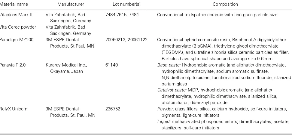
Thirty-two freshly extracted sound human maxillary premo-lars of nearly the same shape and size were collected in a 6-month period from the Department of Oral Surgery, Faculty of Dentistry, Mansoura University, Egypt. The selected teeth were found free of cracks, caries, or developmental defects and extracted for orthodontic puposes. These were sterilized with a gamma irradiation device with a dose of 3.3 kg/y/hr for 12 hours (Gamma Cell 220, Atomic Energy Ltd., Ottawa, Canada).37 The sterilized teeth were then cleaned of any debris, scaled to remove any calculus deposits, then examined under optical microscope to exclude any with major defects. They were then mounted in resin blocks and prepared in a standard manner to receive crowns with 1-mm circumferential all-around shoulder finish line and flat occlusal surface reduced by 2 mm with a 10◦ angle of convergence (Fig 1).
The prepared teeth were divided into two groups of 16 teeth each relative to the composite or porcelain blocks to be used for making the crowns with the CEREC 3D machine. Each group was further subdivided into two subgroups (n=8), relative to the type of resin cement used for crown cementation, either a conventional resin cement with self-etching adhesive (Panavia F 2.0, Table 1) or a self-adhesive universal resin cement (RelyX Unicem, Table 1).
Prepared teeth were uniformly sprayed with reflective pow-der to facilitate the scanning process during optical impression taking (Vita Cerec powder, Table 1). An optical impression was captured for each tooth with the intraoral camera of the CEREC 3D machine. Crowns were then designed, and digital data sent to the milling machine through a wireless connec-tion. The milling unit uses two types of burs (a flat-ended and a round-ended cylindrical diamond) that were changed after every five crown millings. The internal gap distance was set at 25µm to create cement space, while the marginal gap was
adjusted to 0µm.
Figure 1 Mounted and prepared premolar.
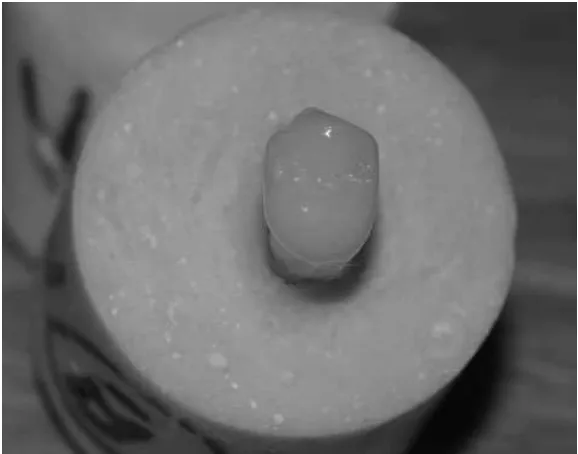
were seated on their corresponding preparations and adjusted when necessary until complete seating was achieved. The com-posite crowns were polished (Soflex discs, 3M ESPE), while the porcelain crowns were glazed at 930◦C without vacuum, following the manufacturer’s recommendations (Fig 2).
The height of each tooth with its corresponding crown was determined with a digital caliper (Digimatic caliper, model CD-6 BS, Mitutoyo Corporation, Kawasaki, Japan) before and after cementation to ensure proper seating. The intaglio surfaces of the composite crowns were grit-etched with 50-µm aluminum
oxide powder in a sandblasting machine microetcher (Micro-cab, Danville Engineering Inc, San Ramon, CA) followed by application of silane coupling agent (Lot number B0DMV, Ul-tradent, South Jordan, UT), while the intaglio surfaces of the porcelain crowns were etched with Porcelain Etch, a 9% hy-drofluoric acid (Lot number B1P6J, Ultradent), followed by application of the same silane coupling agent. For each sub-group crowns were cemented to their corresponding premolars with conventional resin cement with the self-etching adhesive Panavia F2.0; the other subgroup’s crowns were cemented with
Table 1Materials used
Material name Manufacturer Lot number(s) Composition
Vitablocs Mark II Vita Zahnfabrik, Bad Sackingen, Germany
7484,7615, 7484 Conventional feldspathic ceramic with fine-grain particle size
Vita Cerec powder Vita Zahnfabrik, Bad Sackingen, Germany Paradigm MZ100 3M ESPE Dental
Products, St Paul, MN
20060213, 20061122 Conventional hybrid composite resin, Bisphenol-A-diglycidylether dimethacrylate (BisGMA), triethylene glycol dimethacrylate (TEGDMA), and ultrafine zirconia silica ceramic particles as filler. Particles have spherical shape and average size 0.6 mm Panavia F 2.0 Kuraray Medical Inc.,
Okayama, Japan
61140 Base paste:Hydrophobic aromatic (and aliphatic) dimethacrylate, hydrophilic dimethacrylate, sodium aromatic sulfinate,
N,N-diethanolp-toluidine, functionalized sodium fluoride, silanized barium glass
Catalyst paste:MDP, hydrophobic aromatic (and aliphatic) dimethacrylate, hydrophilic dimethacrylate, silanized silica, photoinitiator, dibenzoyl peroxide
RelyX Unicem 3M ESPE Dental Products, St. Paul, MN
236752 Powder:glass fillers, silica, calcium hydroxide, self-cure initiators, pigments, light-cure initiators
Porcelain and Composite Crown Microleakage Ghazyet al
Figure 2 Milled crown seated on prepared tooth.
the self-adhesive universal resin cement RelyX Unicem. All crowns were cemented to their corresponding premolars fol-lowing specific manufacturer’s recommendations under a con-stant load of 5 kg, which was maintained for 10 minutes in a specially designed apparatus (Fig 3).
Cemented crowns were stored in water at 37◦C for 24 hours. They were then subjected to thermocycling between 5 and 55◦C
Figure 3 Cemented crown seated in a device with constant occlusal load of 5 kg maintained until initial setting was achieved.
for a total of 3000 cycles with dwell time of 30 seconds and transfer time 5 seconds. The specimens were then prepared for microleakage testing by coating the root surfaces, except 1 mm from the margin with nail varnish. Specimens were then immersed in a 2.0% basic red fuchsine dye solution for 24 hours at room temperature. The roots of the teeth with their surrounding acrylic base were severed and teeth were split mesiodistally into two halves using a slow-speed Isomet saw (Buehler, Lake Bluff, IL) under constant water cooling.
Specimens were then assessed under magnification (binoc-ular stereomicroscopy, Wild, Heerbrugg, Switzerland) at each margin using the following five-point scale:
Score Description
0 No dye penetration.
1 Dye penetration along the gingival wall. 2 Dye penetration up to 1/2 the axial wall. 3 Dye penetration along more than half to the
full length of the axial wall.
4 Dye penetration extending to the occlusal surface.
A total of 128 readings were available for evaluation of mi-croleakage at the dentin/cement crown interface. These were obtained from 64 specimen halves from all subgroups. Two readings were obtained from each section, and data were sta-tistically analyzed using SPSS (SPSS Inc., Chicago, IL) with 2-way ANOVA and Kruskal-Wallis nonparametric test at the p<0.05 level of significance.
Results
Figure 4 Microleakage overall scores of composite and porcelain crowns with the two cements. Score of 0 indicates no microleakage, while a score of 4 indicates most microleakage that extends throughout the entire tooth/cement interface. No statistically significant differences were detected between the two crown materials (p=0.672).
The use of Panavia F2.0 cement (Fig 5) resulted in signifi-cantly lower microleakage scores when resin composite crowns were used as compared to the use of RelyX Unicem cement (p<0.0001). Similarly, the use of Panavia F2.0 cement (Fig 6)
resulted in significantly lower microleakage scores when porce-lain crowns were used as compared to the use of RelyX Unicem cement (p < 0.015). Figures 7–10 show representative
sec-tioned specimens from the four test groups.
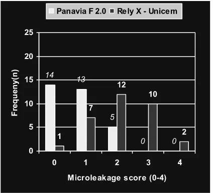
Figure 5 Microleakage scores of Panavia F 2.0 and RelyX Unicem cements with resin composite crowns. Score of 0 indicates no mi-croleakage, while a score of 4 indicates most microleakage that extends throughout the entire tooth/cement interface. Statistically significant dif-ferences were detected between the two cements (p<0.0001).
Figure 6 Microleakage scores of Panavia F 2.0 and RelyX Unicem ce-ments with porcelain crowns. A score of 0 indicates no microleakage, while a score of 4 indicates most microleakage that extends through-out the entire tooth/cement interface. Statistically significant differences were detected between the two cements (p<0.015).
Discussion
Extracted teeth were used in this study in an attempt to approx-imate conditions as close as possible to clinical conditions; however, in the oral environment crowned teeth are exposed to a multitude of challenges ranging from temperature and pH fluctuations related to the food being consumed to mechanical loading applied during mastication in addition to enzymes se-creted to assist digestion. It is difficult in an in vitro set-up to simulate the cumulative effects of these challenges. Therefore, results of this study must be interpreted with caution, as clinical performance of the crowns in terms of microleakage might be worse than what is reported in this study.
The results obtained in the this study showed that there was no statistically significant difference in microleakage scores be-tween crowns milled from Paradigm MZ100 blocks and ones milled from Vita MARK II blocks, irrespective of the cement used. This is not surprising, as both types of crowns were milled with the same machine, and both types of blocks do not undergo any type of further setting after milling. This finding is gener-ally in agreement with findings of Tsitrou,11who concluded that marginal gap of resin composite crowns manufactured with the CEREC 3D system were within the clinical range of accep-tance, regardless of the finish line prepared or the cementa-tion technique used. Furthermore, Behr et al31found marginal
adaptation of crowns cemented with the self-adhesive cement comparable to that achieved with a conventional resin cement, Variolink II; however, they used crowns made with Empress II porcelain, which uses an injection-molding pressing technique rather than the milling technique used in this study for crown fabrication.
Akbar et al12stated that polymer-based materials appear to
Porcelain and Composite Crown Microleakage Ghazyet al
Figure 7Composite resin crown cemented with Panavia F 2.0. This specimen was given a score of 2. The die penetrated along the interface and reached a level limited to the lower half of the axial wall.
microtensile bond strength of conventional resin cement to composite and porcelain blocks, concluded the composite blocks gave a higher bond strength and suggested they could replace porcelain blocks in inlay and crown fabrication.13
Figure 8 Composite resin crown cemented with RelyX Unicem. This specimen was given a score of 2. The die penetrated along the interface and reached a level limited to the lower half of the axial wall.
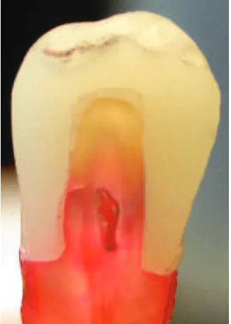
Figure 9Porcelain crown cemented with Panavia F 2.0. This specimen was given a score of 0. There is no evidence of die penetration at any point along the interface.
In this study, microleakage was found to be more pronounced with proprietary self-adhesive cement than that observed with resin cement with a separate self-etch primer/bonding agent. A similar trend was reported by other authors. Ibarra et al
reported that the self-adhesive cement gave a microleakage score more similar to dentin than that recorded with the cement that employed an adhesive in a separate step.33 Yang et al
reported that the bond strength of the self-adhesive cement was lower than that obtained with conventional resin cement.35
Moreover, Escribano and de la Macorra reported that RelyX Unicem had the lowest tensile bond strength among a group of resin cements.34Contrary to this, Piowarczyk et al38stated that microleakage associated with self-adhesive cement was the smallest among a group of tested cements; however, the contrast in the results of that study and the current one may be attributed to differences in test conditions. In Piowarczk et al’s study the crowns were made from a metallic alloy.
Based on the findings of this in vitro study and to reduce the potential for microleakage, dentists should consider use of the resin cement with separate primer/bonding agent when cementing nonmetallic crowns; however, as other self-adhesive resin cements are being made available, more research is needed to explore the potential of these new products in minimizing microleakage. In the oral environment, crowns are subjected to several types of mechanical forces during mastication. Such forces can result, over the long term, in mechanical fatigue, which may have a significant effect on microleakage. Crown material (resin composite vs. ceramic) may play a detrimental role in this respect. Further research is needed to explore the effect of mechanical cyclic loading on microleakage of different nonmetallic crowns.
Conclusions
Under the limitations of this study, the following can be con-cluded:
1. Crown material had no significant effect on microleakage scores.
2. Compared to the self-adhesive cement, RelyX Unicem, the resin cement with a separate primer/bonding agent resulted in significantly lower microleakage scores, irrespective of the crown material.
Acknowledgments
The authors would like to thank the following companies for providing samples of their products: 3M ESPE, Patterson Den-tal, Vident, and Kuraray.
References
1. Otto T, Nisco S: Computer aided direct ceramic restorations: 10-year prospective study of Cerec CAD/CAM inlays and onlays. Int J Prosthodont 2002;15:122-128
2. Thompson JY, Anusavice KY, Naman A, et al: Fracture surface characterization of clinically failed all-ceramic crowns. J Dent Res 1994;73:1824-1832
3. Gu X, Kern M: Marginal discrepancies and leakage of all-ceramic crowns: influence of luting agent and aging conditions. Int J Prosthodont 2003;16:109-116
4. Gernhardt CR, Bekes K, Schaller C: Short term retentive values of zirconium oxide post cemented with glass ionomer and resin
cement: an in vitro study and a case report. Quintessence Int 2005;36:593-601
5. Chang JC, Hart DA, Esty AW et al: Tensile bond strength of five luting agents to two CAD/CAMCAD/CAM restorative materials and enamel. J Prosthet Dent 2003;90:18-23
6. Mormann WH, Bindl A: The Cerec 3-A quantum leap for computer-aided restorations: initial clinical results. Quintessence Int 2000;31:699-712
7. Liu PR: A panorama of dental CAD/CAM restorative systems. Compend Contin Educ Dent 2005;25:507-516
8. Masek R: Designing in 3D—a more visual approach to Cerec correlation. Int J Comp Dent 2003;6:75-82
9. Rusin RP: Properties and applications of a new composite block for CAD/CAM. Compend Contin Educ Dent 2001;22:35-41 10. Giordano R: Materials for chairside CAD/CAM produced
restorations. J Am Dent Assoc 2006;137:145-215
11. Tsitrou E, Northeast S, Van Noort R: Evaluation of the marginal fit of three margin designs of resin composite crowns using CAD/CAM. J Dent 2007;35:68-73
12. Akbar JH, Petrie CS, Walker MP, et al: Marginal adaptation of CEREC 3 CAD/CAM composite crowns using two different finish line preparation designs. J Prosthodont 2006;15:155-163 13. El-Zohairy A, De Geea A, Mohsen M, et al: Micro-tensile bond
strength testing of luting cements to prefabricated CAD/CAM ceramic and composite blocks. Dent Mater 2003;19:575-583 14. Bindl A, Mormann WH: Survival rate of mono-ceramic and
ceramic-core CAD/CAM-generated anterior crowns over 2–5 years. Eur J Oral Sci 2004;112:197-204
15. Blatz M, Sadan A, Kern M: Resin-ceramic bonding: a review of the literature. J Prosthet Dent 2003;89:268-274
16. Albert FE, El-Mowafy OM: Marginal adaptation and
microleakage of Procera AllCeram crowns with four cements. Int J Prosthodont 2004;17:529-535
17. Bindl A, Luthy H, Werner H et al: Strength and fracture pattern of monolithic CAD/CAM-generated posterior crowns. Dent Mater 2006;22:29-36
18. El-Mowafy OM: The use of resin cements in restorative dentistry to overcome retention problems. J Can Dent Assoc
2001;67:97-102
19. Rosenstiel SF, Land MF, Crispin BJ: Dental luting agents: a review of the current literature. J Prosthet Dent 1998;80:280-301 20. Bindl A, Mormann WH: Clinical and SEM evaluation of all
ceramic chair side CAD/CAM generated partial crowns. Eur J Oral Sci 2003;3:163-169
21. Technical product profile: Rely X Unicem 3M/ESPE AG Seefeld, Germany; 2002
22. Gerth H, Dammaschke T, Zuchner H, et al: Chemical analysis and bonding reaction of Unicem and Bifix composite. A comparative study. Dent Mater 2006;22:934-941
23. Kidd EAM: Microleakage: a review. J Dent 1976;4:199-205 24. S¨oderholm KLM: Correlation of in vivo and in vitro performance
of adhesive restorative matherials. Dent Mater 1991;7:74-83 25. Bergenholtz G, Cox CF, Loesche WJ, et al: Bacterial leakage
around dental restorations: its effect on the dental pulp. J Oral Pathol 1982;11:439-450
26. Goldman M, Laosonthorn P, White RR: Microleakage. Full crowns and the dental pulp. J Endod 1992;18:473-475 27. Pilo R, Ben-Amar A: Comparison of microleakage for three
one-bottle and three multiple-step dentin bonding agents. J Prosthet Dent 1999;82:209-213
28. Retief DH: Do adhesives prevent microleakage?. Int Dent J 1994;44:19-26
Porcelain and Composite Crown Microleakage Ghazyet al
30. De Munk J, Varaggs M, Landuyt K, et al: Bonding of an auto adhesive luting material to enamel and dentin. Dent Mater 2004;20:963-971
31. Behr M, Rosenstritt M, Reggnet T, et al: Marginal
adaptation in dentin of a self-adhesive universal resin cement compared with well-tried systems. Dent Mater 2004;20: 191-197
32. Rosenstritt M, Behr M, Lang R, et al: Influence of cement type on the marginal adaptation of all ceramic MOD inlay. Dent Mater 2004;20:463-469
33. Ibarra G, Johnson G, Geurtsen W, et al: Microleakage of porcelain veneer restorations bonded to enamel and dentin with a new self-adhesive resin-based dental cement. Dent Mater 2007;23:218-225
34. Escribano N, de la Macorra JC: Microtensile bond strength of self-adhesive luting cements to ceramic. J Adhes Dent 2006;8:337-341
35. Yang B, Ludwig K, Adelung R, et al: Micro-tensile bond strength of three luting resins to human regional dentin. Dent Mater 2006;22:45-56
36. Rosentritt M, Plein T, Kolbeck C, et al: In vitro fracture force and marginal adaptation of ceramic crowns fixed on natural and artificial teeth. Int J Prosthodont 2000;12:387-391
37. White JM, Goodis HE, Marshall SJ, et al: Sterilization of teeth by gamma radiation. J Dent Res 1994;73:1560-1567
38. Piwowarczyk A, Lauer H, Sorensen L: Microleakage of various cementing agents for full cast crown. Dent Mater