P-ISSN : 1978-225X; E-ISSN : 2502-5600 DOI: https://doi.org/10.21157/j.ked.hewan.v12i1.5428
THE DILATATION OF BRAIN VENTRICLE DUE TO CONGENITAL
TOXOPLASMOSIS IN MICE
CORRELATED WITH APOPTOSIS BUT
NOT WITH TRANSFORMING GROWTH FACTOR BETA
Lucia Tri Suwanti1, 3*, Mufasirin1, 3, and Hani Plumeriastuti2 1
Department of Parasitology, Faculty of Veterinary Medicine, Airlangga University, Surabaya, Indonesia 2
Department of Pathology, Faculty of Veterinary Medicine, Airlangga University, Surabaya, Indonesia 3
Institute of Tropical Disease, Airlangga University, Surabaya, Indonesia *Corresponding author: [email protected]
ABSTRACT
This study aimed to determine the occurences of mice brain ventricles dilatation that congenitally infected with Toxoplasma gondii (T. gondii) as a marker of hydrocephalus and cellular changes in the brain. A total of twenty pregnant mice (11.5 days pregnacy) were divided into two groups, which were control (P1) group and treatment (P2) group. The mice in the treatment group were infected with 101 tachyzoites of T. gondii. All mice were maintained until delivery. The newborn mice were sacrificed and their brain were removed and fixed in 10% buffered formalin to prepare histology slides with HE staining for observation of ventricular width, TUNEL assay for apoptosis observation, and immunohistochemistry for the expression of transforming growth factor beta (TGF-β) observations. The data were analyzed using t test and linear regression. The results showed that ventricular width and apoptosis index significantly increased (P<0.01) in the treatment group compared to control group, but there was no difference in the expression of TGF-β (P>0.05) in both groups. Dilatation of ventricle correlated with the apoptotic index of brain cells but did not correlated with the expression of TGF-β.
____________________________________________________________________________________________________________________ Key words: apoptotic index, brain ventricle, hydrocephalus, Toxoplasma gondii
ABSTRAK
Penelitian ini bertujuan mengetahui pelebaran ventrikel otak anak mencit yang lahir dari induk yang diinfeksi Toxoplasma gondii (T. gondii) sebagai tolok ukur terjadinya hidrosefalus serta untuk mengetahui perubahan seluler pada otak. Dua puluh ekor mencit betina bunting (umur kebuntingan 11,5 hari) dibagi menjadi dua kelompok, kontrol (P1) dan perlakuan (P2). Mencit dari kelompok perlakuan diinfeksi dengan 101
tachyzoite T. gondii. Seluruh mencit dipelihara sampai melahirkan. Anak mencit dikorbankan dan diambil otaknya untuk dilakukan pembuatan preparat dengan pengecatan HE untuk pengamatan lebar ventrikel, TUNEL Assay untuk pengamatan apoptosis, dan imunohistokimia untuk pengamatan ekspresi transforming growth factor beta (TGF-β). Analisis data dilakukan dengan uji t dan regresi linear. Hasil menunjukkan bahwa terjadi peningkatan lebar ventrikel dan indeks apoptosis yang sangat nyata (P<0,01) pada anak mencit yang lahir dari induk yang diinfeksi dibandingkan dengan kontrol, tetapi tidak terjadi perbedaan ekspresi TGF-β (P>0,05) pada kedua kelompok. Ekspresi TGF-β tidak berhubungan dengan lebar ventrikel tetapi lebar ventrikel berhubungan indeks apoptosis pada otak anak mencit.
____________________________________________________________________________________________________________________ Kata kunci: indeks apoptosis, ventrikel otak, hidrosefalus, Toxoplasma gondii
INTRODUCTION
Toxoplasma gondii (T. gondii) are intracellular parasites that can infect warm-blooded animals. Cats usually served as the hosts. This disease is zoonotic and can be transmitted to people. Human is infected through ingestion of sporulated oocysts or eating meat from infected cattle. Infection which occurred in pregnant women can be transmitted to the child at birth and resulted in congenital infection with clinical symptoms ranging from mild to severe symptoms including visual impairment, chorioretinitis, hydrocephalus or microcephaly, intracerebral calcification, seizures, mental retardation and fetal death (Dubey, 2008; Sibley
et al., 2009).
Hydrocephalus defined as the excess of water in the cranial space (Guyton and Hall, 2000) or an increase in the volume of cerebrospinal fluid in the ventricular system of the brain (Wünschmann and Oglesbee, 2001). This situation is a pathological disorder since brain ventricular enlargement is a result of cerebrospinal fluid interrupted flow (Felderhoff-Mueser et al., 2001). In addition to the interrupted flow, the accumulation of fluid in the central nervous system was resulted from disruption of balance between formation and resorption of cerebrospinal fluid
(Johanson et al., 1999). In the mutant mice, hydrocephalus is characterized by dilatation of the fourth ventricle and lateral ventricles.
cerebrospinal fluid also occured in patients with hydrocephalus after subarachnoid hemorrhage (Kitazawa and Tada, 1994; Li et al., 2013).
TGF-ß1 is a multifunctional cytokine involved in the regulation of various biological processes primarily in tissue damage repair (Wyss-Coray et al., 1995; Docagne et al., 2001). In central nervous system,
TGF-β1 regulated damage response. This was proved because long time increased production of TGF-β1 in astrocytes of transgenic mice caused cerebrovascular disorders and degeneration (Wyss-Coray et al., 1995). reveal the correlation of hydrocephalus due to T. gondii
infection with TGF-β expression in the mice brain which was born from infected parent.
MATERIALS AND METHODS
Multiplication of T. gondii Isolates
The isolates which were used in this study were RH strain which were collected from Department of Parasitology, Faculty of Veterinary Medicine, Airlangga University. Isolates multiplication was performed in mice. The isolates were injected intraperitoneally as much as 1x10⁶ tachyzoites and then the mice were maintained for 3-4 days. Mice with symptoms of illness were then sacrificed using head dislocation method. Intraperitoneal fluid was taken by inserting a 3 mL physiological saline into the peritoneal cavity and then the fluid was aspirated. The existence of tachyzoites stadium was examined under microscope and the tachyzoites numbers were counted using improved Neubauerhemocytometer.
Mice Breeding
A total of 20 females pregnant mice were used in this study. In order to make the female mice pregnant, 40 female mice were individually housed adjacent to the male mice. One week after the adaptation, the mice were mated 1:1. The next day, the mice were evaluated for the presence of vaginal plug and if it was positive then the female mice was determined to be pregnant for 0.5 days (Suwanti, 2005). The mice were maintained until gestational age of 11.5 days.
Treatment
Ten of 11.5 days pregnant-mice were divided into two groups: the treatment group (P) consisted of 11.5 days pregnant-mice which were infected with T. gondii
and the control group (K) consisted of 11.5 days pregnant mice which were not infected with T. gondii.
The infectious dose for each mouse was 10 tachyzoites which were dissolved in 100 mL of physiological saline and injected intraperitoneally. The control group was only injected with 100 mL of physiologic saline. The pregnant-mice were maintained until delivery. The
litter was weighed individually and the length and head circumference were measured. The litters were then sacrificed and the cranial bones were taken from half of the litters while the brain was collected from the other half. The cranial bone tissue and the brain were stored in 10% buffered formaline. Brain samples were stained with hematoxilin eosin (HE) and TUNEL to observed ventricular dilatation (as a marker of hydrocephalus) and apoptosis, respectively. The immunohistochemistry test was also carried out to evaluate the expression of TGF-β.
Data Analysis
All data were analyzed using t test with significance
level of α= 0.05 and linear regression.
RESULTS AND DISCUSSION
Hydrocephalus
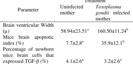
Determination of hydrocephalus was based on ventricle width observation. No particular ventricles were preferred because all ventricles which were seen 58.94±23.51 in newbornmice with uninfected mother to 160.50±11.24 in newborn mice with infected mother (Table 1 and Figure 1).
Table 1. The average of brain ventricular width, brain
apoptotic index, and percentage of brain cells that expressed TGF-β (mean+ SD) from newborn mice
Parameter a,bDifferent superscripts within coulumn indicates significant difference (P<0.05)
Dilatation of the brain ventricles was also found in AIDS patient who experienced ventriculitis and hydrocephalus as the main manifestation of cerebral toxoplasmosis (Sell et al., 2005). A study by Sell et al.
(2005) which observed hydrocephalus using Cranial Computed Tomography with Magnetic Resonance Imaging showed dilatation in lateral and third ventricles.
Indonesia. Although the mice which were born from infected mother did not show hydrocephalus macroscopically, however, after dissection we observed changes in the brain which might indicate the presence of hydrocephalus. Harada et al. (2007) also conduct T. gondii infection in mice using histological observations which were focused on the brain. It was reported that brain ventricular dilation were found on third, fourth, and lateral ventricle. Harada et al. (2007) conducted the research on male mice aged 7 weeks and were observed 4 weeks after infection, which meant that they did the research with acquired infection whereas our study using model of congenital infection. In comparison with previous researches, the results of this research might need to be taken into consideration if the litter born from T. gondii infected mothers to check with cerebral imaging to ascertain the presence of hydrocephalus for early treatment. According to Montoya and Ramington (2008) although most babies
looked normal, after some time (months or years) they could develop symptoms.
Brain Apoptosis
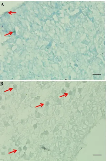
Observations by TUNEL assay showed that an increase in apoptotic index occurred in the brain cells of newborn mice which were born from infected mother compared to the control group. The increase was very significant (P<0.01), nearly 5-fold, from 7.7±2.8 in the group of newborn from uninfected mother to 35.9±12.1 to newborn with infected mother (Table 1 and Figure 2). This suggests that T. gondii
congenital infection caused an increase in apoptotic index of the brain.
The results of this study added information that T. gondii infection either as acquired or congenital infection caused an increase in the apoptotic index of various organs. Previous research also reported an increase in cranial bones apoptotic index in newborn
mice from infected mother (Suwanti and Mufasirin, 2014). Apoptotic cells in the brain as a result of T. gondii infection was likely contributed to the occurred symptoms as describe by Lopes et al. (2007), such as microcephaly and mental disorders in cases of children with congenital toxoplasmosis. Increased apoptosis in brain cells was likely also the explanation for previous research in which a brain from embryo with T. gondii
infection shrunk in size (Suwanti et al., 2010).
After statistical analysis using regression between ventricle width and apoptotic index variable, the ventricular width turned out to be very influential to the
increase of apoptotic index. The wider the ventricular width, the higher the apoptotic index was.
From the analysis, it could be assumed that the mice brain apoptosis occured from ventricular dilatation beside from the result of T. gondii infection as occurred in other organs. In dilated ventricles, the cerebrospinal fluid would push nearby cells and the cells would be stressed and eventually died. According to Sival et al.
(2008), the incidence of hydrochephalus of the newborn mice was associated with an excessive increase of interleukin-18 (IL-18) and interferon gamma (IFN-γ) proinflammatory cytokines in
Figure 2. Mice brain ventricle with TUNEL Assay staining with 1000x magnification. A= Control, B= T. gondii infection, Arrow
cerebrospinal fluid. Previous research showed that the mechanism of apoptosis caused by T. gondii infection was also due to an increase in IFN-γ (Suwanti, 2005; Suwanti and Mufasirin, 2014). The mechanism of brain cell apoptosis due to infection of T. gondii still needed further research.
TGF-β Expression in The Brain
TGF-β expression in the brain was shown by the
percentage amount of the mice brain cells, which in immunohistochemistry staining showed a brownish-black cell cytoplasm. Based on the brain cells that express TGF-β count, either the control or the treatment group showed insignificantly different results after statistical analysis (Table 1). This suggests that T. gondii infection did not lead to increased expression of TGF-β in the brain.
Initially, it was predicted that the expression of
TGF-β in the brain was associated with hydrocephalus
due to T. gondii infection. However, the result found no relationship between two variables. The results were differed from studies on hydrocephalus caused by other factors. However, according to Galbreath et al. (1995), one of the mechanisms of hydrocephalus was due to an increase in production of TGF-β1. In his research, Galbreath et al. (1995) induced hydrocephalus in transgenic mice and proved that the occurrence of hydrocephalus was due to the excessive expression of TGF-β1. This was because TGF-β1 could lead to fibrosis and collagen deposition along the path of cerebrospinal fluid flow (Galbreath et al., 1995; Wyss-Coray et al., 1995). Whitelaw et al. (1999) also reported that increased expression of
TGF-β1 could lead to hydrocephalus. Whitelaw et al.
(1999) showed that in posthemorrhagic
hydrocephalus, which was a complication of premature birth, there was an increase of TGF-β1 in cerebrospinal fluid. In infants who experience mild posthaemorrhagic ventricular dilatation, the levels of TGF-β1 in cerebrospinal fluid was 2.1 ng/mL compared to 0.495 ng/mL in normal infants. In severe cases of permanent hydrocephalus, the TGF- ß1 levels reached 9.7 ng/mL.
CONCLUSION
T. gondii infection in pregnant mice caused
hydrocephalus which was characterized with increase of ventricular width and increase in apoptotic index in new born mice brain cell. Increase in ventricular width would affect brain cell apoptosis. Hydrocephalus caused by T. gondii infection did not correlated with
TGF-β expression in brain.
ACKNOWLEDGMENT
We would like to express our gratitude to The Ministry of Research, Technology, and Higher Education, which had provided the funding for this research through PUPT 2015 and 2016 scheme.
REFERENCES
Docagne, F., N. Colloc'h, V. Bougueret, M. Page, J. Paput, M. Tripier, P. Dutartre, E.T. MacKenzie, A. Buisson, S. Komesli, and D. Vivien. 2001. A soluble transforming growth factor-beta (TGF-beta) type I receptor mimics TGF-beta responses. J. Biol. Chem. 276(49):46243-46250.
Dubey, J.P. 2008. The history of Toxoplasma gondii-the first 100 years. J. Eukaryot. Microbiol. 55(6):467-475.
Felderhoff-Mueser, U., R. Herold, F. Hochhaus, P. Koehne, E. Ring-Mrozik, M. Obladen, and C. Bührer. 2001. Increased cerebrospinal fluid concentrations of soluble Fas (CD95/Apo-1) in hydrocephalus. Arch. Dis. Child. 84:369-372.
Galbreath, E., S.J. Kim, K. Park, M. Brenner and A. Messing. 1995. Overexpression of TGF-beta 1 in the central nervous system of transgenic mice results in hydrocephalus. J. Neuropathol. Exp. Neurol. 54(3):339-349.
Guyton, A.C. and J.E. Hall, 2000. Textbook of Medical Physiology. 10th ed. W.B. Saunders, Philadelphia.
Harada, T., M. Takamoto, D.H. Jin, T. Tada, and K.Sugane. 2007. Young C3H mice infected with Toxoplasma gondii are a novel experimental model of communicating hydrocephalus. Neurol. Res. 29(6):615-621.
Johanson C.E., J. Szmydynger-Chodobska, A. Chodobski, A. Baird, P. McMillan, and E.G. Stopa. 1999. Altered formation and bulk absorption of cerebrospinal fluid in FGF2-induced hydrocephalus.
Am. J. Physiol. 277:263-271.
Kitazawa, K. and T. Tada, 1994. Elevation of transforming growth factor-beta 1 level in cerebrospinal fluid of patients with communicating hydrocephalus after subarachnoid hemorrhage.
Stoke. 25(7):1400-1404.
Li, T., P. Zang, B. Yuan, D. Zhao, Y. Chen, and X. Zhang. 2013. Thrombin-induced TGF-β1 pathway: A cause of communicating hydrocephalus post subarachnoid hemorrhage. Int. J. Mol. Med. 31(3):660-666.
Lopes, F.M.R., D.D. Gonçalves, R. Mitsuka-Breganó, R.L. Freire, and I.T. Navarro. 2007. Toxoplasma gondii Infection in Pregnancy. Braz. J. Infect. Dis. 11(5):496-506.
Montoya, J.G. and J.S. Remington. 2008. Management of Toxoplasma gondii infection during pregnancy. Clin. Infect. Dis. 47:554-566. Sell, M., R. Klingebiel, G. Di Iorio, and S. Sampaolo. 2005. Primary
cerebral toxoplasmosis: A rare case of ventriculitis and hydrocephalus in AIDS. Clin. Neuropathol. 24(3):106-111. Sibley, D., A. Khan, J.W. Ajioka, and B.M. Rosentha. 2009. Review:
Genitic diversity of Toxoplasma gondii in animal and human. Phil. Trans. R. Soc. B. 364:2749-2761. Doi:10.1098/rstb. 2009.0087. Sival, D.A., U. Felderhoff-Mueser, T. Schmitz, E.W. Hoving, C.
Schaller, and A. Heep. 2008. Neonatal high pressure hydrocephalus is associated with elevation of pro-inflammatory cytokines IL-18 and IFN γ in cerebrospinal fluid. Cerebrospinal Fluid Res. 5:21. Doi:10.1186/1743-8454-5-21.
Suwanti, L.T. 2005. Mekanisme Peningkatan Apoptosis Trofoblas Mencit Terinfeksi Toxoplasma gondii melalui Peningkatan Desidua Penghasil IFN- dan TNF- serta Trofoblas Penghasil FAS dan TNFR-1. Thesis. Program Pascasarjana Universitas Airlangga. Surabaya.
Suwanti, L.T. and Mufasirin. 2014. Peranan Sitokin terhadap Kerusakan Tulang Kepala Anak Mencit yang Dilahirkan Oleh Induk yang Diinfeksi Toxoplasma gondii. Research Report. LPPM Universitas Airlangga. Surabaya.
Suwanti, L.T., H. Plumeriastuti, and Mufasirin. 2010. Hambatan Pembentukan Vesikel Otak Embrio sebagai Efek Teratogenik Toxoplasmosis Kongenital pada Ayam. Research Report. LPPM Universitas Airlangga. Surabaya.
Whitelaw,A., S. Christie, and I. Pople. 1999. Transforming growth factor-beta1: A possible signal molecule for posthemorrhagic hydrocephalus?. Pediatr. Res. 46(5):576-580.
Wünschmann, A. and M. Oglesbee. 2001. Periventricular changes associated with spontaneous canine hydrocephalus. Vet. Pathol. 38(1):67-73.