AGRIVITA VOLUME 37 No. 1 FEBRUARY - 2015 ISSN : 0126-0537
http://dx.doi.org/10.17503/Agrivita-2015-37-1-p067-074
HYPOVIRULENT ISOLATES OF
FUSARIUM
COLLECTED FROM CHILI CROPS
IN BOYOLALI REGENCY, CENTRAL JAVA, INDONESIA
Supyani *) and Sri Widadi
Department of Plant Protection, University of Sebelas Maret Jl. Ir. Sutami 36A, Kentingan, Surakarta, Central Java, Indonesia
* ) Corresponding author E-mail: [email protected]
Received: October 3, 2014/ Accepted: March 9, 2015
ABSTRACT
Fusarium, a genus of filamentous fungi, has many species which serving as important pathogens to many diseases in crops. Till today, there have not been effective and efficient control methods for such fungi. Recently, scientists agree that application of biological agents is a tactful choice. Development of hypovirulent strains of fungus as biocontrol agents is very limited. This research was aimed to find hypovirulent isolates of Fusarium from field as biological agents. A hundred isolates of Fusarium from chili were collected in Boyolali, Central Java. Morphological characterization revealed that isolates performed varied colony phenotypes. Based on colony phenotype pattern, isolates were classified into five groups. From each group, one hypovirulent isolate was selected based on colony growth rate on potato dextrose agar media. The selected hypovirulent isolates were used for virulence assay in apple. The result showed that there were four hypovirulent isolates i.e.: B6, C15, D19, and E20 isolates. Total RNA extraction of the identified hypovirulent isolates revealed the existence of viral RNA in C15 isolate. Based on the existence of viral RNA in C15 isolate, the hypovirulent traits were due to mycoviral infection, whereas the hypovirulent traits performed by the other three were due to genetic factors.
Keywords: biological control agent, Fusarium, hypovirulent isolate, virulence assay
INTRODUCTION
A filamentous fungus, Fusarium is the causal agent of many diseases in crops such as vascular wilt, root rot, corm rot, damping-off, yellows, and others. The fungus has a broad host range including rice, horticultural crops, ornamental plants, and others. Overall, the distribution of Fusarium is
known to be cosmopolitan. In Indonesia, Fusarium causes diseases in almost all agricultural commodities. The fungus is found almost in all agriculture land. In general to date, there have not been effective and environmental friendliness control methods for the fungus. Therefore, utilizing a biological control method is a tactful choice (Farr et al., 1989; Agrios, 1997).
In solid media culture, depending on the species, Fusarium colonies can have varying appearances. In general, the aerial mycelium first appears white, and then may change to a variety of colors ranging from white to dark purple. Furthermore, colony phenotypes may also vary according to strain within a species (Alexopoulos et al., 1996).
In the field, there are many strains within a species of fungi. Some of them are virulent whereas the others are hypovirulent. The hypovirulent isolates have at least two possibilities, they are genetically hypovirulent or are infected by mycovirus. From phytopathological perspective, both of them are interesting as they could be developed as biocontrol agents (Ogoshi, 1987; Ghabrial, 2001).
In the biological control practice, the hypovirulent strains of Fusarium has been utilized for controlling the virulence strains which infect fanilla in the field. Here, the hypovirulent strains control the virulent strains via antagonism mechanism (Ogoshi, 1987; Agrios, 1997). On the other hand, to control F. graminearum on wheat, people have developed F. graminearum Virus1- DK21 (FgV1-DK21) (potex-like virus) (Chu et al., 2002; Chu et al., 2004). The other mycovirus, F. graminearum virus from China, 9 isolate (FgV-ch9) has been also developed for controlling F. graminearum on wheat and maize (Darissa et al., 2011). In this case, the mechanism of the hypovirulent strains in controlling the virulent strains is incorporated with viral infection. The hypovirulent
strains are viral infected, and during application, the viruses spread to and infect the virulent strains, and change them into hypovirulent (Lakshman et al., 1998; Ghabrial, 2001; Milgroom and Cortesi, 2004; Nuss, 2005).
In Indonesia, the development of hypovirulent strains of fungus as biocontrol agents is limited. Related to this condition, a preliminary study was needed to find hypovirulent isolates of Fusarium from field as biological control agents. Morphological and virulence traits were used to identify hypovirulent isolates.
MATERIALS AND METHODS
Collection of Fusarium Isolates from Fields Fusarium isolates were collected from endemic areas of the fungus in the center of chili-producing regions in Boyolali regency, Central Java, Indonesia. A hundred pieces of infected trunks were collected from 10 fields (10 samples per 1 hectare of field). Each sample was put into a plastic, and then labelled and kept in an icebox. When samples were arrived in the laboratory, then the samples were transferred to refrigerator prior to fungi isolation.
Isolation and Culture of Fusarium Isolates on Artificial Media
Isolation was done according to the procedure of Streets (1972) with modification. Each infected sample was soaked in 2% NaOCl for 2 min and rinsed in sterile distilled water. Small pieces of the infected samples were transferred to 90 x 15 mm Petri dishes containing 20 ml potato dextrose agar (PDA) (Oxoid Laboratories, Detroit, Mich.), and incubated for 10 days at 25°C. Fusarium isolates, were transferred to new 35 x 10 mm diameter Petri dishes containing 8 ml PDA and incubated at room temperature for 7 days. The isolates were identified to the genera level by the procedures of Nelson et al. (1983). The selected isolates were maintained on regeneration medium at 4oC in a refrigerator as stock cultures.
Morphological Characterization of Fusarium Isolates
Morphological characterization was done as described by Hillman et al. (1990). Experimental cultures were initiated by inoculating 3 x 3 x 3 mm agar cubes, excised from the margins of 7- to 10-day stock cultures, at the center of 90 x 15 mm Petri dishes containing 20 ml of PDA. Plates were
incubated under standard bench top conditions at room temperature. Cultures were observed at 3, 5, and 7 day after inoculation (DAI). The observed and recorded traits were: colony diameter (colony growth rate), colony nature, colony color, amount and color of aerial mycelium.
Virulence Assay of Fusarium Isolates
Virulence assay was done in laboratory as described by Elliston (1985). Mature apples (Rainbow, imported from China) were washed with 5% PURELOX, then five 5-mm diameter x 3-mm-deep plugs of tissue were removed with a burned cork borer and spatula from points equally spaced around each apple. Each apple was inoculated with five isolates of Fusarium sp. One inoculum plug was inserted into each wound, with mycelium facing inward, and pressed with a sterile spatula into complete contact with the tissue. Sites were covered with small pieces of parafilm to retard drying, then the apples were incubated in 35 x 25 x 7 cm plastic boxes under bench top conditions. Lesion diameters were measured at 3, 5 and 7 day after inoculation.
Hypovirulent Isolates of Fusarium Collected From Chili Crops………
RESULTS AND DISCUSSION
Collection of Fusarium Isolates from Fields and Culture in Artificial Media
Fusarium isolates were collected from the center of chili production field in Boyolali regency, Central Java. From this field were collected 100 specimens of infected trunks, and from the specimens were isolated a hundred Fusarium isolates. A hundred Fusarium isolates were cultured on 8 ml PDA plates in 35 x 10 mm diameter Petri dishes at room temperature. On this medium, the cultures of Fusarium isolates showed varied colony morphology from smooth, thin, and cream colored colonies to thick, pink colored colonies with white aerial mycelium. All Fusarium isolates were then used for colony morphology characterization.
Colony Morphology Characterization of Fusarium Isolates
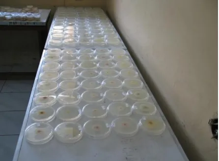
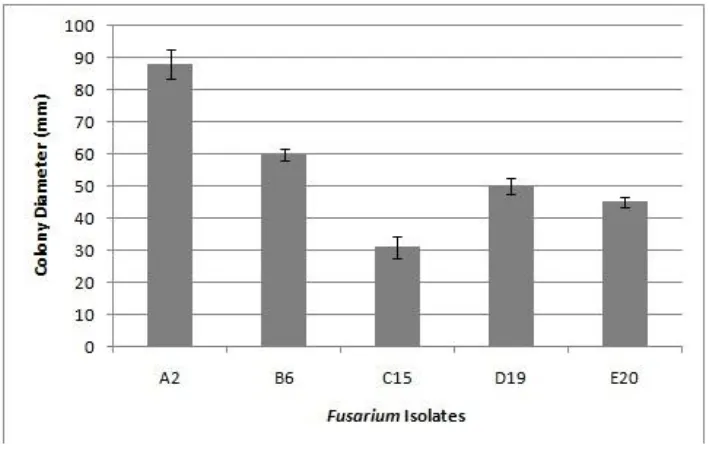
All Fusarium isolates were morphologically characterized. Each isolate was cultured on a 20 ml PDA plate in 90 x 15 mm Petri dish and incubated on the bench top at room temperature for a week (Figure 1). Five days after inoculation (DAI), when the Fusarium colonies performed their best appearance, they were grouped into 5 groups based on their colony morphology pattern (Figure 2). Group A consisted of smooth and thin colonies, was creamy colored, with a few of white aerial myselium; group B consisted of smooth colonies, was purple colored, with much of white aerial mycelium; group C consisted of thick colonies, was pink colored, with white aerial mycelium; group D consisted of rather thick colonies, was white dirty colored, with much of aerial mycelium like cotton; and group E consisted of rather rugged colonies, was white dirty colored, forming a ring pattern, with white aerial mycelium. Then, all the groups of Fusarium isolates were used for colony growth rate assay by culturing them on 20 ml PDA plate in 90 x 15 mm Petri dishes at room temperature, and observed at 5 DAI. The result showed that colony growth rate of isolates within each group was varied. From this assay, one selected isolate showed the fastest colony growth rate from group A, and one another showed the slowest colony growth rate from group B, C, D and E. As a result, there were five selected isolates i.e.: A2, B6, C15, D19 and E20 isolates with colony sizes 88 mm, 60 mm, 31 mm, 50 mm and 45 mm in diameters respectively (Figure 3). Based on these data, from the five
selected isolates, we coined that isolate A2 was virulent, and the other four isolates were hypovirulent.
From mycovirology perspective, phenotype deviations on fungal colonies are the important sign indicating viral infection on them. The deviation could happen on colony profile, colony color, colony growth rate, aerial mycelium quantity, and also sporulation (Ghabrial, 2001). Fusarium graminearum virus-DK21 (FgV-DK21), which causes hipovirulent on its host Fusarium graminearum, changes morphological traits of the fungus including reduction in mycelial growth, increased pigmentation, and decreased (60-fold) production of trichothecene mycotoxins (Chu et al., 2002). In the case of Cryphonectria hypovirus-1 (CHV1), which infects C. parasitica, the virus changed the colony phenotype of the fungus from smooth white yellowish color to rough and white color. Whereas colony of Fusarium isolates indicated the possibility of mycoviral infection. The change of phenotype colony of fungus usually correlates to its virulence. Based on the obtained data of colony profile and colony growth rate, the selected five Fusarium isolates were subjected to virulence assay.
http://dx.doi.org/10.17503/Agrivita-2015-37-1-p067-074
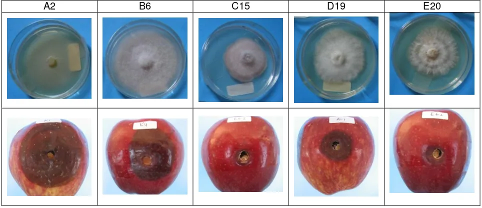
Figure 2. Five groups of Fusarium isolates based on colony morphology. Group A: Smooth and thin colonies, creamy colored, with a few of white aerial mycelium; Group B: Smooth colonies, purple colored, with much of white aerial mycelium; Group C: Thick colonies, pink colored, with white aerial mycelium; Group D: Rather thick colonies, white dirty colored, with much of aerial mycelium like cotton; Group E: Rather rugged colonies, white dirty colored, form a ring pattern, with white aerial mycelium. Fusarium isolates were cultured on 20 ml PDA plates in 90 x 15mm Petri dishes and incubated on the bench top at room temperature for a week
Supyani and Sri Widadi: Hypovirulent Isolates of Fusarium Collected From Chili Crops………
http://dx.doi.org/10.17503/Agrivita-2015-37-1-p067-074 Virulence Assay
Virulence assay was performed by inoculating the selected 5 Fusarium isolates onto apples in five replicates. The result showed that the five Fusarium isolates showed varied virulence level. At 5 DAI, the highest virulence level was shown on isolate A2, in which lesion 60 mm in diameter could be produced, and whereas the lowest virulence level was shown by isolate C15 which could only produce lesion 10 mm in diameter. Furthermore, B6, D19 and E20 isolates produced lesion 40 mm, 24 mm and 15 mm in diameter respectively (Figure 4 and Figure 5).
The other most important indication of viral infection on fungus is the decrease on fungal virulence level (Ghabrial, 2001). Infection of Cryphonectria hypovirus-1 (CHV1) on Cryphonectria
parasitica decreases its virulence level by 25%, whereas the infection of Mycovirus-1 (MyRV1) on the same fungus decreases its virulence level by 80% (Enebak et al., 1994; Hillman et al., 2004; Supyani et al., 2007). In the case of mycovirus, F. graminearum virus-DK21 (FgV-DK21) reduces pathogenicity of the fungus which was proven by reducing its virulence towards wheat (Chu et al., 2002). The other mycovirus, F. graminearum virus from China 9 isolate (FgV-ch9) also reduces pathogenicity of the fungus, by reducing its virulence towards wheat and maize (Darissa et al., 2011).
In this research, based on the phenotype colony traits and virulence, the Fusarium isolate A2 represents virulent isolate, whereas the other 4 selected isolates represent hypovirulent isolates.
A2 B6 C15 D19 E20
Figure 5. Quantified results of virulence assay. Each selected Fusarium isolate was inoculated on apple fruit and incubated under room temperature. Data were collected at 5 day post inoculation (DPI). Each bar represents the average from five replicates with standard deviation
Total RNA Extraction
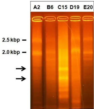
To prove whether the hypovirulent isolates were infected by mycoviruses, total RNA from mycelia of the five selected isolates was extracted. The result of total RNA electrophoresis was shown in Figure 6. From the Figure 6, only C15 isolate showed specific RNA bands. C15 isolate clearly shown had two specific RNA bands sized around 1.0 kbp and 0.8 kbp (pointed by arrows). Here, as a consideration, the ribosomal RNA of the fungus was 28s and 18s ribosomal RNA with size 2.5 kbp and 2.0 kbp respectively (marked by their molecular size). It indicated the existence of mycoviral in C15 isolate. King et al. (2012) reported that to date there are more than 200 mycoviruses identified, and almost all of their genomes were dsRNA. The size of their RNA genomes ranged between 1 and 12.7 kbp. Especially on Fusarium sp, genome size of mycoviral which has been reported to date is 0.8-11.5 kbp dsRNA on F. poae (Compel et al., 1999), 1.4-1.6 kbp dsRNA on F. solani (Nogawa et al., 1996), 2.2-4.0 kbp dSRNA on F. Oxysporum (Kilic and Griffin, 1998) and 2.4 to 7.5 kbp dsRNA on F. Graminearum (Chu et al., 2002; Chu et al., 2004; Darissa et al., 2011).
From this result, it is suggested that the hypovirulent traits performed by C15 isolates of Fusarium in this research were due to mycoviral infection. The hypovirulent traits performed by isolates B6, D19 and E20 of Fusarium in this
research was probably manifestation of fungal genetic variability.
Hypovirulent Isolates of Fusarium Collected From Chili Crops………
CONCLUSIONS
Fusarium isolates infecting chili crops in Boyolali, Central Java showed varied colony phenotype. Based on virulence assay, some of the isolates showed hypovirulency. Total RNA extraction of the hypovirulent isolates revealed the existence of viral RNA in isolate C15. It is suggested that the hypovirulent traits performed by isolates C15 of Fusarium in this research was due to mycoviral infection, whereas the hypovirulent traits performed by the other isolates were due to fungal genetics factor.
ACKNOWLEDGEMENTS
This research was supported by DIPA UNS, under the contract No. 0162.0/023-04.2/XIII/2009, December 31, 2009 and DIPA UNS (PNBP) under the contract No. 501/UN27.11/PN/2014, June 16, 2014.
REFERENCES
Agrios, G.N. 1997. Plant Pathology. 4th Ed. Academic Press. New York. pp. 606. Alexopoulos, C.J., C.W. Mims and M. Blackwell.
1996. Introductory Mycology. 4th Ed. John Wiley and Sons. New York. pp 869. Chu, Y.M., J.J. Jeon, S.J. Yea, Y.H. Kim, S.H. Yun
and Y.W. Lee. 2002. Double-stranded RNA mycovirus from Fusarium graminearum. Applied and Environmental Microbiology. 68 (25): 29–34.
Chu, Y.M., W.S. Lim, S.J. Yea, J.D. Cho, Y.W. Lee and K.H. Kim. 2004. Complexity of dsRNA mycovirus isolated from Fusarium graminearum. Virus Genes 28: 135–143. Compel, P., I. Papp, M. Bibo, C. Fekete and L.
Hornok. 1999. Genetic interrelationships and genome organization of double- stranded RNA elements of Fusarium poae. Virus Genes 18: 49–56.
Darissa, O., P. Willingmann, W. Schäfer and G. Adam. 2011. A novel double-stranded RNA mycovirus from Fusarium graminearum: Nucleic acid sequence and genomic structure. Archives of Virology 156: 647–658.
Elliston, J. E. 1985. Characteristics of dsRNA-free and dsRNA-containing strains of Endothia parasitica in relation to hypovirulence. Phytopathology 82 (2): 151-157.
Enebak, S.A., B.I. Hillman and W.L. MacDonald. 1994. A hypovirulent Cryphonectria parasitica isolate with multiple, genetically unique dsRNA segments. Molecular Plant-Microbe Interactions 7 (5): 590-595. Farr, D.F., G.F. Bills, G.P. Chamuris and A.Y. Hypovirulence-associated suppression of host functions in Chryphonectria parasitica can be partially relieved by high light intensity. Phytopathology 80: 950-956. Hillman, B.I., S. Supyani, H. Kondo and N. Suzuki.
2004. A reovirus of the fungus Cryphonectria parasitica that is infectious as particles and related to the Coltivirus genus of animal pathogens. J. Virol. 78: 892–898.
Kilic, O. and G.J. Griffin. 1998. Effect of dsRNA- containing and dsRNA-free hypovirulent isolates of Fusarium oxysporum on severity of Fusarium seedling disease of soybean in naturally infested soil. Plant and Soil 201: 125–135.
King, A.M.Q., M.J. Adams, E.B. Carstens and E.J. Lefkowitz. 2012. Virus taxonomy: Classification and nomenclature of viruses: Ninth report of the International Committee on Taxonomy of Viruses. San Diego: Elsevier.
Lakshman, D.K., J. Jian and Tavantzis. 1998. A double stranded RNA element from a hypovirulent strain of Rhizoctonia solani occurs in DNA form and is genetically related to the pentafunctional AROM protein of the shikimate pathway. Proc. Natl. Acad. Sci. USA 95: 6425-6429. Milgroom, M.G. and P. Cortesi. 2004. Biological
control of chestnut blight with hypovirulence: A critical analysis. Annual Review of Phytopathology 42: 311-338.
http://dx.doi.org/10.17503/Agrivita-2015-37-1-p067-074 Nogawa, M., A. Nakatani, K. Gonda, M. Shimosaka
and M. Okazaki. 1996. Replication of double-stranded RNA in mycovirus from the plant pathogenic fungus, Fusarium solani. FEMS Microbiology Letters 137: 45–49.
Nuss, D. L. 2005. Hypovirulence: Mycoviruses at the fungal-plant interface. Nature 3: 632-642.
Ogoshi, A. 1987. Ecology and pathogenicity of anastomosis and intraspecific groups of Rhizoctonia solani Kühn. Ann. Rev. Phytopathol 25: 125-143.
Streets, R.B. 1972. Diagnosis of plant diseases.The University of Arizona Press.USA
Supyani, S, B.I. Hillman and N. Suzuki. 2007. Baculovirus expression of all the mycoreovirus 1 genome segments and identification of the guanylyltransferase- encoding segment. J. Gen. Virol. 88: 342-350.