RISK FACTORS AND OUTCOME DIFFERENCES BETWEEN
ISCHEMIC AND HEMORRHAGIC STROKE
Ivica Bilic, Gordan Diamanja, Iva Lusic, Meri Matijaca and Kresimir Caljkusic University Department of Neurology, Split University Hospital Center, Split, Croatia SUMMARY - The aim of the study was to justify the hypothesis that risk factors do not differ between ischemic and hemorrhagic stroke. This retrospective study included 1066 stroke patients. The prevalence of risk factors and hospital-based survival were compared between patients with ischemic and hemorrhagic stroke. Data were retrieved from patient records. Statistical analysis was done by use of i-test and t-test for dependent samples. The group of hemorrhagic stroke con-sisted of70 (47.9%) female and 76 (52.1%) male patients. The group ofischemic stroke included 450 (48.9%) female and 470 (51.1%) male patients. Ischemic stroke patients had a higher prevalence of hypertension (79% vs. 72%), atherosclerotic diseases (50% vs. 34%) and atrial fibrillation (15.5% vs. 4.2%), and were statistically significantly older (72.5±10.4 VS. 65.7±12.8) than those with hemor-rhagic stroke, however, fatal outcome was more common in the latter (26% vs. 17%). In conclusion, data analysis pointed to differences between hemorrhagic and ischemic stroke according to both risk factors and stroke outcome.
Key words: Cerebrovascular disorders - classification; Cerebrovascular disorders - diagnosis; Cerebrovascular disorders - risk foctors; Incidence; Age - distribution
Introduction
Stroke is the second leading cause of mortality worldwide and remains the leading cause of adult physical disability. Almost 1 300 000 people die from cerebrovascular diseases in Europe every year. In 2007, some 6 million people died from stroke and millions suffer disabling sequels of stroke. The costs of stroke include medical care, rehabilitation and lost productivity of stroke survivors. It is measured in bil-lions of euros every year. The costs in terms of pain and suffering of its victims and their families cannot be measured. Stroke is a highly heterogeneous disor-der with distinct subtypes, each presenting specific clinical and epidemiological aspects!·2 Risk factors for stroke include systolic or diastolic hypertension, Correspondence to: Ivica Bilic, MD, MS, University Department of Neurology, Split University Hospital Center, Spinuceva 1, HR-21000 Split, Croatia
E-mail: ibilic@kbsplit.hr
Received October 27,2008, accepted in revised form June 1, 2009
diabetes, atrial fibrillation, hypercholesterolemia, cig-arette smoking, heavy alcohol consumption, and oral contraceptive use.
By definition, stroke produces neurologic deficits that persist for at least 24 hours. Stroke produces focal symptoms and signs that correlate with the area of the brain supplied by the affected blood vessel.
Ischemic stroke has many causes with differ-ent clinical presdiffer-entations, risk factors, courses and outcomes3,4. The prognosis and management of isch-emic stroke are directly related to the specific mecha-nism of ischemic lesion. Comparison of functional outcome and survival and recurrence rates can allow clinicians to identify patients at a higher risk of stroke recurrence and death. Early classification of isch-emic stroke subtype is of substantial practical clini-cal value. The classification of ischemic stroke used in this study was The Oxfordshire Community Stroke Project (OCSP) classification, which defines four clinically identifiable subgroups of cerebral infarction: total anterior circulation infarction (TACI), partial
1. Bille et al. Risk factors and outcome differences between ischemic and hemorrhagic stroke
anterior circulation infarction (PACI), posterior cir-culation infarction (POCI), and lacunar infarction (LACI). OCSP has the ability to predict the prog-nosis and shows good correlation with the underly-ing pathophysiology and imagunderly-ing findunderly-ings on cranial computed tomography'·6.
There is another stroke classification system used in some recent stroke studies, and it is named TOAST (Trial ofORG 10172 in Acute Stroke Treat-ment) classification, which denotes five diagnostic subgroups of ischemic stroke: large-artery atheroscle-rosis, cardioembolism, small-vessel occlusion, i.e. la-cunar, stroke of other determined etiology, and stroke of undetermined etiology. One argument against the use of this type of subclassification system is the dif-ficulty of performing the necessary technical exami-nations in all patients; so we decided to use OCSP in this particular survey.
Hemorrhagic stroke was classified as intracerebral hematoma or subarachnoid hemorrhage.
Correct classification of stroke subtypes in patients with acute stroke is crucial for early management and for predicting the prognosis7•s
The objective of this hospital-based study was to evaluate outcomes of patients with stroke (according to risk factors and stroke type) treated at Split University Hospital Center during a one-year period. The hypoth-esis was that ischemic and hemorrhagic strokes do not differ in outcome when risk factors are considered. Patients and Methods
The study was retrospective and was conducted by three neurologists and two neurology residents. It included all patients treated for stroke at University Table 1. Stroke type and outcome analysis
Department of Neurology, Split University Hospi-tal Center, Split, Croatia, during a one-year period
(2006). Stroke was defined according to the World Health Organization definition as "rapidly develop-ing clinical signs of focal (or global) disturbance of cerebral function lasting for more than 24 hours (un-less interrupted by surgery or death), with no apparent cause other than of vascular origin"9.
The study included first ever stroke, recurrent stroke, all ischemic strokes, and all types of hemor-rhagic stroke as mentioned above. Recurrent stroke was defined as a new neurological deficit fitting the definition of ischemic stroke, occurring after a period of unequivocal neurological stability or improvement lasting for ,,24 hours and not attributable to edema, mass effect, brain shift syndrome, or hemorrhagic transformation of the incident cerebral infarction.
Medical chart of every stroke patient was surveyed for the following risk factors: sex, age, atherosclerotic disease, diabetes, hyperlipidemia, cigarette smoking, previous stroke, atrial fibrillation, and alcoholism, and for stroke outcome. The hospital-based study included
1066 patients.
Statistical analysis was performed using the Sta-tistica 7.0 for Windows software, t-test for dependent samples and Pearson's xl-test. The level of significance was set at P<0.05.
Results
The study included 1066 stroke patients treated at University Department of Neurology, Split Uni-versity Hospital Center, in 2006. Hemorrhagic and ischemic stroke are presented by subtypes in Table 1.
There were 70 (47.6%) women and 76 (52.4%) men
Outcome
Stroke type Incidence (%) Deceased (%) p
400
Intracerebral hematoma Subarachnoid hemorrhage Lacunar stroke (LACI) Partial anterior circulation infarction (PACI)
Total anterior circulation infarction (TACI) Posterior circulation infarction (POCI) 111/146 (76.0) 37 (25.3) 35/146 (24.0) 3 (2.0) 153/920 (16.6) 22 (2.4) 109/920 (11.8) 16 (1.7) <0.001 475/920 (51.6) 94 (10.2) 183/920 (19.9) 24 (2.6)
Table 2. Risk foctor analysis
Risk factor
Sex (n~546; 51.1 %) Male
(n~520; 48.9%) Female
Mean± SD
Age (yrs) Median
Range
H}1lertension (n~849; 80%) yes
Atherosclerotic disease (n~525; 49%) yes
H}1lerlipidemia (n~382; 36%) yes
Atrial fibrillation (n~153; 14%) yes
Diabetes (n~465; 44%) type 1
type 2
Smoking (n~179; 17%) yes
Previous stroke (n~212; 20%) yes
Alcohol consumption (n~197; 18%) yes Mean± SD
Hospital stay (days) Median
Range • x} -test; ** t - test
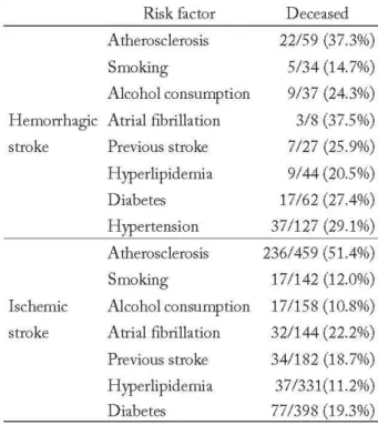
Table 3. Risk factors according to stroke type and lethal outcome
Risk factor Deceased
Atherosclerosis 22/59 (37.3%)
Smoking 5/34 (14.7%)
Alcohol consumption 9/37 (24.3%) Hemorrhagic Atrial fibrillation 3/8 (37.5%)
stroke Previous stroke 7/27 (25.9%)
H}1lerlipidemia 9/44 (20.5%)
Diabetes 17/62 (27.4%)
Hypertension 37/127 (29.1%)
Atherosclerosis 236/459 (51.4%)
Smoking 17/142 (12.0%)
Ischemic Alcohol consumption 17/158 (10.8%) stroke Atrial fibrillation 32/144 (22.2%) Previous stroke 34/182 (18.7%) H}1lerlipidemia 37/331(11.2%)
Diabetes 77/398 (19.3%)
Acta Clin Croat, Vol. 48, No.4, 2009
Stroke t}1le Hemorrhagic Ischemic (N~l46) (N~920) P 76 470 0.753' 70 450 65.7±12.8 72.5±10.4 68 74 ,0.001" 28-93 35-95 105 744 0.045' 57 468 ,0.001' 43 339 0.026' 7 146 ,0.001' 5 22 0.15' 55 383 34 145 0.11' 26 186 0.195' 36 161 0.27 11.9±8.2 10.9±11.6 0.84" 13 10 1-39 1-31
with hemorrhagic stroke, and 450 (48.9%) women and 470 (51.1%) men with ischemic stroke. The mean age was 72.9±10.5 (range 33-95) years in female patients and 69.8±11.9 (range 25-94) years in male patients. Statistical analysis showed the women having suffered stroke to be significantly older than men (P,O.OOl).
Risk factor analysis is presented in Table 2.
Atherosclerosis was more common in patients suf-fering from ischemic stroke than in those with hem-orrhagic stroke. Atrial fibrillation and dyslipidemia were also more common as risk factors in ischemic than in hemorrhagic stroke. Patients with ischemic stroke were older than those with hemorrhagic stroke. Lethal outcome was more common in patients with hemorrhagic stroke, the difference being statisti-cally significant (P~0.004). Table 3 shows differences in risk factors and lethal outcome between different types of stroke.
Discussion
Analysis and comparison of the data collected clearly showed the hemorrhagic and ischemic stroke
1. Bille et al. Risk factors and outcome differences between ischemic and hemorrhagic stroke
to differ according to some risk factors and outcome. Comorbidity has a significant impact on stroke out-come!O Prognosis depends on the type of stroke, the degree and duration of obstruction or hemorrhage, and the extent of brain tissue death. The location of hemorrhagic stroke is an important factor in the outcome, and this type generally has a worse progno-sis than ischemic stroke. The 30-day mortality from hemorrhagic stroke ranges from 35 to 52 percentl l
-15;
one-half of these deaths occur within the first two d ays13.!'. Prognosis is generally poor when compared with ischemic stroke. Age, Glasgow Coma Score less than 8 at presentation, hematoma volume of greater than 60 mL, and intraventricular blood are predic-tors of high mortality13,17,18. Considering risk factors, atherosclerosis was more frequently found in ischemic stroke patients because atherosclerotic plaques narrow the inner diameter of the vessel, resulting in inade-quate tissue vascularization.
Results of this hospital-based study confirmed that there was no sex difference in the type of stroke (P=0.7S3).
Considering risk factors for ischemic stroke, there is a consensus in population and hospital-based stud-ies that hypertension is the most common risk fac-tor predisposing patients for all subtypes of ischemic stroke. Our study confirmed this statement. Results of previous hospital-based studies suggest that isch-emic stroke is a polyetiologic disturbance with clear differences in the risk factor profile among particu-lar ischemic stroke subtypes. According to previous investigations, the synergistic action of hypertension, diabetes and hyperlipidemia predisposes patients for lacunar stroke'. Of the known controllable risk fac-tors, hypertension is most important19. Hypertension accelerates the atherosclerosis process, therebyincreas-ing the risk of atherothrombotic cerebral infarction. It also increases the risk of cerebral hemorrhage in part by promoting the development of cerebral vascular micro aneurysms (Charcot-Bouchard aneurysms)20,21.
Hemorrhagic and ischemic stroke differ according to outcome and risk factors. Atherosclerosis, atrial fi-brillation and hyperlipidemia are the most powerful risk factors, which contribute to differences in mani-festation of the resulting stroke type and outcome. These risk factors are by far more common in ischemic than in hemorrhagic stroke.
402
Ischemic stroke can be predisposed by excessive alcohol intake and by intracerebral and subarachnoid hemorrhage via multiple mechanisms (e.g., via hy-pertension, atrial fibri11ation, rebound thrombocytosis and platelet aggregation and clotting disturbances)22.
Our data confirmed the increased risk of primary intracerebral hemorrhage to be associated with low cholesterol, a relationship that may apply specifically to hemorrhages from hypertensive vasculopathy'3
Since treatment measures for stroke are sti11 rather limited, and knowing the high number of patients suffering stroke every year, it is important to be famil-iar with the stroke risk factor profile for each patient, and to be aware that prevention, i.e. timely identifica-tion and therapy for stroke risk factors, is the most efficacious method of stroke treatment. The individu-als with a relatively high risk profile can take steps to modify other risk factors through lifestyle changes and/or medical treatment. Similarly, public awareness programs aimed at increasing the recognition of stroke warning signs and altering modifiable risk factors can be designed to address the high-risk groups.
Although this study did not produce any new or surprising results, the value of small hospital-based studies like this one lies in strengthening the aware-ness of the important role of stroke prevention while influencing risk factors. Every person, not only health care professionals, can contribute to stroke prevention by promoting healthy lifestyle and avoiding known risk factors for stroke.
References
1. SUDLOW CL, WARLOW CPO Comparable studies of the incidence of stroke and its pathological subtypes: results from an international collaboration. Stroke 1997;28:491-9. 2. DEMARIN V. Moidani udar - rastuCi medicinski i
soci-jalno ekonomski problem. Acta Clin Croat 2004;43:9-13. 3. SACCO RL, TONI D, MOHR
JP.
Classification ofisch-emic "mh. In, BARNETT HJM, MOHR JP, BENNT
MS, YATSU FM, editors. Stroke: pathophysiology, treat-ment and prognosis. 3rd ed. Philadelphia: Churchill Living-stone; 1998:341-55.
4. BOGOUSSLAVSKY J, MELLE GV, REGL! F. The
Lau-sanne Stroke Registry: analysis of 1000 consecutive patients with first stroke. Stroke 1988;19:1083-92.
5. ROVIRA A, GRIVE E, ROVIRA A, ALVAREZ-SABIN
J.
Distribution territories and causative mechanisms of isch-emic stroke. Eur RadioI2005;15:416-26.6. TEl H, UCHIYAMA S, OHARA K, KOBAYASHI M,
UCHIYAl\1A Y, FUKUZAWA M. Deteriorating ischemic stroke in four clinical categories classified by Oxfordshire Community Stroke Project. Stroke 2000;31:2049-54.
7. GODOY DA, PINERO G, Di NAPOLI M.
P"dict-ing mortality in spontaneous intracerebral hemorrhage: can modification to original score improve the prediction? Stroke
2005;37(SuppI4);1038-44.
8. MURAT SUMER M, ERTURK O. Ischemic stroke sub-types: risk factors, functional outcome and recurrence.
Neu-wI Sci 2002;22;449-54.
9. HATANO S. Experience from a multicenter stroke register: a preliminary report. Bull WHO 1976;54:541-53.
10. FISCHER U, ARNOLD M, NEDELTCHEV K, SCHO-ENEENBERGER RA, KAPPELER L, HOLLINGER P,
et al. Impact of comorbidity on ischemic stroke outcome. Acta
Neurol Scand 2005;113:108-13.
11. ANDERSON C, CHAKERA T, STEWARD-WYNNE
EG, JAMROZIK KD. Spectrum of primary intracere-bral hemorrhage in Perth, Western Australia, 1989-1990: incidence and outcome. J Neurol Neurosurg Psychiatry
1994;57;936-40.
12. COUNSELL C, BOONYAKARNUKUL S, DENNIS M.
Primary intracerebral hemorrhage in the Oxfordshire Com-munity Stroke Project. Cerebrovasc Dis 1995;5:26-31.
13. BRODERICK J, BROTT T, DULDNERJE, TOMSICK
T, HUSTER G. Volume of intracerebral hemorrhage: a pow-erful and easy-to-use predictor of 30-day mortality. Stroke
1993;24;987-93.
14. FOGELHOLM R, MURROS K, RISSANEN A,
AVIKAINEN S. Long term survival after primary intrac-erebral haemorrhage: a retrospective population based study. J Neurol Neurosurg Psychiatry 2005;76:1534-8.
15. FLAHERTY ML, HAVERBUSCH M, SEKAR P, KISSELA BM, KLEINDORFERD, MOOMAW C], d ai.
Long-term mortality after intracerebral hemorrhage.
Neurol-ogy 2006;66;1182-6.
16. FRANKE CL, SWIETEN JC, ALGRA A, van GIJN
J.
Prognostic factors in patients with intracerebral hemorrhage. J Neurol Neurosurg Psychiatry 1992;55:653-7.
17. LISK DR, PASTEUR W, RHOADES H, PUTNAM RD,
GROTTA Jc. Early presentation of hemispheric intracere-bral hemorrhage: prediction of outcome and guidelines for treatment allocation. Neurology 1994;44:133-9.
18. MAYER SA, SACCO RL, SHI T, MOHRJP. Neuwlogic
deteriorations in noncomatose patients with supratentorial intracerebral hemorrhage. Neurology 1994;44:1379-84.
19. GVOZDENOVIC S, RABI ZIKIC T, ZARKOV M,
BOZIC K, ZIKIC M. Podtipovi ishemijskog moidanog udara: ucestalost i profil faktora rizika. Zbornik saietaka 2. kongres neurologa Bosne i Hercegovine s medunarodnim sudjelovanjem, Mostar, 2006.
20. WOLF PA, D'AGOSTINO RB, O'NEAL MA, SYT-KOWSKI P, KASE CS, BELANGER AJ, d al. Secul"
trends in stroke incidence and mortality. The Framingham
Study. Stwke 1992;23;1551-5.
21. WILLIAMS GH, BRAUNWALD E. Cerebrovascular dis-eases. In: BRAUNWALD E, et al., editors. Harrison's
prin-ciples of internal medicine, 1rh ed. New York: McGraw Hill,
1987;1024.
22. GORELICK PE. Alcohol and stroke. Stroke 1987;18(Suppl
1);268-71.
23. SEGAL AZ, CHIU RI, EGGLESTON-SEXTON PM,
BEISER A, GREENBERG SM. Low cholesterol as a risk factor for primary intracerebral hemorrhage: a case-control study. Neuroepidemiology 1999;18:185-93.
Saietak
RAZLIKE U CIMBENICIMA RIZIKA I ISHODU IZMEDU ISHEMIJSKOG I HEMORAGIJSKOG MOZDANOG UDARA
L Bilii, G. Dzamonja, L Lufit, M. MatiJaca i K. Caljkufit
Cilj studije bio je provjeriti opravdanost hipoteze kako se cimbenici rizika ne razlikuju izmedu ishemijskog i hemora-gijskog moidanog udara. Ova retrospektivna studija ukljucila je 1066 bolesnika s moidanim udarom. Ucestalost rizicnih cimbenika i bolnicko preiivljenje usporedeni su izmedu bolesnika s ishemijskim i hemoragijskim moidanim udarom. Podatci su izvedeni iz bolesniCkih kartona. Statisticka analiza je provedena pomocu x2-testa and t-testa za zavisne uzor-ke. Skupina bolesnika s hemoragijskim moidanim udarom imala je 70 (47,9%) iena i 76 (52,1%) muskaraca, a skupina s ishemijskim moidanim udarom 450 (48,9%) iena i 470 (51,1%) muSkaraca. Bolesnici s ishemijskim moidanim udarom imali su vecu ucestalost hipertenzije (79010 prema 72%), aterosklerotske bolesti (50% prema 34%) i atrijske fibrilacije (15,5% prema 4,2%) i bili su statistiCki znaeajno stariji (72,5±1O,4 prema 65,7±12,8) od bolesnika s hemoragijskim moidanim udarom, ali je smrtni ishod bio cdCi kod ovih potonjih (26% prema 17%). Dakle, analiza prikupljenih podataka ukazala je na razlike izmedu hemoragijskog i ishemijskog moidanog udara u rizicnim cimbenicima i ishodu bolesti.
Kljucne rijeci: Cerebrovaskularne bolesti klasifikaciJa; Cerebrovaskularne bolesti diJagnostika; Cerebrovaskularne bolesti -rizicni Cimbenici; IncidenciJa; Dob - raspo4fela