Corresponding author: [email protected]
Prevalence and distribution of thalassemia
trait screening
Nailil Husna, Imanuel Sanka, Ahmad Al Arif, Chintya Putri, Elory Leonard, Niken Satuti Nur Handayani*
Laboratory of Genetic and Breeding, Faculty of Biology, Universitas Gadjah Mada, Yogyakarta, Indonesia
DOI: http://dx.doi.org/10.19106/JMedSci004903201702
ABSTRACT
Thalassemia is an inherited disorder of autosomal recessive gene caused by decrease or absent production of one or two type of globin chain. This disorder will affect the quality and quantity of blood production. In Indonesia, thalassemia is not concerned as
urgency, although it lies in thalassemia belt area. Thalassemia is classiied according
to the particular globin chain which affected such as α-thalassemia and β-thalassemia. Besides thalassemia, there are variant hemoglobinopathy called HbE. The aim of this study was to assess the prevalence of thalassemia carriers among the volunteer of screening in Yogyakarta Special Region from 2012 until 2015. The thalassemia carrier screening was conducted by collaborating with Indonesian Association of Parents of Children with Thalassemia (APCT) Yogyakarta. The hematological measurement and High-Performance Liquid Chromatography (HPLC) were performed on Prodia Laboratory Yogyakarta. The analysis of carriers prevalence was conducted in Laboratory of Genetics and Breeding, Faculty of Biology, Universitas Gadjah Mada, Yogyakarta. Among 241 volunteers, we found 44 volunteers was diagnosed as β-thalassemia carrier, 30 volunteers as α-thalassemia carrier as well as HbE disorder carrier, and 1 volunteer was diagnosed as α−β-thalassemia
carrier. The number of thalassemia carrier shows no signiicant difference each year. The
prevalence of thalassemia carrier is high, even though the distribution is limited by the location where the screening took place.
ABSTRAK
Thalassemia adalah kelainan bawaan pada gen resesif autosomal akibat penurunan atau tidak diproduksinya satu atau dua jenis rantai globin. Kelainan ini mempengaruhi kualitas dan kuantitas produksi darah. Di Indonesia, thalasemia belum menjadi masalah penting
walaupun lokasinya termasuk dalam daerah sabuk thalasemia. Thalasemia diklasiikasikan
β-thalasemia dan HbE serta satu pembawa α−β-thalasemia. Jumlah pembawa thalasemia
tidak menunjukkan perbedaan yang signiikan setiap tahunnya. Prevalensi pembawa
thalasemia tinggi, meskipun distribusinya dibatasi oleh lokasi dimana skrining dilakukan.
Keywords: Thalassemia trait screening - α-thalassemia - β-thalassemia – HbE – HPLC
INTRODUCTION
Thalassemia is an inherited disorder of autosomal recessive gene caused by decrease or absent production of one and/ or two type of globin chain. This disorder will affect the quality and quantity of blood production.1 Thalassemia is classiied
according to the particular globin chain
which affected. The α-globin is involved in α-thalassemia and theβ-globin chain is affected in β-thalassemia.2,3 Approximately
400 mutations have been reported to cause this disorder in the world population.4,5 The
mutation can cause hereditary anemias due to decrease hemoglobin synthesis and red blood survival.6 Beside, structural hemolgobin
variant known as hemoglobinophaty are also caused by abnormality in globin chain such as HbS, HbC and HbS.7
Previous studies reported that the distribution of this disorder were limited to the areas called “thalassemia belt” (lies from the Mediterranean basin through the Middle East and Indian subcontinent up to Southeast Asia), but recent migration of people have spread thalassemia genes throughout the world.6,8 About 7% of the population was
recorded inherit the disorder throughout Africa and Asia.1 In Indonesia, thalassemia is not
considered as major health problems, although it lies in thalassemia belt area. Furthermore, thalassemia carrier in Indonesian population was recorded and shown high prevalence,
for detail 3-20% was α-thalassemia carrier, 3% was β-thalassemia carrier and 1-33% was
HbE carrier.9
The thalassemia trait which inherits an individual only has mutation from one of the parents.1 The thalassemia trait does not suffer
thalassemia, but if the trait marry another trait, there would be a chance to have thalassemia major disorder or severe anemia.10 Refer to the
population which reported above, this study was conducted to to assess the prevalence of thalassemia carriers among the volunteer of screening in Yogyakarta Special Region from 2012 until 2015.
MATERIALS AND METHODS
Volunteers
The material of this research was the hematological database as the result of screening from Prodia Clinical Laboratory, including 241 sheets of cellular medical test which contain quantity of cellular blood components. Thalassemia screening was conducted annually and collaborated with Indonesian Association of Parents of Children with Thalassemia (IAPCT) Yogyakarta, the Indonesia Thalassemia Foundation (ITF), Prodia Clinical Laboratory, Yogyakarta and Faculty of Biology Universitas Gadjah Mada, Yogyakarta. From 2012 until 2015, there was 241 volunteers who attended the screening.
Procedure
Blood collection was conducted in Faculty of Biology, Universitas Gadjah Mada, Yogyakarta. The hematological
measurement and High-Performance Liquid
Prodia Clinical Laboratory, Yogyakarta. To
deine the thalassemia prevalence, the analysis
was conducted in Laboratory of Genetics and Breeding, Faculty of Biology, Universitas Gadjah Mada, Yogyakarta. Informed of consent was obtained from the volunteer who observed.
Analysis
The data was analyzed using formula of Microsoft Excel 2010 due to the assigned ranged from Prodia Clinical Laboratory, Yogyakarta. The analysis was conducted in Laboratory of Genetics and Breeding, Faculty of Biology, Universitas Gadjah Mada, Yogyakarta.
RESULTS
There were 241 volunteers which measured on the thalassemia screening.
From the screening we found 75 volunteers who have thalassemia trait, meaning that
almost 45% of the volunteer were diagnosed disorder. FIGURE 1 shows the prevalence of thalassemia among volunteers screened. The
β-thalassemia trait was recorded most often
which reached out 44 volunteer, while it was
founded that α-thalassemia carrier and HbE
disorder carrier had the same number of 30 volunteers. However, we found one volunteer
who had α-β-thalassemia carrier phenotype.
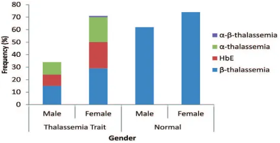
FIGURE 2 shows that the prevalence for female of thalassemia carrier and HbE disorder carrier were higher than male.
FIGURE 1. The prevalence of thalassemia among volunteers screened.
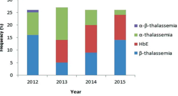
FIGURE 3 shows the prevalence of thalassemia trait in four different years from 2012 to 2015. The total number of thalassemia
carrier showed no signiicant difference from
2012 to 2015. For α-thalassemia carrier, in 2012 the number for volunteer with the
carrier was lower than β-thalassemia carrier
(9 volunteers detected). But, different with 2013, the number of volunteers detected with
α-thalassemia carrier was the highest among
other thalassemia carrier (13 volunteers detected). In 2014 and 2015, the number of
α-thalassemia carrier was the lowest among
others, which were 6 and 2 volunteers
detected respectively. The number of carrier
prevalence for β-thalassemia carrier in 2012
and 2015 was the highest comparing to other thalassemia carriers. In 2013, its prevalence was the lowest among others, and in 2014,
number of prevalence for β-thalassemia
carrier was ranked number two. Also in 2012,
we obtained a volunteer with α-β-thalassemia
carrier phenotype. HbE disorder carrier was
irstly found in 2013 screening with stable
number in the next year of screening and it
luctuated between 9-11 volunteers detected
with the carrier.
FIGURE 3. The prevalence of thalassemia trait in four different years.
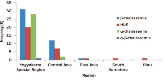
FIGURE 3 shows the distribution of thalassemia carrier according to their respective
native region detected in ive provinces, which
are Yogyakarta Special Region, Central Java, East Java, South Sumatra and Riau. Based from the screening, the most often thalassemia
FIGURE 4. Distribution of thalassemia trait in ive provinces of Indonesia
DISCUSSION
The major study of this research was to determine the thalassemia carrier including its prevalence and distribution. Thalassemia carrier can be found when an individual have one or two mutation(s) on a chromosome number 11 (α-thalassemia) and one mutation on a chromosome number 16 (β-thalassemia). In accordance to the diagnostic, we used several hematological measurement which were: Hemoglobin (Hb), Mean Corpuscular Volume
(MCV), Mean Corpuscular Hemoglobin
(MCH), Mean Corpuscular Hemoglobin Concentration (MCHC), Hemoglobin AA (HbA2) and Hemoglobin F (HbF).11-15 The
α-thalassemia trait can be diagnosed by the
low quantity of MCH and MCV.15 With HbA
reducing and HbA2 and HbF increasing,
β-thalassemia carrier can be predicted.11-14,16
On this research, we also determined the HbE disorder carrier which had similar phenotype
with β-thalassemia carrier but had low MCV and MCH also the peripheral blood ilm had
a normal red cell morphology or minimal morphological abnormalities.7,17
Based on FIGURE 1, the prevalence of thalassemia carrier was considered high in Indonesia. The high prevalence can be caused by the gene pool which occurs in some region
in Indonesia.18,19 In addition, the HbE disorder
carrier also showed the same trend and recorded on every province (FIGURE 4). Furthermore, according to gender depicted in FIGURE 2, the prevalence for female of thalassemia carrier and HbE disorder carrier were higher than male. Rather than having any kind of relevance to each other, thalassemia carrier on the other hand is not affected by gender since this carrier is inherited as recessive autosomal disorder.15 This phenomenon, could be
happened because there was more female than male participant for the whole screening. Thus, the probability to thalassemia carrier in female was higher than male, as well as shown in number of normal volunteers.
On FIGURE 3, thalassemia carrier was
always found in each year of the screening. Total number of thalassemia carrier recorded
showed no signiicant difference in each
year. It was related with thalassemia carrier prevalence and distribution.Each thalassemia carrier showed different pattern in each years of screening. For α-thalassemia, there were two forms which diagnosed silent carrier of α-thalassemia and α-thalassemia carrier. The carrier formmainly has two deleted copies of the α-globin gene, whereas the silent carrier only lacking one α gene.3 Silent carrier of
phenotype,9 so in this study the carrier which
analysed only the α-thalassemia carrier. The number of the α-thalassemia carrier in 2012 was lower than β-thalassemia carrier
which was 9 volunteers. But in 2013, the
α-thalassemia carrier was the highest number
for the screening, in which 13 volunteers
detected with the carrier. In 2014 and 2015, the number of α-thalassemia carrier was the lowest than others, in which 6 and 2 volunteers respectively.
The β-thalassemia, compared to other disorder, was the most common single gene disorder found in the world. It results from variety of moleculardefects that reduce
(β+-thalassemia) or abolish (β0-thalassemia)
of the β-globin chains of hemoglobin.20
Inheritance of one β-thalassemia gene results in the thalassemia carrier (thalassemia minor), inheritance of two defective β-globin genes, can result in a wide ranged spectrum of clinical conditions depending on the type of b-thalassemia mutation (β0 or β+-thalassemia) and several other known and uncertain genetic
factors.21 Number of carrier prevalence for
β-thalassemia carrier in 2012 and 2015 was the highest comparing to other thalassemia carriers. In 2014, number of prevalence for β-thalassemia carrier was ranked number two and in 2013, its prevalence was the lowest among others. In 2012, we obtained a volunteer with α-β-thalassemia carrier phenotype. The α-β-thalassemia carrier is a rare disorder because the carrier has a mutation on each
chromosome (chromosome 11 and 16).16
Furthermore, HbE is the most common variant hemoglobinopathies in South East Asian.22 It has a nucleotide substitution in
β-globin chain at codon 26 causing glutamic acid substitute into lysine.17 This mutation
causes a cryptic splice site and makes an abnormal mRNA processing. HbE may be present in homozygous state, heterozygous
state, and variety of compound heterozygous states such as hemoglobin E/β-thalassemia
and sickle cell/hemoglobin E disorder.17
On FIGURE 3, HbE disorder carrier was irstly found in 2013 with stable number in the next year of screening, it luctuated in 9-11 volunteers. The stable number of the prevalence may be explained by the speciic mutation site mentioned above, compared to large range mutation of other thalassemia.
As it can be seen, there was a decreasing number of α-thalassemia carrier and increasing number of β-thalassemia from 2013 to 2015. Since the screening was voluntary basis, it couldn’t be termed that the prevalence pattern was as described from the diagram (FIGURE 3). The screening conducted with voluntary basis which was before screening programes, education session and information was provided so attendant were given time to provide consent and able to undergo for the testing.23-25 This kind of screening was less suitable to determine prevalence of disorder because the overall screening result is affected by the number of volunteers in each year. However, from the result at least we can know the prevalence and distribution of particular disorder from the screening attendant, which can be used as basic for recommending
mandatory screening26 or doing further
analysis.
On FIGURE 4, we showed that the
volunteer came from ive province in
the prevalence of thalassemia carrier in Sumatra Island and Medan city. It was reported that more than 7% of the population of Palembang, Melayu Sumatra was positively
β-thalassemia carriers, approximately 20%
of Sunda Kecil population and 30% of East Sumba also positively HbE disorder carrier.18
The newest research gained in 2008 informed
that the prevalence of α-thalassemia and β-thalassemia in Medan population were
3.35% and 4.07% respectively.19 This data
may complete the basis data of thalassemia carrier distribution. From the paper, we obtained over 241 volunteer, we found 75 volunteers who had thalassemia carrier. Even the distribution is limited by the location where the screening took place, we can conclude that the prevalence for thalassemia carrier is high.
In addition, Indonesia urgently needed more kind of research. There still many province and area in Indonesia which the thalassemia carrier prevalence and distribution remains unknown. Furthermore, through this paper we can know whereabouts the thalassemia and other hemoglobinopathy exist. This used as prevention strategy from thalassemia. Before the disorder seriously become national problem in Indonesia.
CONCLUSION
From the screening in 2012 until 2015 there are 75 volunteers or almost 45% are recorded with thalassemia carrier. Most of the disorder
volunteers are diagnosed with β-thalassemia
carrier. Total number of thalassemia carrier
prevalence shows no signiicant difference
from 2012 to 2015. The highest number of thalassemia carrier prevalence detected in the
screening from 2012 to 2015 is β-thalassemia, α-thalassemia, thalassemia HbE, and β-thalassemia carrier respectively. Most of
thalassemia carrier distribution is recorded
on Yogyakarta Special Region, followed by Central Java and East Java.
ACKNOWLEDGEMENTS
We would like to thank IAPCT Yogyakarta, the ITF, the volunteer on the IAPCT Thalassemia Screening 2012 until 2015 and researchers from Faculty of Biology, Universitas Gadjah Mada, Yogyakarta who
supported the research, speciically thank
Thalassemia Research Team. We also acknowledge Prodia Clinical Laboratory Yogyakarta for obtaining samples and providing us the hematological data sheets.
REFERENCES
1. Weatherall DJ, Clegg J. The thalassaemia
syndromes. Oxford: Blackwell Scientiic. 2001; p:289, 422-439.
2. Weatherall DJ. The Thalassemias. BMJ 1997;
314(7095):1675-8.
3. Langlois S, Ford JC, Chitayat D, Désilets VA, Farrell SA, Geraghty M, et al. Carrier screening for thalassemia and hemoglobinopathies in Canada. J Obstet Gynaecol Can 2008;
30(10):950-71.
4. Thein SL. Genetic modiier of beta-thalassemia. Hematologica 2005;
90(5):649-60.
5. Moosa MM, Ayub MI, Bashar AE, Sarwardi G, Khan W, Khan H, et al. Combination of
two rare mutations causes β-thalassaemia
in a Bangladeshi patient. Genet Mol Biol
2011; 34(3):406-9. http://dx.doi.org/10.1590/ S1415-47572011005000026
6. Ikehara K. Advances in the study of genetic disorders. Europe: InTech, 2011; p:101.
7. Fucharoen S, Winichagoon P.
Hemo-globinopathies in Southeast Asia: molecular biology and clinical medicine. Hemoglobin
8. Rahimi Z, Rezaei M, Nagel RL, Muniz A. Molecular and hematologic analysis of
hemoglobin Q-Iran and hemoglobin Setif
in Iranian families. Arch Iran Med 2008;
11(4):382-6. http://dx.doi.org/08114/AIM.
008
9. Viprakasit V, Lee-Lee C, Chong QT, Lin
KH, Khuhapinant A. Iron chelation therapy in the management of thalassemia: the Asian
perspectives. Int J Hematol 2009; 90(4):435-45. http://dx.doi.org/10.1007/s12185-009-0432-0
10. Steinberg M. Disorders of hemoglobin. Cambridge: Cambridge University Press,
2001; p:431-69.
11. Jandl J. Blood. Boston: Little, Brown: 1987. 12. Hoyer JD, Kroft SH. Color atlas of
hemoglobin disorders, a compendium based
on proiciency testing. Northield: College of
American Pathologists; 2003.
13. Old JM. Screening and genetic diagnosis of hemoglobinopathies. Scand J Clin Lab
Invest 2007; 67(1):71-86. http://dx.doi.
org/10.1080/00365510601046466
14. Galanello R, Origa R. Beta-thalassemia.
Orphanet J Rare Dis 2010; 5(1):11. http://
dx.doi.org/ 10.1186/1750-1172-5-11
15. Vichinsky EP. Clinical manifestations of
alpha-thalassemia. Cold Spring Harb Perspect
Med 2013; 3(5):011742. http://dx.doi. org/10.1101/cshperspect.a011742
16. Muncie HL Jr, Campbell J. Alpha and beta thalassemia. Am Fam Physician 2009;
80(4):339-44.
17. Bachir D, Galacteros F. Hemoglobin E disease. Creteil: Orphanet Encyclopedia,
2004; p:2-4.
18. Lanni F, Sofro ASM, Ismadi M, Marzuki
S. ISVI-5 (GÆC): the most common
β-thalassemia mutation found in the
Island of Sumatera. Indonesian Journal of
Biotechnology 2004; 6:571-7.
19. Ganie RA. Distribution of (alpha and beta) thalassemia trait and hemoglobin E in Medan. Majalah Kedokteran Nusantara 2008;
41(2):117-22.
20. Angastiniotis M, Modell B. Global
epidemiology of hemoglobin disorders.
Annals NY Acad Sci 1998; 850:251-69.
21. Weatherall DJ, Hoffbrand AV, Catovsky D, Tuddenham EGD. Hemoglobin and inherited disorders of globin chain synthesis in postgraduate hematolog 5th edition. New
Jersey: Blackwell Scientiic Publishing, 2005; p:85-90.
22. Clarke GM, Higgins TN. Laboratory
investigation of hemoglobinopathies and thalassemias: review and update. Clin Chem
2000; 46(8 Pt 2):1284-90.
23. Mitchell JJ, Capua A, Clow C, Criver CR.
Twenty-year outcome analysis of genetic screening programs for Tay-Sachs and b-thalassemia disease carriers in high schools. Am J Hum Genet 1996; 59(4):793-8.
24. Zlotogora J, Carmi R, Lev B, Shalev SA. A targeted population carrier screening program for severe and frequent genetic diseases in
Israel. Eur J Hum Genet 2009; 17(5):591-7.
http://dx.doi.org/10.1038/ejhg.2008.241 25. Cousens NE, Gaff CL, Metcalfe SA, Harper
MB. Carrier screening for beta-thalassaemia:
a review of international practice. Eur J Hum
Genet 2010; 18(10):1077-83. http://dx.doi.
org/10.1038/ejhg.2010.90
26. Alhamdan NA, Almazrou YY, Alswaidi FM, Choudhry AJ. Premarital screening for thalassemia and sickle cell disease in Saudi
Arabia. Genet Med 2007: 9(6):372-7. http://