Corresponding author: : [email protected]
Basic principles and diagnostic of
colposcopy
Heru Pradjatmo
Gynecologic Oncology Division, Department of Obstetric and Gyneclogy,
Faculty of Medicine/Dr. Sardjito General Hospital, Universitas Gadjah Mada, Yogyakarta
DOI: http://dx.doi.org/10.19106/JMedSci004702201506
ABSTRACT
The colposcope is an optical system that offers illumination and magniications between 10 and 16 times. Colposcopy is the diagnostic test to evaluate patients with an abnormal cervical cytological smear, abnormal VIA (visual inspection with acetic acid application) or VILI (visual inspection with Lugo’s Iodine application), abnormal appearing cervix and directing biopsies. The colposcopic examination of the cervix starts with “General Assessment” to immediately recognize the level of examination reliability. Examination should assessed for three variables: 1) adequate or inadequate, with the reason given; 2) squamocolumnar junction visibility; and 3) transformation zone type. Colposcopic features and patterns will correspond with underlying speciic histological features. The greater the expertise and experience of the colposcopist, the greater the conidence in the assessment of the atypical transformation zone (TZ). For practical purposes, the most important aspect is always the recognition or exclusion of underlying actual invasive disease. The presence or absence of precancerous lesions can conirm with colposcopy.
ABSTRAK
Kolposkop adalah sistem optik yang memberikan iluminasi cahaya dan perbesaran antara 10 dan 16 kali. Kolposkopi adalah tes diagnostik untuk mengevaluasi pasien dengan sitologi Pap’s smear abnormal, abnormal VIA (visual inspection with acetic acid application) atau abnormal VILI (visual inspection with Lugo’s Iodine application), atau serviks yang tampaknya abnormal dan membantu biopsi terarah. Pemeriksaan kolposkopi serviks dimulai dengan “Penilaian Umum” yaitu segera mengenali tingkat keandalan pemeriksaan. Pemeriksaan harus menilai tiga variabel: 1) pemeriksaan adekuat atau tidak adekuat, dengan diberikan alasan 2) visibilitas batas skuamokolumnar dan 3) jenis zona transformasi. Pola gambaran kolposkopi akan sesuai dengan gambaran spesiik histopatologi yang mendasarinya. Semakin besar keahlian dan pengalaman ahli kolposkopi, semakin besar kesesuaian dalam penilaian zona transformasi atipi. Untuk tujuan praktis, aspek yang paling penting adalah selalu mengenali atau dapat menyingkirkan adanya penyakit invasif. Jadi ada atau tidak adanya lesi prakanker dapat dikonirmasi dengan kolposkopi.
INTRODUCTION
The Papanicolaou smear (Pap smear) is a commonly used screening test for uterine cervix to detect potentially pre-cancerous and cancerous processes in the cervix. However, lately Naked-eye visual inspection of the uterine cervix, after application of 5% acetic acid (VIA) or visual inspection of Lugol’s iodine (VILI), provides simple tests for early detection of precancerous cervical lesions and early invasive cancer.1-4 The VILI is similar to the Schiller’s iodine test, which used for early detection of cervical neoplasia in the third and fourth decades of the 20th century, but it discontinued after the advent of cervical cytology testing. The potential dificulties in implementing cervical cytology-based screening in low resource settings have prompted the investigation of the accuracy of alternative low-technology tests such as VIA and VILI in the early detection of cervical neoplasia.
Colposcopy is the diagnostic test to evaluate patients with an abnormal cervical cytological smear, abnormal VIA or VILI and abnormal appearing cervix. It entails the use of a ield microscope to examine the cervix for acetic acid, and Lugol’s iodine are applied to stain the cervix temporarily. The colposcope is an optical system that offers illumination and magniications between 10 and 16 times. The cervix and vagina examined under magniication, and all abnormal areas identiied. The characteristics of acetowhite changes, if any, on the cervix and vagina following the application of dilute acetic acid are useful in colposcopic interpretation and in directing biopsies. It is important to document the indings of colposcopic examination carefully, immediately after the procedure, in a colposcopic record.
BASIC PRINCIPLES OF COLPOSCOPY The method of the investigation a speculum should be placed without previous vaginal exploration. It should not be completely open until the cervix has been visualized in order not to damage the unseen cervix. The speculum that used should have a dull-coated inner surface to prevent the relection of the colposcope light that will interfere with the examination. The irst procedure should be a naked eye examination of the cervix to evaluate its shape and size, the existence of any laceration, and the presence of leukoplakia or erythroplakia.
The direct colposcopic examination can then performed after one has cleaned the mucus that usually coats the surface of the cervix. Diagnosis of a lesion by colposcopy at this time can be taken as meaningful. Flooding the cervix with an aqueous solution of 3–5 % acetic acid followed in 15 to 20 seconds by the disappearance of the cervical mucus and improvement in the clarity of the colposcopic image. Differences in cellular density, thickness, and keratinization of the mucosa, breaks in the epithelium, and even the columnar papillae may clearly visible after the acetic acid has applied. However, the vascular bed becomes less evident, perhaps due to arteriolar spasm. The effect of acetic acid disappears after 2 minutes, and reapplication becomes necessary. Various techniques have been proposed to obtain better visualization of the cervical lesion and blood vessels appearances, as follow:5-10
1) Ethyl chloride to differentiate typical vessels, which contract on application of that chemical and the atypical ones of carcinoma, do not contract;
3) Using luorescent lamp;
4) Using green ilter provide the visualization of the vascular bed;
5) If any doubt regarding the integrity of the epithelium using 1-2 percent silver nitrate solution is useful to visualize it, the reagent will produce whitish color of the denuded subdermis and permitting to differentiate erosion and a red congested area;
6) In vivo staining method to apply the acetic acid solution. It consists of hematoxylin solution for 1 to 2 minutes and then 0.5 % hydrochloric acid for one minute; a deep blue stain is as positive test implying malignancy, and a light blue color is as negative;
7) Schiller’s test use Lugol solution, it is useful as an adjunct in the differentiation of some different image which looks alike supericially, such as ground structure found with vaginitis, also makes it possible to deine the exact limits of a lesion and
to study its degree of maturation. The fundamental principle of Schiller’s test is that only mature tissue containing of glycogen take up iodine, the atypical or carcinomatous epithelium is iodine-negative also young epithelium, columnar epithelium and involuted epithelium.
In the colposcopic examination report, there are several simple abbreviations and symbols (TABLE 1 and FIGURE 1). It would be just useful when the examiner and the gynecologist in charge of the patient are not the same people. The inal report of the colposcopist should include a description of the colposcopic image using the symbols or a simple set of abbreviations or initial.11 Then a diagnosis taking into account the peculiarities of the cervix being examined, recommendations as to follow-up and treatment of symptoms should be added as well.
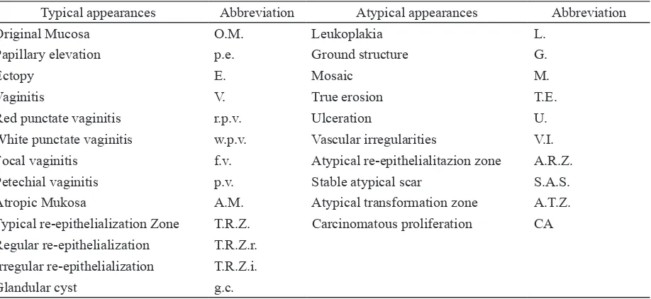
TABLE 1. Several simple abbreviations in colposcopic examination12, 13
Typical appearances Abbreviation Atypical appearances Abbreviation Original Mucosa O.M. Leukoplakia L.
Papillary elevation p.e. Ground structure G.
Ectopy E. Mosaic M.
Vaginitis V. True erosion T.E. Red punctate vaginitis r.p.v. Ulceration U. White punctate vaginitis w.p.v. Vascular irregularities V.I. Focal vaginitis f.v. Atypical re-epithelialitazion zone A.R.Z. Petechial vaginitis p.v. Stable atypical scar S.A.S. Atropic Mukosa A.M. Atypical transformation zone A.T.Z. Typical re-epithelialization Zone T.R.Z. Carcinomatous proliferation CA Regular re-epithelialization T.R.Z.r.
FIGURE 1. The schema of colposcopic examination report
INDICATION FOR COLPOSCOPY
Indications for colposcopy as follow: 14-16 1) Papanicolaou smear consistent with CIN
2 or CIN 3, persistent CIN I or cancer; 2) Papanicolaou smear with evidence of
HPV infection (don’t assume it is only HPV);
3) Papanicolaou smears with ASCUS or repeated ASCUS;
4) Abnormal-appearing cervix or suspicious-looking cervix;
5) Acetopositivity on visual inspection with acetic acid (VIA);
6) Acetopositivity on visual inspection with acetic acid using magniication (VIAM); 7) Positive on visual inspection with Lugol’s
iodine (VILI);
8) Persistent unsatisfactory quality on cytology;
9) Lesions that are more likely missed or under-read by colposcopic examination include endocervical lesions, extensive lesions that are dificult to sample, and necrotic lesions.
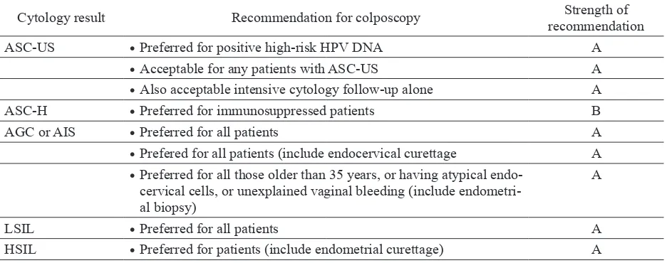
TABLE 2. Recommendation for colposcopy, American Society for Colposcopy and Cervical Pathology
Cytology result Recommendation for colposcopy Strength of
recommendation
ASC-US • Preferred for positive high-risk HPV DNA A
• Acceptable for any patients with ASC-US A
• Also acceptable intensive cytology follow-up alone A
ASC-H • Preferred for immunosuppressed patients B
AGC or AIS • Preferred for all patients A
• Prefered for all patients (include endocervical curettage A • Preferred for all those older than 35 years, or having atypical
endo-cervical cells, or unexplained vaginal bleeding (include endometri-al biopsy)
A
LSIL • Preferred for all patients A
HSIL • Preferred for patients (include endometrial curettage) A
CLASSIFICATION
The current nomenclature committee established at 2008. International Federation of Cervical Pathology and Colposcopy World Congress in Auckland were replaced by new colposcopy terminology of the International Federation of Cervical Pathology and
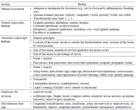
TABLE 3. 2011 International Federation of Cervical Pathology and Colposcopy Clinical and Colposcopic Terminology of the Vagina
Section Pattern
General assessment • Adequate or inadequate for the reason (e.g., cervix obscured by inlammation, bleeding, scar)
• Squamocolumnar junction visibility: completely visible, partially visible, not visible • Transformation zone types 1, 2, 3
Normal colposcopic
indings • • Original squamous epithelium: mature, atrophicColumnar epithelium; ectopy/ectropion
• Metaplastic squamous epithelium; nabothian cysts; crypt (gland) openings • Deciduosis in pregnancy
Abnormal colposcopic
indings • • General principlesLocation of the lesion: Inside or outside the transformation zone; location of the lesion
by clock position
• Size of the lesion: number of cervical quadrants the lesion covers • Size of the lesion as percentage of cervix
• Grade 1 (minor):
• Fine mosaic; ine punctation; thin acetowhite epithelium; irregular, geographic border • Grade 2 (major):
• Sharp border; inner border sign; ridge sign; dense acetowhite epithelium; coarse mosaic; coarse punctuation; rapid appearance of acetowhitening; cuffed crypt (gland) openings • Nonspeciic:
• Leukoplakia (keratosis, hyperkeratosis), erosion • Lugol’s staining (Schiller’s test): stained or nonstained Suspicious for
invasion
• Atypical vessels
• Additional signs: fragile vessels, irregular surface, exophytic lesion, necrosis, ulceration (necrotic), tumor or gross neoplasm
Miscellaneous ind -ings
• Congenital transformation zone, condyloma, polyp (ectocervical or endocervical),
in-lammation, stenosis, congenital anomaly, posttreatment consequence, endometriosis
COLPOSCOPIC APPEARANCES
The 2011 colposcopic terminology of the cervix starts with “General Assessment.” of the cervix to immediately recognize the level of reliability of the examination. The popular terms “satisfactory colposcopy” and “unsatisfactory colposcopy” abandoned because they have the connotation of an inadequate examination that needs to repeat. The colposcopic examination should be assessed for three variables: 1) adequate or inadequate, with the reason given; 2)
squamocolumnar junction visibility; and 3) transformation zone type.16,19
inside the endocervical canal or when a lesion covers the squamocolumnar junction with its inner border in the endocervical canal. The squamocolumnar junction may be not visible when all or most of the squamocolumnar junction cannot be seen because it is in the endocervical canal, then mentioned as transformation zone types 1, 2, 3. The reason that the visibility and site of the squamocolumnar junction are so important is that it dictates both the ability to do a satisfactory examination and when treatment is
FIGURE 2. A method of identifying outer and inner borders of the transformation zone (SCJ: squamocolumnar junction) 16
indicated, what the extent and type of excision. In the “abnormal colposcopic indings,” we added the localization of the lesion to either inside or outside the transformation zone.20
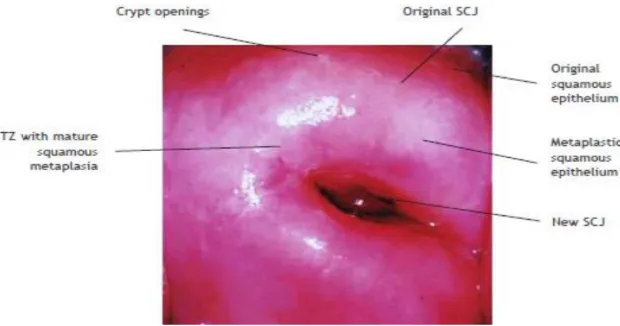
Normal cervix (normal colposcopic inding)
Three epithelial types cover the normal cervix i.e. 1) original squamous epithelium: mature or atropic; 2) columnar epithelium: ectopy/ectropion; 3) transformation zone (TZ): metaplastic squamous epithelium, nabothian cyst, crypt opening (FIGURE 2-4).
FIGURE 3. Normal colposcopic inding, entire new SCJ is visible, the TZ is fully visualized. The metaplastic
FIGURE 4. Normal colposcopic inding of the cervix12,14,16
The original squamous epithelium is a featureless, smooth, pink epithelium. No features are suggesting columnar epithelium such as gland openings or Nabothian cysts. The epithelium is considered “always” squamous and not transformed from columnar to squamous. The epithelium will not stain white after application of acetic acid; it will stain brown after application of Lugol’s iodine. The columnar epithelium is a single-cell layer, mucous producing, tall epithelium that extends between the endometrium and the squamous epithelium. Columnar epithelium appears red and irregular with stromal papillae and clefts. With acetic acid application and magniication, columnar epithelium has a grape-like or “sea-anemone” appearance. It is found in the endocervix, surrounding the cervical os, or (rarely) extending into the vagina. Squamocolumnar junction a clinically visible line was seen on the ectocervix or within the distal canal (e.g., post-cryotherapy), which demarcates endocervical tissue from squamous (or squamous metaplastic tissue). The SJ represents the cranial extension of the TZ and, therefore, most CIN but not necessarily the metaplastic process. Congenital or acquired factors determine the substantial extent of exposed columnar epithelium; such a inding is pathologic and is known to colposcopist as ectopy. The physiologic process of the changes in the columnar
epithelium to form squamous epithelium as metaplastic squamous epithelium, this part of the metaplastic squamous epithelium also mentioned as TZ.
usual pink color of the original mucosa pales a bit, even though it remains homogeneous. After a few minutes, it is possible to see the terminal capillaries in a manner suggesting a head of delicate hair. With Iodine staining (Schiller’s test) the cervix takes an intense, homogenous chestnut color. The colorization is less remarkable at the level of the vaginal vault. At the intraepithelial junction the color disappears because the columnar epithelium does not ix iodine. Colposcopic features suggestive of metaplastic change: a smooth surface with subtle, uniform-calibre vessels, mild acetowhite change, negative or partial positivity with Lugol’s iodine.
Abnormal colposcopic inding
Cervical intraepithelial neoplasia (CIN) is an asymptomatic condition suspected following cytological screening, usually no obvious characteristic signs with naked eye. CIN originates as a result of atypical metaplasia (dysplasia) mostly in the transformation zone. It is associated with alteration in cell contacts, decreased deposition of glycogen and increase cell turnover rates, which are important in the recognition. Cytology recognizes the possibility of CIN being present and histology allows diagnostic conirmation. However, colposcopy provides the important information which includes conirmation of the presence of a lesion, its topography; location of the lesion inside or outside the transformation zone, size, number of cervical quadrants and grade; suggest biopsy technique and appropriate treatment, and even the possibility of unsuspected cancer as well. Colposcopy has a reported sensitivity ranging from 87% to 99% to diagnose cervical neoplasia, but its speciicity is lower, between 23% and 87%.2,21 Atypical TZ is area of transformation zone, which can be recognized with the colposcope as having undergone morphologic change
into squamous metaplasia, different to the normal pattern of physiologic transformation of columnar epithelium. Histology sampling from this area may reveal a spectrum of change varying from immature metaplasia to dysplasia or deinitive precancerous change, culminating in preclinical invasive cancer. Location of the lesion is relative to the original squamocolumnar junction. “Inside” location means medial to the original squamocolumnar junction and ‘Outside’ is vice versa.
Pattern of atypical transformation zone Color and opacity
Acetowhite epithelium
After application of a dilute solution of acetic acid areas of high nuclear density appear white (FIGURE 5). Dilute of 3 – 5 % acetic acid has a differential effect on the cervical epithelium. After dissolving mucous and demonstrating the SJ, it has a peculiar reversible effect on metabolically active squamous and columnar epithelium as white, or oyster grayish color in the transformation zone referred to as acetowhitening. The acetowhitening is thought related to the presence of increased nucleoprotein. The acetowhite common with CIN and the degree of change usually relects the lesion grade. By logistic regression analysis the risk for a higher histologic grade when assessed by colposcopy was greatest in women with variation of the acetowhite color (odds ratio/OR =16.0; 95% CI: 10.0-26.0).22
FIGURE 5. Uterine cervix with CIN before (left) and after (right) application of acetic acid 12
Angioarchitecture
The blood supply of squamous epithelium arises from stromal vessels and usually arranged in a regular pattern. When there is increasing metabolic activity, such as in CIN, the vessels become more prominent and can be readily visible colposcopically. Variation in intraepithelial capillary pattern and caliber of vessels occurs to produce characteristic patterns in the acetowhite epithelium. The grosser pattern more likely associated with the more severe grades of histology changes. By logistic regression analysis the risk for a higher histologic grade when assessed by colposcopy was greatest in women with coarse vessels (OR = 10.0; 95% CI: 3.2-34.0).22
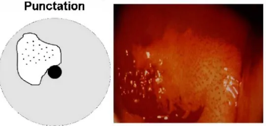
Punctuation
Single loop capillaries lying within the stromal papillae are seen end on, as dots or punctuation coursing obliquely or perpendicularly toward the surface of the epithelium (FIGURE 6). The pattern may vary from regular one of extremely ine, closely spaced, loop capillaries of narrow caliber with increased and irregular intercapillary spacing. A focal colposcopic pattern in which capillaries appear in a tippled pattern. The iner the punctuation appearance, the more likely the lesion is to be low grade or metaplasia. The coarser the punctuation the more likely the lesion is to be of major grade.
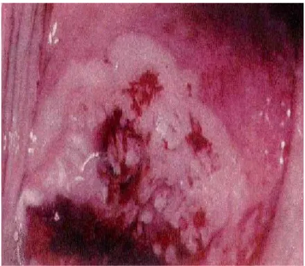
Mosaic pattern
A terminal vessel running in thin ibrous septa may cut the epithelium into abnormal pattern described as a mosaic (FIGURE 7). This mosaic pattern will vary from the minimal form showing ine caliber vessels delineating a small area of regular size and shape, through varying stages to that of coarse, prominent surface vessels surrounding blocks which may be grossly irregular in size and shape with obvious increased intercapillary
distance. A focal colposcopic appearance in which the new vessel formation appears as a rectangular pattern like a mosaic. The smaller the mosaic, the more likely the lesion is to be a low grade. The coarser, wider, and more irregular the mosaic, the more likely the lesion to be a major grade. The combination of both punctuation and mosaic structure will frequently intermingle and represent underlying abnormal vascular growth and activity.
FIGURE 7. Punctuation and mosaic pattern of blood vessel14
Atypical vessels
Non-speciic changes showing bizarre, irregular branching with considerable variation in caliber and coarse more frequently associated with the feature of invasive carcinoma. These are the result of new vessel formation and are usually seen only in high-grade lesion where invasion may have already taken place (FIGURE 8). A focal abnormal colposcopic pattern in which the blood vessel pattern appeared not as punctuation or mosaic or as the inely branching capillaries of normal epithelium, but rather as irregular vessels with an abrupt and interrupted coarse-appearing as commas, corkscrew capillaries, or spaghetti-like forms.
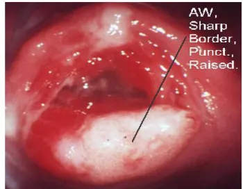
Surface contour or coniguration changes Irregular surface contour with excres-cences, particularly with abnormal vascular changes is more likely associated with a feature of high-grade CIN or early invasive lesions (FIGURE 9).22 However, its can be seen in lesser grades, especially when HPV involvement is prominent. Other causes of an irregular surface may be cystic inclusions and even persistence of the villous-like columnar structure still undergoing metaplastic change. Location of the lesion inside or outside the TZ. Location of the lesion is relative to the original squamocolumnar junction. “Inside” location means medial to the original squamocolumnar junction (toward the cervical os) and vice versa. The border of a lesion is a sharp border that is a straight edge of an acetowhite cervical lesion.
FIGURE 9. Show the surface contouror coniguration
changes14
TABLE 4. Colposcospic features suggestive abnormal changes25
Low-grade disease (minor change).
A smooth surface with an irrgular outer border. Slight acetowhite change, slow to appear and quick to
disappear. Fine punctation and ine regular mosaic. Mild, often speckled iodine partial positivity.
High-grade disease (major change).
A generally smooth surface with a sharp outer border. Dense acetowhite change, which appears early and is slow to resolve; it may be oyster white. Coarse punctation and wide irregular mosaics of differing size. Dense acetowhite change within columnar epithelium may indicate glandular disease. Iodine negativity, a yellow appearance in a previously densely white epithelium
Suggestive of invasive cancer
Irregular surface, erosion, or ulceration. Dense acetowhite change. Wide irregular punctation and mosaic, atypical vessels irregular mosaics of differing size. Dense acetowhite change within columnar epithelium may indicate glandular disease. Iodine negativity, a yellow appearance in a previously densely white epithelium.
Other edge deinitions are feathered or geographical margin, usually associated with a low-grade lesion, and rolled peeling edges that associated with a high-grade lesion.7 The inner border sign is a sharp demarcation between a thin and a dense acetowhite area within the same.23 The Ridge sign is an opaque
Other colposcopic indings
Vaginocervicitis
Cervicitis may cause abnormal Pap smears and make colposcopic assessment
more dificult. Many authorities recommend treatment before biopsy when a STD is strongly suspected.
FIGURE 10. The appearance of Strawberry spot in Trichomoniasis infection (left), and purulent discharge and Nabothian cyst (b) in cervicitis (a) (right)14
Traumatic erosion
Traumatic erosions are most commonly caused by speculum insertion and over vigorous Pap smears but can also result from such irritants as tampons, diaphragms, and intercourse (FIGURE 11).12
FIGURE 11.Traumatic erosion of the cervix12
Atrophic epithelium
Atrophic vaginal or cervical epithelium may also cause abnormal Papanicolaou smears (FIGURE 12). Colposcopists will often prescribe estrogen for 2 to 4 weeks before a colposcopy in order to “normalize” the epithelium before the examination. This is generally felt to be safe even if dysplasia or cancer is present because the duration of therapy is short and these lesions do not express any more estrogen receptors than a normal cervix.
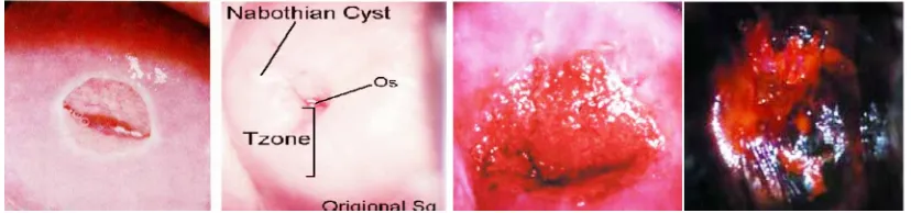
Nabothian cysts
Nabothian cysts are normal. They are areas of mucus producing epithelium that is “roofed over” with squamous epithelium. They do not require any treatment. They provide markers for the transformation zone since they are in squamous areas but are remnants of columnar epithelium.
Congestion (erythroplakia)
This relects epithelial thickness and blood supply (FIGURE 13). Full thickness mature squamous epithelium will not be as red as columnar epithelium. CIN, particularly of an advanced grade, will appear more congested than normal inactive metaplasia.
FIGURE 13. Nabothian cyst and congestion (erythroplakia) of blood vessel12
Leukoplakia (keratosis)
These are the areas of white elevation visible before the application of acetic acid. Leukoplakia is a sign of disordered keratin production and the possibility of underlying abnormal epithelium. It is a particular feature of HPV affected tissue and condylomata.
Colposcopic Assessment of topography
The management of suspected CIN would depend on the site of the lesion.
The transformation zone must be assessed completely as successful treatment will depend on removal of the area within which precursors of cancer develop. Focal lesion-entirely visible, this should not pose any therapeutic problems. Lesion extend into the cervical canal, it is often possible to see the upper extremity with manipulation of the cervix, where this is not possible, the colposcopic indings are “TZ type 2 or 3” and other management techniques will be necessary. Lesion not visible, caution is required to avoid missing disease high in the cervical canal. False positive Pap smear is possible but unusual. Colposcopic assessment not possible, there may be technical dificulties in exposing the cervix. The co-existence of signiicant inlammation or atrophy can interfere with the examination. Speciic treatment should allow later adequate assessment. Lesion extends onto vagina, although this is uncommon, it is well recognized as a reason for recurrent abnormal Pap smears. HPV affected epithelium not uncommonly involves the vagina.
Colposcopic grading
Cytology has alerted the possibility of CIN being present. Colposcopy has conirmed the presence of a lesion and its topography. It is important to assess the lesion’s signiicance and colposcopic grading. Such factors as color, vascularity, margins and contour need to be assessed. The grade will depend on interpretation of: a). degree of acetowhitening, b). caliber, shape, and pattern of punctuation and mosaic, c). intercapillary distance, d). presence of atypical vessels, e). surface contour, f). margin of abnormal epithelium.
CIN. Richard Reid has introduced his concept of the combined Colposcopic Index, which is a widely accepted technique used in the assessment of the degree of concurrent HPV involvement.17
Interpretation of Atypical Features of TZ
Interpretation of atypical features of transformation zone can be assessed in the following ways:17
HPV atypia
“Low grade lesions” HPV - borderline CIN
CIN I
with or without features of HPV
CIN II
“High grade lesions”
CIN III ESI (early stromal invasion)
Frank invasive carcinoma
Subjective assessment of the overall morphology and pattern
The fundamental practical principles involved are to decide: a). Is there normal or abnormal TZ? b). Is the lesion in or out of range? c). Can conident exclusion of invasive disease be made?. Colposcopic features can then be expressed in the term “consistent with an underlying histologic diagnosis” of:
Grading (Coppleson)
Grade I (insigniicant not suspicious) HPV
up to CIN I
Flat, acetowhite epithelium, borders not so sharp; semi-translucent with or without ine caliber, regular shaped vessels, patterns are ill deined with short intercapillary distance.
Grade II (signiicant, suspicious) CIN II - III
Flat, whiter epithelium sharply bordered, increased opacity; regular shaped vessels,
varying caliber, absence of atypical vessels; deined patterns, increased intercapillary distance.
Grade III (highly signiicant, highly
suspicious) CIN III or ‘microinvasive’
Colposcopic Sign Zero point One point Two points Colour Low-intensity acetowhitening (not completely
opaque); indistinct acetowhitening; transparent or translucent acetowhitening.
Acetowhitening beyond the margin of the transformation zone. Pure snow-white color with intense surface shine (rare
Intermediate shade - grey/white color and shiny surface (most lesions should be scored in this category)
Dull, opaque, oyster white; grey
Lesion margin and
Surface conigu -ration
Microcondylomatous or micropapillary. Contour
Flat lesions with indistinct margins. Feathered
or inely scalloped margins. Angular, jagged
lesions.Satellite lesions beyond the margin of the transformation zone edges. Internal de-marcations between areas of differing colposcopic appearance—a central area of high-grade change and peripheral area of low-grade change Vessels Fine/uniform-calibre vessels- closely and
uniformly placed
Poorly formed patterns of ine
punctation and/or mosaic
Vessels beyond the margin of the transforma-tion zone
Fine vessels within microcondylomatous or micropapillary lesions
Absent vessels Well deined coarse punctation or mosa-ic, sharply demarcat-ed – and randomly and
widely placed
Iodine staining Positive iodine uptake giving mahogany brown color
Negative uptake of insigniicant lesion,
i.e., yellow staining by a lesion scoring three
points or less on the irst three criteria
Partial iodine uptake - variegated, speckled appearance
Negative iodine
uptake of signiicant
lesion, i.e., yellow staining by a lesion already scoring four points or more on
the irst three criteria TABLE 3. The modiied Reid colposcopic index (RCI)16,26
Areas beyond the margin of the
transformation zone, conspicuous on colposco-py, evident as iodine-negative
areas (such areas are frequently due to paraker-atosis)
“Colposcopic Score” 0-2 = Likely to be CIN 1
Colposcopy has a role in the prevention of cervical cancer by identifying preinvasive or invasive lesions. Very strongly that the heart of colposcopic practice is the identiication of the most abnormal area for biopsy.27 However, colposcopy is subjective and is responsible for 52% of screening failures.28 Technology is constantly evolving, which is a subjective examination method continue to pursue to more objective.28,29 Pretorius et al. studied for determining the relative importance of colposcopically directed biopsy, random biopsy, and endocervical curettage (ECC) in diagnosing cervical intraepithelial neoplasia (CIN) II or worse. They concluded that random biopsies of the cervix may be helpful in the detection of CIN II or worse in patients with HGSIL or cancer cytology and negative colposcopy. Endocervical curettage increases detection of CIN II or worse regardless of referral cytology and should be included in routine colposcopic evaluation of patients with abnormal cytology.30 Soutter et al. performed research with dynamic spectral imaging based on objectivity, quantitative determination and acetowhitening effect. Showed that the method of dynamic spectral imaging more sensitive than colposcopy in detecting high-grade lesions and can provide improved guidance for biopsy.28
CONCLUSION
In summary, colposcopy is the diagnostic test to evaluate patients with an abnormal cervical cytological smear or abnormal-appearing cervix. Colposcopic features and patterns will correspond with underlying speciic histological features. The greater the expertise and experience of the colposcopist, the greater the conidence in the assessment
of the atypical TZ. For practical purposes, the most important aspect is always the recognition or exclusion of underlying actual invasive disease. The presence or absence of precancerous lesions can conirm with colposcopy.
ACKNOWLEDGEMENTS
This paper was presented on The Training of Management of Pre-cancerous Lesion, Indonesian Society of Obstetrics & Gynecology Yogyakarta at December 12-14, 2014 in Yogyakarta.
REFERENCES
1. Denny L, Kuhn L, Pollack A, Wainwright H, Wright TC Jr. Evaluation of alternative methods of cervical cancer screening for resource-poor settings. Cancer 2000; 89(4):826-33. http:// dx.doi.org/10.1002/1097-0142(20000815) 89:4<826::AID-CNCR15>3.0.CO;2-5
2. Belinson JL, Pretorius RG, Zhang WH, Wu LY, Qiao YL, Elson P. Cervical cancer 111 screening by simple visual inspection after acetic acid. Obstet Gynecol 2001; 98(3):441- 4.
h t t p : / / d x . d o i . o rg / 1 0 . 1 0 9 7 / 0 0 0 0 6 2 5 0 -200109000-00014
h t t p : / / d x . d o i . o r g / 1 0 . 1 0 1 6 / S 0 0 2 9 -7844(01)01454-5
3. Sankaranarayanan R, Budukh AM, Rajkumar R. Effective screening programmes for cervical cancer in low-and middle-income developing countries. Bull Word Healt Organ 2001; 79(10):954-62.
5. Burke L, Antonioli DA, Ducatman BS. Coposcopy text and atlas. Connecticut: Appleton & Lange, 1990.
6. Soutter, P. Practical colposcopy. Oxford: Oxford University Press, 1993.
7. Wright VC, Lickrish GM, Michael Shier R. Basic and advanced colposcopy. Part 1: a practical handbook for treatment, 2nd ed. Texas: Houston, 1995.
8. Anderson M, Jordan J, Morse A, Sharp F. Integrated colposcopy. 2nd ed. London: Chapman Hall Medical, 1996.
9. Burghardt E, Pickel H, Girardi F. Colposcopy cervical pathology. Textbook and Atlas. New York: Thieme, 1998.
10. Singer A, Monaghan J. Lower genital tract precancer colposcopy, pathology and treatment. 2nd ed. Oxford: Blackwell Science, 2000. http://dx.doi. org/10.1002/9780470760093
11. Cartier R, Cartier I. Practical colposcopy. 3rd ed. Paris: Laboratoire Cartier, 1993.
12. Dexeus S, Carrera JM, Coupez F. Colposcopy. 10th ed. Toronto: W.B. Saunders Co. 1977. 13. Stal A, Wilbanks GD. An international
terminology of colposcopy: Report of the nomenclature committee of the international federation of cervical pathology and colposcopy. Obstet Gynecol 1991; 77(2):313- 4. http://dx.doi.org/10.1097/00006250-199102000-00032
14. Mayeaux EJ Jr, Newkirk G. Introduction to colposcopy. [cited 2015, December 5] Avaible from: URL: http://lib-sh.lsumc.edu/ fammed/ atlases/colpoat.html, 2000
15. Campion M, Ferris D, di Paola F, Reid R. Modern colposcopy : a practical approach. Augusta: Educational System Inc., 1991. 16. Sellors W, Sankaranarayanan R. Colposcopy
and treatment of cervical intraepithelial neoplasia: a beginners manual. IARC, 2003.
17. Shai MI, Luesley DM, Jordan JA, Dunn JA, Rollason TP, Yates M. Randomised trial of immediate versus deferred treatment strategies for the management of minor cervical cytological abnormalities. Br J Obstet Gynaecol 1997; 104(5):590-4.
http://dx.doi.org/10.1111/j.1471-0528.1997. tb11538.x
18. Wright TC Jr, Cox JT, Massad LS, Twiggs LB, Wilkinson EJ. 2001 Consensus guidelines for the management of women with cervical cytological abnormalities. JAMA 2002; 287(16):2120-9. http://dx.doi.org/10.1001/ jama.287.16.2120
19. Bornstein J, Bentley J, Bösze P, Girardi F, Haefner H, Menton M, et al. 2011 Colposcopic terminology of the international federation for cervical pathology and colposcopy. Obstet Gynecol 2012; 120(1):166-72. http://dx.doi. org/10.1097/AOG.0b013e318254f90c
20. Walker P, Dexeus S, De Palo G, Barrasso R, Campion M, Girardi F, et al. International terminology of colposcopy: an updated report from the international federation for cervical pathology and colposcopy. Obstet Gynecol 2003; 101(1):175-7.
h t t p : / / d x . d o i . o r g / 1 0 . 1 0 1 6 / S 0 0 2 9 -7844(02)02581-4
h t t p : / / d x . d o i . o rg / 1 0 . 1 0 9 7 / 0 0 0 0 6 2 5 0 -200301000-00032
21. Mitchell MF, Schottenfeld D, Tortolero- Luna G, Cantor SB, Richads-Kortum R. Colposcopy for the diagnosis of squamous intraepithelial lesions: a meta-analysis. Obstet Gynecol 1998; 91(4):626-31.
22. Kierkegaard O, Byralsen C, Hansen KC, Frandsen KH, Frydenberg M. Association between colposcopic indings and histology in cervical lesions: the signiicance of the size of the lesion. Gynecol Oncol 1995; 57(1):66- 7. http://dx.doi.org/10.1006/gyno.1995.1100 23. Scheungraber C, Glutig K, Fechtel B, Kuehne-
a speciic and signiicant colposcopic sign for moderate or severe dysplasia (cervical intraepithelial neoplasia 2 or 3). J Low Genit Tract Dis 2009; 13(1):1-4. Doi: 10.1097/ LGT.0b013e31817ff92a.
24. Scheungraber C, Koenig U, Fechtel B, Kuehne-Heid R, Duerst M, Schneider A. The colposcopic feature ridge sign is associated with the presence of cervical intraepithelial neoplasia 2/3 and human papillomavirus 16 in young women. J Low Genit Tract Dis 2009; 13(1):13-6. http://dx.doi.org/10.1097/ LGT.0b013e318180438a.
25. Nazeer S, Shai MI. Objective perspective in colposcopy. Best Pract Res Clin Obstet Gynaecol 2011; 25(5):631-40. http://dx.doi. org/10.1016/j. bpobgyn.2011.04.008.
26. Reid R, Scalzi P. Genital warts and cervical cancer. VII. An improved colposcopic index for differentiating benign papillomaviral infections from high-grade cervical intraepithelial neoplasia. Am J Obstet Gynecol 1985; 153(6):611-8.
h t t p : / / d x . d o i . o r g / 1 0 . 1 0 1 6 / S 0 0 0 2 -9378(85)80244-1
27. Jeronimo J, Schiffman M. Colposcopy at a crossroads. Am J Obstet Gynecol 2006; 195(2):349-53. http://dx.doi.org/10.1016/j. ajog.2006.01.091
28. Soutter WP, Diakomanolis E, Lyons D, Ghaem-Maghani S, Ajala T, Haidopoulos D,
et al. Dynamic Spectral imaging: improving
colposcopy. Clin Cancer Res 2009; 15(5): 1814-20. Doi: 10.1158/1078-0432.CCR-08- 1636.
29. Li W, Gu J, Ferris D, Poirson A. Automated image analysis of uterine cervical images. Medical Imaging 2007; 651465142:P-2. 30. Pretorius RG, Zhang WH, Belinson JL, Huang