Indo. J. Chem., 2006, 6 (2), 117 - 120
Edy Giri Rachman Putra & Abarrul Ikram
117
Corresponding author. Tel. +62 21 7560922,
Fax +62 21 7560926; Email: [email protected] (E.G.R. Putra)
NANOSIZE STRUCTURE OF SELF-ASSEMBLY SODIUM DODECYL SULFATE:
A STUDY BY SMALL ANGLE NEUTRON SCATTERING (SANS)
Edy Giri Rachman Putra
*, and Abarrul Ikram
Neutron Scattering Laboratory, National Nuclear Energy Agency (BATAN) Kawasan Puspiptek Serpong, Tangerang 15314, Indonesia
Received 9 March 2006; Accepted 5 May 2006
ABSTRACT
Small Angle Neutron Scattering (SANS) investigation on the self-assembly sodium dodecyl sulfate (SDS) molecules as a function of concentration and additives has been carried out. SANS spectrometer which has been completely installed at the neutron scattering laboratory (NSL) BATAN in Serpong, Indonesia has played most important role to determine the growth (size) and also the shapes of a micelle structure. In this works we report that spherical micelle structure with a radius of 16.7 Å will transform to ellipsoidal or rod-like micelle structure with the long axis extends up to 50 Å by increasing the concentration of SDS. Similar to that the micelle structures change by addition of salt in SDS micellar solutions.
Keywords:nanostructure, micelle, self-assembly.
INTRODUCTION
Surfactants are amphiphilic molecules with one part of the molecule is hydrophilic (likes water) and another part is hydrophobic (dislikes water or likes oil). In aqueous solution the surfactant molecules form a micelle structure by self-aggregation or self-assembly to minimize the free energy of the solution. The micelles formed at low concentration are spherical and their structure changes with the concentration, temperature, and the present of additives, such as electrolytes or salts[1-5]. Depending on the particular molecular architecture of the surfactant molecule, a variety of nano or microstructures can form. Beside spherical possible micelle structures are ellipsoid, worm-like, spherical vesicle, and lamellar sheets.
Small Angle Neutron Scattering (SANS) is a technique for obtaining structural information of material in length scale between approximately 1 – 100 nm [6]. It can be easily used to study liquid, amorphous and crystalline samples. Information about size, shape or distribution of inhomogeneities can be extracted from data of scattered neutron. The experiment involves scattering of monochromatic beam of neutron from the sample and measuring the intensity of elastically scattered neutron as a function of scattering angle. The momentum transfer, Q is the modulus of the resultant between incident, ki and scattered ks, wave vectors which is given by
s i 4 sin
Q k k (1)
where 2 is scattering angle and is neutron wavelength. By substituting equation (1) into Bragg’s Law of diffraction, a very useful expression is given as
2
d
Q (2)
wheredis a distance.
The intensity (Q) of small angle scattering is defined as the coherent differential cross section (d/
d) and as a function of scattering vector Q for a monodisperse interacting micelles system can be expressed as
2 2
m s
d
n V P Q S Q
d (3)
wherendenotes the number density of the micelles,m andsare the scattering length densities of the micelle and the solvent, respectively. The term of (m - s) is called contrast factor.Vis the volume of a micelle. The aggregation number N of the micelle is related to the micellar volume V by the relation V = N, where is the volume of a surfactant monomer. P(Q) is the intraparticle structure factor and depends on the shape and size of the particles. S(Q) is the interparticle structure factor and is determined by the interparticle distance and the particle interaction. In a simple case of monodisphere system of homogenous particle with a radiusR,P(Q) is given by
2 1
3
3 ( ) 3(sin cos
( ) J QR QR QR QR
P Q
QR QR (4)
whereJ1is spherical Bessel function of first order. Here, V can be expressed as (4/3)R3 for spherical volume of micelle. For another shape, the expression of P(Q) in Eq. (4) will be different and given from another standard geometries form such as ellipsoid, cylinder, chain, core-shell, etc.
Indo. J. Chem., 2006, 6 (2), 117 - 120
Edy Giri Rachman Putra & Abarrul Ikram
118
where g(r) is the radial distribution function, i.e. the probability to finding another particle at a distancerfrom a centered reference particle.
In this work, we use SANS in order to investigate the various nanostructure formed in aqueous, water and also electrolyte solutions of anionic surfactant, i.e. sodium dodecyl sulfate (SDS). SDS is one of the most widely used surfactants in colloid or microemulsion system. It has been known that the SDS molecules aggregate to form a spherical micelle in water (micellar solution) at critical concentration, at 0.008M. This concentration is called critical micelle concentration (CMC). From SANS measurement of micellar solution systems it is possible to obtain quantitative information about geometrical dimension of surfactant SDS aggregate. The changes in nanostructure is observed, when the surfactant composition is varied on different salts were added. These are due to inter-aggregate interaction and free energy contributions related to counterions [5].
EXPERIMENTAL SECTION
SDS, sodium bromide (NaBr), potassium bromide (KBr), cesium bromide (CsBr), and D2O (98% atom D) were obtained from Aldrich. These chemicals were used as supplied without any further purification. The micellar solutions were prepared by dissolving a certain amount of surfactant and salt in D2O. The use of D2O instead of H2O for preparing micellar solutions provides a better contrast in SANS experiments [6]. The salts were dried over night at 100 C to release the hydrate water content. Concentration dependence samples of SDS solution were made with concentration of 0.01, 0.04, 0.17, 0.34, 0.70, 1.2, 1.5, and 1.7 M. For varying salts solution, for each of 0.3 M of SDS solutions, NaBr, KBr and CsBr salt was added. The final salt solutions are 0.1 M.
SANS measurements were carried out using SANS spectrometer at neutron scattering laboratory (NSL) BATAN in Serpong, Indonesia. The wavelength of the neutron beam was 4.32 Å and the experiments were performed at two different samples to detector distances of 1.5 m and 3 m to cover a momentum transferQrange of 0.002 to 0.3 Å-1. The scattered neutrons were detected using a two-dimensional position sensitive detector (2D-PSD) with 60mm diameter of a beam stopper.
The measurements were made both for the concentration and salts-type dependence using 5 mm thickness of a quartz cell. Each sample has been to exposed neutron beam for 1 hour. Meanwhile background, noise and detector efficiency
measurements were carried out for 2, 16 and 12 hours, respectively. During the experiment, the temperature was maintained at room temperature.
RESULT AND DISCUSSION
All SANS experiments have been done by using Small Angle Neutron Scattering (SMARTer) facility at neutron scattering laboratory (NSL), BATAN in Serpong and some results were obtained. The scattering length density must be calculated first due to obtaining a good contrast in SANS experiment. D2O was used as a solvent which gives a scattering length density about 6.38 x 1010 cm-2, compare to 3.0 x 1010 cm-2for SDS micellar solution.
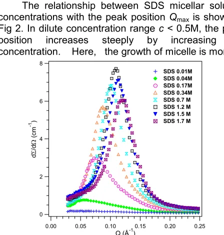
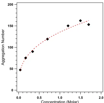
Fig 1 shows the scattering distribution profiles from SDS micellar solution with varying concentration. The peak position which occurs atQmax2/dwhered is the average distance between the micelles is related to the concentration. When the concentration of the surfactant molecules increases, the peak shifts to higher Q value due to the decrease of average distance between the micelles. The peak position is also related to the aggregation number or the size of micelle. At the lowest concentration, 0.01M and just above the critical micelle concentration the scattering distribution profile shows something similar as a background. It can be explained that the distance between micelles which formed in small number is quite large. Consequently the interparticle interaction will give a very weak coherent differential cross section. The relationship between SDS micellar solution concentrations with the peak position Qmaxis shown in Fig 2. In dilute concentration rangec < 0.5M, the peak position increases steeply by increasing the concentration. Here, the growth of micelle is more
8
Fig 1. SANS distribution profiles of SDS micellar
Indo. J. Chem., 2006, 6 (2), 117 - 120
Edy Giri Rachman Putra & Abarrul Ikram
119
Fig 2. The peak position of SDS micellar solution in
D2O as a function of SDS concentration
200
Fig 3. The relationship between the aggregation
number or the number of SDS molecules in micelle with SDS concentration.
dominant in dilute concentration range as the repulsive interaction between micelles is much less rather than in higher concentration.
By increasing SDS concentration further, Qmax will appear at higherQvalue (is not shown in the picture) [8]. This is not related to average distance between the micelles, but related to thickness of bilayer SDS molecules. Since at higher concentration SDS micellar solution will form a lamellar structure.
The growth of the length or size of micelle is measured to determine the growth of micelle. Fig 3 and Fig 4 show the relationship between the length (major axis) and the size (number of micelle or aggregation number) of micelle with concentration. The scattering distribution profile which is shown in Fig 2 can be calculated by assuming that S(Q) 1. Therefore the scattering distribution profile is mainly determined by
200
Fig 4. The major axis increment of SDS micellar
solution in D2O as a function of SDS concentration. The minor axis was 16.7 Å
3.0
Fig 5. SANS distribution profiles from 0.3M SDS
micellar solution and in presence of 0.1 M NaBr, 0.1 M KBr, and 0.1 M CsBr
intraparticle factor P(Q). By applying Eq. (3) and (4), the size and shape of micelle can also be determined. Detail of data analysis in small angle neutron scattering is described in another reference [9].
Fig 3 shows that the major axis, a increases by increasing the concentration with a constant minor axis,
b=c= 16.7 Å. Minor axis was determined first by fitting the scattering distribution profile at large Q as a function of concentration. SDS micellar solution which has a spherical structure at the lowest concentration,c
= 0.04 M will form ellipsoidal structures with the a/b
ratio up to 3 by increasing the concentration. The a/b
Indo. J. Chem., 2006, 6 (2), 117 - 120
Edy Giri Rachman Putra & Abarrul Ikram
120
also increases by increasing the concentration, Fig 4. Once again, the aggregation number increases significantly in a very narrow concentration range, i.e. c
< 0.05 M.
In case of SDS micellar solutions in presence of 0.1 M NaBr, 0.1 M KBr, and 0.1 M CsBr, the scattering distribution profiles are shown in Fig 5. These salts have a same co-ion (Br-), but different counterions. All the scattering distribution profiles show a correlation peak which is an indication of repulsive interaction between ionic micelles. The 0.3 M SDS data has a peak at Q
0.08 Å-1 and it is in quite good agreement with the literature [4,5]. It is observed that the measured distributions and thus P(Q) are independent of the salt concentration for Q> 0.1 Å-1. This is possible if micelles are ellipsoidal and the minor axis is independent of the salt concentration. The data analysis of 0.3M SDS micellar solution in the presence of 0.1 M salts is given in Table 1.
Pure 0.3 M SDS micellar solution shows that the SDS micelles are ellipsoidal with the dimensiona= 29.8 Å, b = 16.7 Å and the aggregation number N = 86, as shown in Table 1. When the salts, such as NaBr, KBr and CsBr are added into the SDS micellar solution, it is observed that the correlation peaks shifted and broadened. The shifting of correlation peak to lower Q
indicates the micelle grows on the addition of salt. The broadening of peak is due to the screening of the repulsive interaction between the micelles in the presence of salts. The effect becomes more important as the salts goes from NaBr to CsBr.
Table 1 shows that the minor axis does not change on addition of salts, meanwhile the aggregation number in presence of salts increases. These results show that the growth depends on the type of counterions, Na+, K+ and Cs+. From previous studies the interaction of ion with water most certainly plays important role in deciding the growth of micelle in aqueous electrolyte solution [10]. Therefore if the hydrated size of counterion is smaller, it has tendency to cover the charge on the micelle. The hydrated size of the Na+, K+ and Cs+ ions are 3.6,
Table 1. Micellar parameter for 0.3M SDS + 0.1M ABr
(A = Na, K, Cs) solutions. Qmax (Å-1)
Minor axis (Å)
Major axis (Å)
Aggregation Number
SDS 0.3 M 0.087 16.7 29.8 86
SDS 0.3 M +
NaBr 0.1 M 0.081 16.7 36.7 106
SDS 0.3 M +
KBr 0.1 M 0.076 16.7 43.3 125
SDS 0.3 M +
CsBr 0.1 M 0.072 16.7 46.7 135
3.3, and 3.3 Å, respectively [11]. Thus, from our studies show that Na+counterion has a larger hydrated size, due to the aggregation number of SDS micelle is smaller than the other counterions, i.e. K+and Cs+.
CONCLUSION
Small angle neutron scattering (SANS) has shown to be a powerful technique in determining SDS micelle nanostructure. The size and shape of micelle change with concentration and also variation of counterions, i.e. NaBr, KBr and CsBr. By increasing the concentration, the spherical micelle changes into ellipsoidal micelle, where the a/b ratio extends from 1 to 3. At dilute concentration range, the parameters of micelle, such as major axis, aggregation number and peak position increase steeply due to the repulsive interaction between micelles is much less. This effect is also found by addition of a smaller hydrated size counterion.
ACKNOWLEDGEMENT
The authors would thank to Drs. P. S. Goyal and V. K. Aswal from Bhabba Atomic Research Centre, India for providing the data analysis software and also for beneficial discussions. The work was supported by National Nuclear Energy Agency (BATAN) on financial year of 2005.
REFFERENCES
1. Wennerstrom, H. and Lindman, B., 1987, Top. Curr. Chem. 87, 1.
2. Chen, S. H., 1986, Annu. Rev. Phys. Chem. 37, 351.
3. Chevalier, Y., and Zemb, T., 1990, Rep. Prog. Phys. 53, 279 .
4. Goyal, P.S., 1990,Phase Transit.50, 143.
5. Aswal, V.K., and Goyal, P.S., 2000, Phys. Rev. E
61, 3.
6. Goyal, P. S., 1995, Small Angle Neutron Scattering, IAEA/RCA Workshop on SANS, Bhabba Atomic Research Center, Mumbay, April 1995.
7. Putra, E.G.P, 2006,in preparation. 8. Putra, E.G.P, 2006,in preparation.
9. Feigin, L.A., and Svergun, D.I., 1987, Structure Analysis by Small-Angle X-Ray and Neutron Scattering, G. W. Taylor, Editor, Plenum Press, New York.
10. Kumar, S., Aswal, V. K. Singh, H. N. Goyal, P. S. and Kabir-ud-Din, 1994,Langmuir10, 4069. 11. Cotton F. A. and Wilkinson, G. , 1980, Advanced