P-ISSN.2089-1180, E-ISSN.2302-2914
CrossMark
Published by DiscoverSys
INTRODUCTION
Women living almost about half of their lives with estrogen deficiency condition. One of the symp-toms that can degrade the quality of life at the age of menopause is a decline in cognitive function and this symptom will be increased with age.1,2
At least 10% of the population aged 65 years or more experience cognitive impairment and increased by about 50% at age 85 years. Epidemiological studies have shown that women have a higher incidence of dementia, particularly Alzheimer’s disease than men.3
The ageing-related decrease of cell plasticity that is normally maintained by the continuous addition of new neurons brought about by neuro-genesis and contribute to senescence-dependent impairments of brain function. In aging, a dysreg-ulation of the HPA system leads to elevated levels of circulating corticosteroid levels that will inhibit
precursor cell proliferation in the brain. While neurotropic factors and growth factors are decline with age, transforming growth factor β1 (TGFβ1) might increase and in old mice inhibited the prolif-eration of early precursor cells.2,4
Many studies reported that estrogen hormone replacement ther-apy reduces the risk of Alzheimer’s and cognitive function in postmenopausal women and elderly.5
In vivo studies reported the effects of estrogen on the areas in the brain such as cerebral cortex and hippocampus.6
The hippocampus is involved in learning and memory processes, that hippocampal glutamatergic neuron express ER.7
The neuropro-tective effect of estrogen in physiologic doses is well establish, but the molecular and cellular mecha-nisms remain controversial.8
Most studies show that the cerebral cortex is most strongly protected
ABSTRACT
Background: Estrogen deficiency condition can degrade the quality of life, decline in cognitive function will be more severe trough age. Phytoestrogen compounds can be found in pegaga leaf extract, tomatoes, and papaya is an easy and inexpensive way to increase estrogen levels in post menopause women through extra gonadal estrogen induction. Therefore, the aims of this study were to examine the effect of tomato juice, physical exercise, and combination of these treatments on promoting neurons and ERβ expression in somatosensory cortex that contribute to cognitive function of post-ovariectomy rats.
Method: Twenty-eight female healthy Wistar rats (Rattus novergicus), 8-10 weeks old, from Laboratory of Biochemistry, Faculty of Medicine Airlangga University include in this experiment. The animals were housed in the animal-care facility with ad libitum food and water. The temperatur was maintained at 18°C-24°C. The treatments were done 2 weeks after ovariectomy. Tomato were made in Laboratory of Pharmacognocy and Phytochemistry, Faculty of Pharmacy, Airlangga University, from inner part of the tomato fruits (mucous like substance) with freeze dry method (-40°C).
Results: The weight of white rat Rattus norvegicus post ovariectomy in this study was between 133-170 gram with a mean weight 154.32 ± 9.72 gram. Hematoxylin/eosin staining showed neuronal deficit in the control rats brain. In figure 1, the tomato group showed the largest of neurons number (145.43 ± 17.728), followed the combination group (140.57 ± 22.449), the exercise group (136.86 ± 23.104) and the smallest number in the control group (96.43± 28.965). Four weeks after treatments the number of neurons increased significant in the tomato group (p=0.001), exercise group (p=0.004) and combination group (p=0.002) from the control group. This study showed no significant different between tomato and exercise group (p=0.500), tomato and combination group (p=0.701) and between exercise and combination group (p=0.769).
Conclusions: In conclusion, our data demonstrated that post ovariectomy rats showed deficit numbers of neurons and decreased ERβ in the somatosensory cortex. Treatment with physical exercise, tomato juice, and combination of these treatments increased the number of neurons and ERβ expression in the somatosensory cortex.
Keywords: Neurons, ERβ expression, Post-Ovariectomy, Rats.
Cite This Article: Laswati, H., Subadi, I., Andriana, M., Kurniawati, P., Pangkahila, J. 2016. Tomato (Lycopersicum Commune) juice and physical exercise increase number of neurons and ERβ expression in post-ovariectomy rats brain. Bali Medical Journal 5(3): 84-91. DOI:10.15562/bmj. v5i3.309
*Correspondence to: Patricia Maria Kurniawati, Department of Physical Medicine and Rehabilitation, Faculty of Medicine, Airlangga University, Surabaya-Indonesia
1Department of Physical Medicine and Rehabilitation, Faculty of Medicine, Airlangga University, Surabaya-Indonesia
2Department of Andrology and Sexology, Faculty of Medicine, Udayana University, Bali-Indonesia
Tomato (
Lycopersicum Commune
) juice and physical
exercise increase number of neurons and ERβ
expression in post-ovariectomy rats brain
Hening Laswati,1 Imam Subadi,1 Meisy Andriana,1 Patricia Maria Kurniawati,1*
by estrogen, followed by the striatum and hippo-campus region.9
During menopause, the decline in estrogen may increase the risk of diseases and affect quality of life. Dietary soy phytoestrogens have been shown to improve memory function in postmenopausal women.5
Due to the use of estrogen replacement therapy increases the risk of malignancy in the breast and endometrial, then phytoestrogens that have estrogenic effects as an alternative to estrogen replacement therapy.10
Phytoestrogens are a component of the plant with the structure and effects similar to mamalian 17β-estradiol.10,11
Epidemiological data describing how they could be used as a way the symptoms of menopause and associated diseases, but the bene-fits for brain function and behavior has not been known.12
Estrogenic activity of phytoestrogens depend on binding affinity to the estrogen recep-tor, which determined the aromatic ring and the hydroxyl group at a specific place. The effects of phytoestrogens can directly by occupying ER, but also can indirectly through sex hormone binding globulin (SHBG). Phytoestrogen signals on brain by activation of classical estrogen receptors (ERs), there are ERα and ERβ.10,11,13-16
Estrogen exerts chronic or genomic effects and rapid signaling or non-genomic effects via the regulation of the activa-tion of kinase signaling pathways.7,10, 11,17,18
In vivo study reported that estrogen also bind G-protein-coupled transmembrane-bound receptor, leading to effects on cell proliferation and survival in the adult brain.19,20
In the human brain, ERβ appears to be the predominant receptor in areas the cere-bral cortex, hippocampus, anterior olfactory nucleus, cerebellum, dorsal raphe, substantia nigra, midbrain and several brain stem nuclei. In the macaque monkey brain, ERβ is widely express in the adult hippocampus, that a key region regulating cognitive and emotion function.13,20
Phytoestrogen compounds can be found in pegaga leaf extract, tomatoes and papaya. Examination radioimmunoassay solid phase in the tomato fruit mucilage obtained phytoestro-gen content of 1037.0 ± 37.7 pg/g.22
Tomato (Lycopersicum commune) included in the Solanaceae family are often found in parts of Indonesia. Tomato is a fruit daily consumed by many people, in addition to cheap, easy to take it. Physical exercise of moderate intensity (60-75% maximum heart rate) is an easy and inexpensive way to increase estrogen levels in post menopause women through extra gonadal estrogen induction. The combination of physical exercise and phytoestro-gens (Marsilea crenata Presl) reported to yield an increase in estrogen levels higher than just physi-cal exercise or consumption of phytoestrogen.23
In vivo study reported that tomato juice increase bone density of menopause rats.24
Has never been reported to influence the consumption of tomatoes as phytoestrogens on cognitive function and there was still little information of the effects physical exercise and combination of physical exercise and phytoestrogen on the cognitive function. Therefore, the aims of present study were to examine the effect of tomato juice, physical exercise and combination of these treatments on promoting neurons and ERβ expression in somatosensory cortex that contrib-ute to cognitive function of post-ovariectomy rats. Because samples were taken from the brain, there-fore this study using post-ovariectomy Wistar rats (Rattus norvegicus).
MATERIALS AND METHODS
Animals, Tomato Juice, and Tissue Preparation
Twenty-eight female healthy Wistar rats (Rattus novergicus), 8-10 weeks old, from Laboratory of Biochemistry, Faculty of Medicine Airlangga University include in this experiment study. The animals were housed in the animal-care facility with ad libitum food and water. The temperatur was maintained at 18°C-24°C. The treatments were done 2 weeks after ovariectomy. Tomato juice was done in Laboratory of Pharmacognocy and Phytochemistry, Faculty of Pharmacy, Airlangga University, was made from inner part tomato fruits (mucous like substance) with freeze dry method (-40°C). This process was continue with sublima-tion in the vacuum freeze dried chamber with pres-sure of 0.036 psi (0.0025 bar), and the temperature was increased until reached 38°C. The tomato juice dose was calculated from conversion human dose to animal dose,25
whereas the average human dose of tomato fruits 400-600 gram/day.26
Subjects were randomized divided into 4 groups, there are : the control group just got aqua 2cc / day per scoop, the administration of tomato juice group ( 220mg / kg / day dissolved in aqua 2cc per scoop), the physi-cal exercise group ( swimming without load for 30 minutes, 3 times / week), and the group of combi-nation of tomato juice administration and physical exercise. After four weeks treatments, animals were decapitated, and brains were removed and put into 4% paraformaldehyde for histology evaluation and immunohistochemistry analysis.
Histology and Cell Counting
paraffin embedding. Sagittal 4-μm serial section were made and directly mounted on gelatin-coated slides. The sections were deparaffinized in xylene and rehydrated, then stained with hematoxylin/ eosin (HE). Neuronal counting from the somato-sensory cortex (postcentral gyrus) were deter-mined histology by light microscopy, magnifying 1000 times.
Immunohistochemistry
Paraffin sections were deparaffinized in xylene, and rehydrated. Sagittal sections (4-μm-thick) of the somatosensory cortex of postcentral gyrus were rinsed with PBS for 3 minutes, then incubated in citrate buffer, rinsed again with phosphate buffer saline (PBS) 2 times for 3 minutes. Process follow by section were incubated with H2O2 3% for10 minutes, follow by washed in PBS 2 times for 3 minutes. Sections were then incubated in monoclo-nal rat anti-ERβ for 1 hours at room temperature, rinsed with PBS 2 times for 3 minutes, and then incubated with secondary Ab (biotinylated) for 30 minutes, follow by washed with PBS 2 times for 3 minutes. Sections were incubated with strepta-vidin-HRP for 30 minutes, rinsed with PBS for 3 minutes, and then incubated with 3,3’-diamino-benzidine chromogen for staining for 3-5 minutes. After this step, rinsed with PBS for 3 minutes, and with aquadestilata 2 times for 3 minutes’ follow by incubated with Meyer’s Hematoxylin for 10 minutes. Sections were then mounted on slides and cover-slipped with Permount.Calculation of ERβ expression was observed as a brown color in the nuclei of neurons, each slide on a microscope field of view with a magnification of 1000 times.
Statistical Analysis
All data were presented as mean ± SD. Statistical significant differences were determined using one-way ANOVA and post hoc test to determine differences between groups. Correlation was deter-mined with Pearson test. All statistical test carried out using SPSS 23 and statistical significance was set at p<0.05 for all analysis.
RESULTS
Histological Changes in The Post Ovariectomy Rats Brain
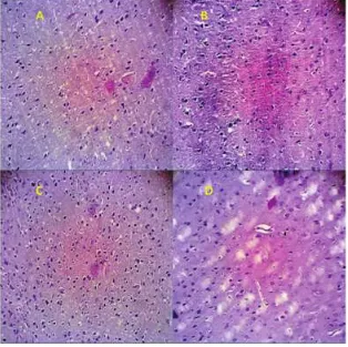
The weight of white rat Rattus norvegicus post ovariectomy in this study between 133-170 gram with a mean weight 154.32 ± 9.72 gram. Hematoxylin/eosin staining showed neuronal defi-cit in the control rats brain. In figure 1, the tomato group showed the largest of neurons number (145.43 ± 17.728), followed the combination group (140.57 ± 22.449), the exercise group (136.86 ±
Figure 1 Sagittal section (4μm) of the
soma-tosensory cortex show histology com-parison of post ovariectomy rats brain in the control group and the treatment groups. Note the increased of mean number of neurons in the treatment groups. The tomato group, exercise group and combination group are not significant between groups
Figure 2 Sagittal section (4μm) of the somatosensory cortex post
23.104) and the smallest number in the control group (96.43± 28.965).
Four weeks after treatments the number of neurons increased significant in the tomato group (p=0.001), exercise group (p=0.004) and combina-tion group (p=0.002) from the control group.
This study showed no significant different between tomato and exercise group (p=0.500), tomato and combination group (p=0.701) and between exercise and combination group (p=0.769)
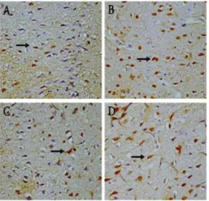
Immunoreactivity in The Post Ovariectomy Rats
The combination group showed the largest of ERβ expression (18.71 ± 1.380), followed by the tomato juice group (12.86 ± 2.193), the physical exercise group (8.14 ± 1.345) and the smallest expression in the control group (5.29 ± 1.113) (Figure 3).
In this study we obtained significant increased ERβ expression in tomato group (p=0.000), exer-cise group (p=0.011) and combination group (p=0.000) after 4 weeks’ treatments. LSD Post Hoc test showed a significant difference between tomato and exercise group (p=0.000), tomato and combi-nation group (p=0.000), exercise and combicombi-nation group (p=0.000). Pearson test showed correlation between the number of neurons and ERβ expres-sion (r=0.514; p=0.005).
DISCUSSION
In this study, we found significant neurons deficit and decreased ERβ expression on somatosensory cortex in post ovariectomy rats. Study on ERβ knout out mice (BERKO) showed morphological abnormalities in the brain, neuronal hypo cellular-ity with severe neuronal deficits in somatosensory cortex area.27 In vivo studies reported that loss of
estrogen caused may lead loss of synaptic connec-tions in hippocampus or decline in basal forebrain cholinergic function or choline acetyl transferase activity.17
Other studies reported that in very old rats and post ovariectomy rats demonstrated decreased levels of ERβ expression on the brain.15,16,28 Study in
ERβ KO revealed an abnormal neuronal migration and increase of apoptotic neuronal death. There was a severe neuronal deficit in the somatosensory cortex.29 This suggest that ERβ is the functional ER
within the region of the cortex and have an import-ant role in the maintenance of number of neurons in rat’s brain. In ovariectomized rats, dendritic spine density in ventromedial hypothalamic nucleus (VMN), CA1 region on hippocampus, and medial prefrontal cortex is decreased as compared to gonadal intact rats.30 The nervous system is
Figure 3 Mean of ERβ expression in the sagittal
section (4μm) of the somatosensory cortex show immunoreactivity compar-ison. Note the increased number of ERβ expression in the treatment groups. The combination group show the highest ERβ expression, followed by the tomato group and the exercise group.
Figure 4 ERβ expression in sagittal sections (4μm-thick) of the
capable of plasticity and that the dendritic spine is the major site of this activity.
Statistical analysis showed that there was signifi-cantly increase the number of neurons after treat-ment in all groups (p<0.05), but the effect of tomato juice administration, physical exercise (swimming without resistance) and combination of these treat-ments showed the same effect (p>0.05) There was significant increase of ERβ expression after ment in all groups (p<0.05) and each of the treat-ment was have the different effects (p<0.05).
The effects of tomato juice (phytoestrogen) on neurons and ERβ expression of somatosensory cortex in our study were consistent with previous studies. In vitro studies reported that when neuron cultures from the cortex and hippocampus were exposed to neurotoxic substances, phytoestrogen demontrated neuroprotective antioxidant effects. For ovariectomized rats, phytoestrogens have associated with increased expression of BDNF that involved in neurogenesis and preservation of neurons.16
In vitro study with cultured of H19-7/ IGR-IR neural cell line reported that genistein, daidze, and 17β-estradiol elevated the expression of BDNF mRNA and the effects BDNF-Trk path-way have the important role in the regulation of neuronal cells proliferation.5
Endogenous BDNF and Tsk signaling pathways mediate cortical progenitors’ survival and neurogenesis. Brain-derived neurotrophic factor (BDNF) is a polypep-tide, a neurotrophins produced by the neurons is crucial in neuronal development, survival and plasticity, an important role in memory formation. Phytoestrogen increased choline acetyltransferase and nerve growth factor messenger RNA in the frontal cortex and hippocampus in the female rats.10
Neuron signals are known to regulate the effects of estradiol on astrocytes. Astrocytes also express ERβ, and these cells are involved in the neuroplastic and neuroprotective actions of the hormone.6,31,32
The physiological functions of astrocytes include control of hormonal release by neuron, neuropro-tection and modulation of neuronal regeneration.9,27
Astrocyte secrete neurotrophic factors BDNF that can induce cell proliferation.19
In vivo studies reported that treatment of ovariectomized rats with estradiol 17β induces certain hippocampal neurons to form new synaptic connections with other nerve cells.6
The intracellular signaling pathways leading to neuroprotection involve PI3K, AKT and mito-gen-activated protein kinase (MAPK).7,8,18,33
Estrogen is a powerful inducer of MAPK. In adult rats, estrogen increases serotonin receptor mRNA and IGF-1 mRNA expression in the brain to influence brain cells proliferation through stimulate precursor cell proliferation.2,19
Estradiol as a negative regulators of cell death, has been
shown to promote the expression of Bcl-2 in NT2 neurons and Bcl-XL in PC12 cells and cultured hippocampal neurons.33
Many in vitro studies in several neuronal-culture model system reported that estradiol (E2) protects against toxicities caused by serum-deprivation, β-amyloid, excitotoxins and oxidative stress. In the animal models of cerebral ischemia, estradiol increased ERβ expres-sion and has been shown to attenuate neuronal death through inhibition of nuclear factor-кB (NF- кB).8,27
In this study, physical exercise increased the number of neurons and ERβ expression. Previous studies in animal models reported that physi-cal exercise has an acute up-regulation effect on precursor cell proliferation and neurogenesis. It has been suggested mediated by vascular endothelial growth factor (VEGF) or insulin growth factor 1 (IGF-1). In vivo studies reported that IGF-1, FGF mRNA and BDNF mRNA were elevated in rodents by exercise.34
Physical exercise induced hippo-campal neurogenesis in adult and aged mice and running increases cell proliferation and neurogen-esis in adult mouse dentate gyrus.2,35
Individuals with traumatic brain injury (TBI), exercise-induced improvements in cognitive function after participa-tion in vigorous aerobic exercise training.36
Brain-derived neurotrophic factor (BDNF) activity may mediate these effects. This neurotrophic factor is a protein found in high concentration primarily in the hippocampus, cerebral cortex, hypothalamus and cerebellum and have a role in neurogenesis, dendritic growth, and long-term potentiation of neurons.37
The combination group showed the highest ERβ expression and increased the number of neurons. This suggest that phytoestrogen and IGF-1 have synergistic effects on ERβ expression in the promo-tion of neuronal survival and neuroprotecpromo-tion.18
While the effects of each treatment on ERβ was significantly different, all of each treatment have been the same effects on number of neurons. There was correlation between the number of neurons and ERβ expression (p<0.05). This suggest the impor-tance of the role of ERβ for the development and plasticity of neurons. Adult neurogenesis requires a specific molecular and cellular microenvironment such as neurogenic niche. Within this niche, gap junctions, paracrine effects of neurotransmitters, neurotrophic factors and growth factors control sequential steps in neurogenesis.2
and crosstalk with other intracellular signaling pathways.
CONCLUSION
In conclusion, our data demonstrated that post ovariectomy rats showed deficit numbers of neurons and decreased ERβ in the somatosen-sory cortex. Treatment with physical exercise, tomato juice and combination of these treatments increased the number of neurons and ERβ expres-sion in the somatosensory cortex. There was correlation between the number of neuron cells and ERβ expression. This study providing information regarding the potential implication of phytoestro-gen and physical exercise for the prevention and treatment against neurodegenerative diseases in estrogen deficiency condition. Further studies are needed to elucidate the mechanism of neuronal loss in post menopause and age-related neurodegener-ation, the neuroprotective effects of phytoestrogen and physical exercise.
REFERENCES
1. Verhaeghen P and Cerella J. “Aging, executive control, and
attention: a review of meta-analyses”. Neuroscience and Biobehavioral Reviews 2002; 26(7): 849-57.
2. Klempin F and Kempermann G. Adult hippocampal
neu-rogenesis and aging. Eur Arch Psychiatry Clin Neurosci 2007; 257: 271-280.
3. Amer DAM, Kretzschmar G, Muller N, Stanke N, Lindemann D and Vollmer G. Activation of transgenic estrogen receptor–beta by selected phytoestrogens in sta-bly transduced rat serotonergic cell line. Journal of Steroid Biochemistry & Moleular Biology 2010; 120: 208-217.
4. Cerbai P, Lana D, Nosi D, Petkova-Kirova P, Zecchi S,
Brothers HM, Wenk GL and Giovannini MG.The neu-ron-astrocyte-microglia triad in normal brain ageing and in a model of neuroinflammation in the rat hippocampus. PLOS one 2012; 7(9): e45 250.
5. Pan M, Han H, Zhong C and Geng Q. Effects of Genistein and Daidzein on Hippocampus Neuronal Cell Proliferation and BDNF Expression in H19-7 Neural Cell Line. The Journal of Nutrition. Health and Aging 2012; 16(4): 389-394.
6. McEwen B. Estrogen actions throughout the brain. Recent
Prog Horm Res 2002; 57: 357-384.
7. Hojo Y, Munetomo A, Mukai H, Ikeda M, Sato R, Hatanaka
Y, Murakami G, Komatsuzaki Y, Kimoto T and Kawato S. Estradiol rapidly modulates spinogenesis in hippocampal dentate gyrus: Involvementof kinase networks. Hormones and Behavior 2015; 74; 149-56.
8. Elzer JG, Muhammad S, Wintermantel TM, Regnier-Vigouroux A, Ludwig J, Schutz G and Schwaninger M. Neuronal estrogen receptor-α mediates neuroprotec-tion by 17β-estradiol. Journal of cerebral Blood Flow & Metabolism 2010; 30: 935-942.
9. Dhandapani KM and Brann DW. Estrogen-Astrocyte interactions: Implications for neuroprotection. BMC Neuroscience 2002; 3: 6.
10. Ososki AL and Kennelly EJ. Phytoestrogens: a Review of the Present State of Research. Phytother. Res. 2003; 17: 845-869.
11. Patisaul HB. Phytoestrogen Action in the Adult and Developing Brain. Journal of Neuroendocrinology 2005; 17: 57-64.
12. Yang T-S, Wang S-Y, Yang Y-C, Su C-H, Lee F-K, Chen S-C, Tseng C-T, Jou H-J, Huang J-P and Huang K-E. Effects of standarized phytoestrogen on Taiwanese menopausal women. Taiwanese Journal of Obstetrics & Gynecology 2012; 51 : 229-235.
13. Fan X, Xu H, Warner M and Gustafsson JA. ERβ in CNS: new roles in development and function. Progress in Brain Research 2010; 181: 233-250.
14. Lee H-R, Kim T-H and Choi K-C. Functions and physio-logical roles of two types of estrogen receptors, ERα and ERβ, identified by estrogen receptor knockout mouse. Lab Anim Res 2012; 28(2):71-76.
15. Mott NN and Pak TR. Review Article. Estrogen Signaling and the Aging Brain: Contex-Dependent Considerations for Postmenopausal Hormone Therapy. ISRN Endocrinology 2013; Article ID 814690.
16. Sony M, Rahardjo TBW, Soekardi R, Sulistyowati Y, Lestariningsih, Yesufu-Udechuku A, Irsan A and Hogervorst E. Phytoestrogens and cognitive function: a review.Maturitas 2014; 77(3): 209-20.
17. McEwen BS and Alves SE. Estrogen Actions in the Central Nervous System. Endocrine Reviews 1999; 20(3): 279-307.
18. Behl C. Oestrogen as a neuroprotective hoemone. Nature Reviews /Neuroscience 2002; 3: 433-442.
19. Fowler CD, Johnson F and Wang Z. Estrogen regulation of cell proliferation and distribution of estrogen receptor-α in the brains of adult female prairie and meadow voles. The Journal of Comparative Neurolgy 2005; 489: 166-179. 20. Woolley CS. Acute effects of estrogen on neuronal
physiol-ogy. Annu. Rev. Pharmacol.Toxicol 2007; 47: 657-680. 21. Amer DAM, Kretzschmar G, Muller N, Stanke N,
Lindemann D and Vollmer G. Activation of transgenic estrogen receptor–beta by selected phytoestrogens in sta-bly transduced rat serotonergic cell line. Journal of Steroid Biochemistry & Moleular Biology 2010; 120: 208-217. 22. Putra HL and Mahaputra L. Natural Phytoestrogen
Contents in Severals Fruit and Leafs; The Future Replacement Hormone Therapy in Menopause Women. Jurnal Cakrawala 2011; 6 (1): 88-95.
23. Laswati H. The effect of combination of physical exercise and semanggi leaves administration on bone remodeling process in postmenopausal women. In: Sutarjadi H, Agil M, Avanti C and Surjadhana A (Eds). Women’s Health
and Traditional Medicine. 1st ed. Surabaya, Airlangga
University Press 2007: pp 53-69.
24. Laswati H, Hendarto H, Irawati D and Mahaputra L. Tomato juice increase bone density of menopause rats. Jurnal Veteriner 2015; 16(3): 457-462.
25. Gosh MN. Fundamentals of Experimental Pharmacology.
1st ed. Calcutta: Scientific Book Agency 1971: pp 84-88.
26. Heber D. 2004. Review article vegetables, fruits and phy-toestrogens in the prevention of diseases. Journal Postgrad Med 50:145-149.
27. Wang L, Andersson S, Warner M and Gustafson J-A. Morphological abnormalities in the brains of estrogen receptor β knockout mice. PNAS 2001; 98 (5): 2792-2796. 28. Weiser MJ, Foradori CD and Handa RJ. Estrogen receptor
beta in the brain: From form to function. Brain Res Rev 2008; 57(2): 309-320.
29. Habauzit D, Fluoriot G, Pakdel F and Saligaut C. Effects of estrogen and endocrine-disrupting chemicals on cell dif-ferentiation-survival-proliferation in brain: Contributions of neuronal cell lines. Journal of Toxicology and Environmental Health 2011; 14: 300-327.
30. Frankfurt M and Luine V. The evolving role of dendritic spines and memory: Interactions with estradiol. Hormones and Behavior 2015; 74: 28-36.
31. Chaban VV, Lakhter AJ and Micevych P. A membrane estrogen receptor mediates intracellular calcium release in astrocytes. Endocrinology 2004; 145(8): 3788-3795. 32. Azcoitia I, Sierra A, and Garcia-Segura LM.
33. Garcia-Segura LM, Azcoita I and DonCarlos LL. Neuroprotection by Estradiol. Progress in Neurobiology 2011; 63: 29-60.
34. Carro E, Nunez A, Busiguina S and Torres-Aleman I. Circulating insulin-like growth factor 1 mediates effects of exercise in brain. J Neurosci 2000: 20: 2926-33.
35. van Praag H, Kempermann G, and Gage FH. Running increases cell proliferation and neurogenesis in adult mouce dentate gyrus. Nat Neurosci 1999; 2: 266-270. 36. Chin LM, Keyser RE, Dsurney J and Chan L. Improved
Cognitive Performance Following Aerobic Exercise Training in People With Traumatic Brain Injury.
Archives of Physical Medicine and Rehabilitation 2015; 96:754-9.
37. Szuhany KL, Bugatti M, and Otto MW. A meta- analytic review of the effects of exercise on brain-derived neuro-trophic factor. Journal of Psychiatric Research 2015; 60: 56-64.