1 International Journal of Health Sciences
Vol. 11, Issue 2 (April - June 2017)
Burned-out metastatic testicular tumor: Choriocarcinoma
Introduction
Testicular carcinoma represents only 1% of all neoplasm in men. The peak incidence is in the third and fourth decades, between 25 and 35 years of age. The second peak of prevalence occurs in the 71-90-year-old age group, with metastasis and lymphoma most common. The third smaller peak occurs in children where yolk sac tumors and teratoma occur.
Of testicular tumors, germ cell tumors (GCTs) account for 95% of the malignant tumors. 40-50% of GCTs are seminomas. Non-seminomatous GCTs include embryonal cell carcinoma (20-25%), teratoma (5-10%), choriocarcinoma (1-3%), and mixed tumors (20-40%; a mixture of virtually all histological types).1
The term “burned-out testicular tumor” refers to a regressed testicular tumor which presents with its metastases. Since it was irst described by Prim in 1927, around 70 cases were reported in the literature, mostly in literature related to pathology and urology. However, the radiologic description of this rare entity is infrequent.2
Case Report
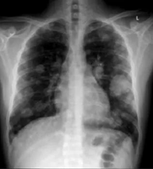
A 22-year-old male complained of hemoptysis, weight loss, and abdominal pain for the past 2 weeks. The patient had no relevant past medical history. Chest X-ray showed bilateral, multiple, variable size opacities suggestive of metastasizes (Figure 1). At King Khalid University Hospital, workup to assess for the primary source was performed which include scrotal ultrasonography that revealed bizarre-shaped macrocalciication (focal clump of calciication) is present in the central aspect of the right testis (Figure 2). Computed
tomography (CT) scan of abdomen and pelvis showed large para-aortic complex soft tissue lesions with central necrosis representing enlarged lymph nodes as a result of secondary deposits (Figure 3). The patient’s serum β-human chorionic gonadotropin (β-hCG) level was elevated, i.e., 9020 IU/L. Other laboratory investigations including α-fetoprotein were within normal ranges.
Trucut biopsy was obtained from one of the lung nodules, which showed microscopic features of hemorrhagic necrotic tissue with scattered mono- and multinucleated malignant cells. These cells have smudgy nuclei with some intranuclear inclusions and abundant eosinophilic opaque cytoplasm. A panel of immunohistochemical stains was performed and shows that the tumor cells are positive for cytokeratin pan, epithelial membrane antigen, and β-hCG. They are negative for CD30, CD117, and alpha-fetoprotein. The inal pathological diagnosis was concluded as “choriocarcinoma” metastatic testicular tumor.
A right radical orchiectomy was performed. Histopathology showed subtotal atrophy of the testis with scarred and ibrotic tissues. There was no evidence of GCT.
Four months later, the patient present with slurred speech and decrease attention, for which he undergo magnetic resonance imaging brain showed hemorrhagic left temporoparietal space occupying lesion with adjacent vasogenic edema highly suggestive of hemorrhagic metastasis.
Discussion
The term “burned-out testicular tumor” refers to a regressed testicular tumor which presents with its metastases.2 The Burned-out testicular tumor is a very rare clinical entity. There is no clinical inding in the testicle because it regresses spontaneously without any treatment and generally presents with metastases. Clinical examination of the testis and scrotal sonography are pivotal in the initial diagnosis of such neoplasms. We present a case of a 22-year-old male with hemoptysis, weight loss, and abdominal pain for the past 2 weeks and no palpable lesion on testicular examination. No relevant past medical history.
Keywords: Burned-out tumor, germ cell tumor, testicular tumor
Mohammed S. El-Sharkawy,
Abdulaziz S. Al-Jibali
Department of Radiology, King Khalid
University Hospital, King Saud University,
P.O. Box 7805, Riyadh 11472, Saudi Arabia
Address for correspondence:
Abdulaziz S. Al-Jibali,
Department of Radiology, King Khalid
University Hospital, King Saud University,
P.O. Box 7805, Riyadh 11472, Saudi Arabia. Phone: 00966504883250.
E-mail: Dr.aziz@msn.com
ABSTRACT
WEBSITE: ijhs.org.sa
ISSN: 1658-3639
PUBLISHER: Qassim University
El-Sharkawy and Al-Jibali: Burned-out metastatic testicular tumor: Choriocarcinoma
2 International Journal of Health Sciences
Vol. 11, Issue 2 (April - June 2017)
pathogenesis of this phenomenon may be that the high metabolic rate of the tumor causes it to rapidly outgrow its blood supply. The patients may present with widespread metastases, but no primary tumor except for an area of calcification within the testis.3 They often demonstrate
little or no remaining viable tumor, with mostly scarring and ibrosis found at histologic analysis after orchiectomy. The syncytiotrophoblasts involved in these lesions with choriocarcinoma produce β-hCG which is raised in these tumors. With early widespread metastasis, patients may present with symptoms referable to their metastases rather than a palpable testicular mass. Sites of metastases include the lung, liver, gastrointestinal tract, and brain. The primary tumor and metastases are often hemorrhagic.4
Patients with mixed GCTs with choriocarcinoma fair better than those with pure choriocarcinoma tumors, but a very high level of β-hCG (50,000 IU/L) has a poor prognosis with
a 5-year survival rate of 48%.4 High-resolution sonography
of the scrotum with linear high-frequency transducers allows the detection of small, highly echogenic foci, hypoechoic zones, microlithiasis, or microcalcifications. At scrotal US, they appear as a hypoechoic or ill-defined, intratesticular calcified lesion. During CT scan of the abdomen, when large retroperitoneal tumors or lymph nodes are detected in young men, ultrasound examination of bilateral testis is recommended, even if a mass is not palpable in the scrotum.5
Conclusion
Scrotal sonography is very important for the detection of intratesticular lesions, especially in patients with extragonadal metastatic involvement and normal palpation for the testis. A burned-out testicular tumor should be considered when punctuate echogenic foci are seen without any evidence of hypoechoic mass lesions. Metastatic disease secondary to burned-out lesion has the same prognosis as a primary testicular malignancy.
References
1. Sidhu PS, Sriprasad S, Bushby LH, Sellars ME, Muir GH. Impalpable testis cancer. BJU Int 2004;93:888.
2. Perimenis P, Athanasopoulos A, Geraghty J, Macdonagh R. Retroperitoneal seminoma with ‘burned out’ phenomenon in the testis. Int J Urol 2005;12:115-6.
3. Comiter CV, Renshaw AA, Benson CB, Loughlin KR. Burned-out primary testicular cancer: Sonographic and pathological characteristics.
J Urol 1996;156:85-8.
4. Horwich A, Shipley J, Huddart R. Testicular germ-cell cancer. Lancet 2006;367:754-65.
5. Kebapci M, Can C, Isiksoy S, Aslan O, Oner U. Burned-out tumor of the testis presenting as supraclavicular lymphadenopathy. Eur Radiol 2002;12:371-3.
Figure 1: Chest X-ray showing bilateral, multiple, variable size, pulmonary nodules
Figure 2: Ultrasound of right testis showing a focal clump of
calciication present in central aspect of testis. No evident mass
effect or surrounding masses. A hypoechoic irregular halo was noted