Open access: www.balimedicaljournal.com or www.ojs.unud.ac.id 5
TUBERCULUM SELLAE MENINGIOMA Vs. MACROADENOMA:
How to differentiate preoperatively?
Mardjono, I., Arifin, M. Z., Sidabutar, R., Singh, A., and Sevline, E. O.
Department of Neurosurgery, Faculty of Medicine Universitas Padjadjaran–Dr. Hasan Sadikin General Hospital, Bandung-Indonesia
Objective: Sellar region tumors may origin from a various number of structures and each of them have a specific clinical and radiological appearance. Among these pathological processes, one of the most challenging is to distinguish between tuberculum sellae meningioma (TSM) and macroadenoma hypophysis (MH). Differentiating these two entities preoperatively is very important to decide which approach will be most suitable and beneficial. The purpose of this study is to produce a simple preoperative scoring system to differentiate these two that can be applied in specific conditions where MRI is not available or could not be performed. Methods: This analytical retrospective cohort study contains data obtained from patients treated in Neurosurgery Department of Dr. Hasan Sadikin General Hospital-Bandung from 1 January 2008 until 31 December 2010. There were 34 patients enrolled in this study, in which 15 of them were diagnosed with MH and 19 patients diagnosed with TSM confirmed with pathology examination. Results: From clinical presentation we found that the event of endocrinopathy occurs significantly in macroadenoma hypophysis (p=0.002). Whereas from radiological evaluation there were 7 parameters that significantly distinguish these two entities including hyperostosis, sellar floor configuration, homogeneity of mass, contrast agent enhancement, waist configuration, peritumoral edema, and dural attachment. From these findings, we propose a simple scoring system to differentiate macroadenoma hypophysis and tuberculum sellae meningioma with a 84.2% sensitivity and 100% specificity. Conclusion: although MRI is the modality of choice in differentiating macroadenoma hypophysis and tuberculum sellae meningioma but our scoring system can be used as an aid in choosing best surgical approach.
Keywords: sellae, region tumour, macroadenoma hypophysis, tuberculum sellae meningioma.
INTRODUCTION
Neurosurgery came to developing countries over a half a century ago, yet the vast majority of population in these countries do not have equity in access to it, owing to the cost of neurosurgical care and geographical isolation of patients. Many biomedical equiments such as CT (computed tomography) scan and MRI (magnetic resonance imaging) are not available to most of the population in these countries.1
The examination of choice in pituitary and sellar region tumors is MRI, because it depicts the complex anatomy around the sellar wall. Almost 30 pathologic entities occur in this region, and most can be distinguished using MRI.2 Sellar region tumors are one of the most challenging tumor cases
Correspondence: Mardjono, I.
Address: Department of Neurosurgery, Faculty of Medicine Universitas Padjadjaran–Dr. Hasan Sadikin General Hospital. Jl. Pasteur No, 38, Bandung 40161, West Java, Indonesia
Phone: +6222 2041694, Fax : +6222 2041694, Email: joy_mardjono@yahoo.com
for neurosurgeons. Two of the most common entities that should be distinguished because of their similarities especially in imaging studies are tuberculum sellae meningioma and macroadenoma hypophysis.3 Preoperative distinction of these tumors is important for best surgical approach. Tuberculum sellae meningioma is usually operated by craniotomy approach, wheras macroadenoma hypophysis uses trans sphenoidal approach.4,5
We propose a simple scoring system based on clinical and radiological evaluation using CT scan that can be used as an aid for determining surgical strategy in cases where MRI could not be performed.
METHOD
This retrospective cohort study consists of 34 patients with sellar region tumors treated and operated in neurosurgery ward of Dr. Hasan Sadikin General Hospital Bandung from 1 Janury 2008 until 31 December 2010. After the patients received a full verbal and written explanation of this procedure, all provided informed consent.
Open access: www.balimedicaljournal.com or www.ojs.unud.ac.id 6
and 19 patients with tuberculum sellae meningioma. Data collected included clinical presentation and various characteristics based on CT scan imaging.
Data were processed on a personal computer by using commercially available statistic software. These variables were compared using t test with p value ≤0.05. Only significant variables were then summarized into a scoring system and then tested for its specificity and sensitivity.
RESULTS
From 34 patients in our study, there were 12 male and 22 female patients with average age slightly higher in tuberculum sellae meningioma group (Table 1). As shown below, there is significant correlation of endocrine abnormalities
(p=0.002) in macroadenoma hypophysis. Whereas there is no significant correlation between sex, age, duration of symptoms, tumor size, chief complaint and visual field defect.
Based on radiological findings shown on CT scan, there were certain characteristics that we analyzed to differentiate these two entities (Figure 1 and 2). There is significant correlation of various radiological presentations such as homegeneity on CT scan (p=0.017), contrast enhancement (p=0.001), hyperostosis (p=0.002), thinning of sellar (p<0.001), presence of edema (p=0.004), size of the sellar waist (p=0.007) and alsodural attachment (p<0.001) of tumors originating as macroadenoma hypophysis or tuberculum sellae meningioma (Table 2).
Table 1
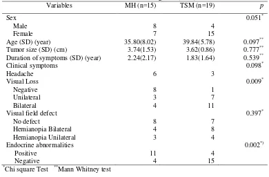
Clinical Presentation of Patients Diagnosed with Macroadenoma hypophysis (MH) and Tuberculum Sellae Meningioma (TSM)
Variables MH (n=15) TSM (n=19) p
Sex 0.051*
Male 8 4
Female 7 15
Age (SD) (year) 35.80(8.02) 39.84(5.78) 0.097** Tumor size (SD) (cm) 3.74(1.53) 3.62(0.86) 0.777** Duration of symptoms (SD) (year) 2.24(2.17) 1.83(1.64) 0.539**
Clinical symptoms 0.098*
Headache 6 3
Visual Loss 0.009*
Negative 8 1
Unilateral 3 7
Bilateral 4 11
Visual field defect 0.397*
No defect 8 7
Hemianopia Bilateral 4 8 Hemianopia Unilateral 3 4
Endocrine abnormalities 0.002*) Positive
Negative
11 4
4 15
*
Chi square Test **Mann Whitney test
A Tuberculum sellae meningiomas B macroadenoma hypophysis
Figure 1
Tuberculum sellae meningiomas appear distinctively homogeneous and enhance entirely after application of contrast. On the contrary, macroadenoma hypophysis have various
Open access: www.balimedicaljournal.com or www.ojs.unud.ac.id 7
Figure 2
CT Scan of a 44 Year Old Lady Diagnosed with Tuberculum Selae Meningioma.
Note the homogenous enhancement and lobulated configuration but no hiperostosis of the bone is present.
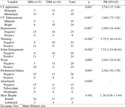
Table 2
Comparison of Radiological Presentation on CT Scan between Macroadenoma Hypophysis (MH) and Tuberculum Sellae Meningioma (TSM) Variable MH (n=15) TSM (n=19) Total p PR (95%CI)
CT appearance 0.001* 2.74(1.37-5.48) Homogen 2 13 15
Inhomogen 13 6 19
CT Enhancement 0.001** 3.60(1.75-7.42) Minimal 9 1 10
Bright 6 18 24
Hyperostosis 0.002** 2.50(1.54-4.04) Negative 15 10 25
Positive 0 9 9
Thinning <0.001** 5.75 (2.36-14.01) Negative 4 19 23
Positive 11 0 11 Sellar Enlargement
Negative Positive Waist
4 11
16 3
20 14
<0.001**
0.008*
7.53 (1.15-48.84)
2.63(1.10-6.24) Negative 5 15 20
Positive 10 4 14
Peritumoral Edema 0.004* 2.36(1.50-3.70) Negative 15 11 26
Positive 0 8 8
Attachment <0.001* -
Negative 15 0 15 Tuberculum 0 13 13 Diaphragm 0 6 6
Mass Shaped 0.442 1.38 (0.56 – 3.44) Round 16 11 27
Lobulated 3 4 4
*
Chi square Test **Mann Whitney test
Table 3
Scoring System of Various Variables to Differentiate between Macroadenoma Hypophysis and Tuberculum Sella Meningioma
Variable PA (n=15) TSM (n=19) p Se Sp PPV NPV
Score <0.001*
>3 0 16 84.2% 100% 100% 83.3%
≤3 15 3
*
Open access: www.balimedicaljournal.com or www.ojs.unud.ac.id 8
Figure 3
ROC curve showing cut off point of 3 in determining our scoring system.
DISCUSSION
The sellae tursica which resembles a Turkish saddle if viewed from the side, forms a semicircular, central depression within the sphenoid bone. The antero-superior edge of the sella is marked by a horizontal ridge, the tuberculum sellae.6 Two of the most frequent pathological process found in this region are macroadenoma hypophysis and tuberculum sellae meningioma.3,7Although according to previous reports the incidence varies according to age, gender and ethnic group. In our series we found predilection for menigioma higher in female patients with average age of 40 years old.7-9
There were three most common symptoms in our report; headache, visual disturbance and endocrinopathy (Table 1). Although almost 70% diabetes, we observed that insipidus occurrence was the highest endocrine abnormality present in macroadenoma hypophysis. This stalk compression preoperative planning, particularly in regard to pneumatization and the anatomy of the sphenoid sinus as not all neurological centres have MRI facilities.2
In our study, we found seven radiological criteria to help recognize these two entities. Macroadenomas have variable appearances
because they tend to have necrosis, cyst formation, and hemorrhage that appear as mixed attenuation. Curiously there were 6 patients with tuberculum sellae meningioma that show inhomegeneous of mass and 1 patient showing minimal enhancement after contrast administration. Macroadenoma hypophysis are soft tumors which usually indent at diaphragm sellae, giving them a ‘snowman’ configuration. This is one feature that can help to distinguish between a pituitary macroedema and a meningoma.2,9 In our report, 10 out of 15 patients showed a positive waist configuration and also 4 patients of tuberculum sellae meningiomas also had feature.
There was more than 50% of tuberculum sellae meningioma that did not show signs of hyperostosis (10 patients) nor peritumoral edema (11 patients) which are usually characteristics for this type of tumor (Table 2). Adjacent hyperostosis, is the best seen on CT, is present in more than one third of cases and is a helpful sign in meningiomas.12,13 In some previous reports, the sellae turcica is usually not expanded or only slightly enlarged in tuberculum sellae meningiomas. This is in accordance to our report where 3 patients with tuberculum sellae meningiomas had sellar enlargement, in contrast to macroadenoma hypophysis (11 patients).9,11
Sellar floor thinning or erosion are other criteria that could be useful in diagnosing macroadenoma hypophysis. Eleven patients with macroadenoma hypophysis showed sellar floor thinning but there were 4 cases that did not have this feature. Obtuse dural margins and dural tail enhancement of lesions involving the sella, are helpful in the preoperative diagnosis.12 Most of the specific CT scan features that we analyze in our series, showed significance in helping to diagnose macroadenoma hypohysis and tuberculum sellae meningioma.
differentiate tuberculum sellae meningioma from the macroadenoma hypophysis, because craniotomy is done for meningioma, whereas a transsphenoidal route is preferred for most macroadenoma hypophysis.3,4,11,14 Transsphenoidal surgery is the approach of choice for macroadenoma hypophysis.15 Tuberculum sellae meningiomas usually have a firm, rubbery consistency and often require sharp dissection rather than simple suctioning for their removal.11 Based on our preference, all of our patients diagnosed with tuberculum sellae meningioma were operated using a pterional approach.
CONCLUSSION
In conclusion, the superiority and usefullnes of MRI is unquestionable as it is the gold standar imaging to distinguish macroadenoma hypophysis and tuberculum sellae meningioma but this modality is often not available in many countries. A simple scoring system can be useful as a tool for preoperative surgical strategy in differentiating these two entities. A score of more than 3 is most likely to be diagnosed as tuberculum sellae meningioma whereas less than 3 is representative for macroadenoma hypophysis.
REFERENCES
1. Black, P. 2007. The future of neurosurgery: a call to leadership. Clin. Neurosurg. 54.
2. Khan-Ali. Pituitary Adenoma Imaging. 2011. (Downloaded 4th April 2012) In :http:// Diaphragma sellae meningioma mimicking pituitary macroadenoma: a case report. IJCRIMPH. 2 (11) : 404.
5. Taylor, S. L., Barakos, J. A., Harsh, G. R., and Wilson, C. B. 1992. Magnetic resonance imaging of tuberculum sellae meningiomas: preventing preoperative misdiagnosis as pituitary macroadenoma. Neurosurgery. 31(4):621.
6. Schubiger, O. 1996. Radiology of pituitary adenomas in: Landolt, A. M., Vince, M. L., and Reilly P.L, eds. Pituitary Adenomas. UK: Churchill Livingstone. p. 177-219,
7. Jagannathan, J., Kanter, A. J., Sheehan, J. P., Jane, J. A. Jr., and Laws, E. R. Jr. 2007. Benign brain tumors: sellar/parasellar tumors. Neurol Clin. 25 (4):1231-.
8. Al-Mefty, O., Abdulrauf-Saleem, I., and Haddad-Geoeges, F. 2012. Meningiomas. In: Winn, H. R., ed. Youman’s Neurological Surgery. 6th edition. Philadelphia: Elsevier Saunders;. pp 1426-1449.
9. Mc-Vallo, B., and Mc-Cormack, B. 2007. Surgical management of tubeculum sellae and sphenoid ridge meningiomas. In: Schmidek, ed. Schmidek & Sweet’s Operative Neurosurgical Techniques Indications, Methods, and Results. 4th edition . USA :W.B.Saunders;. pp 305-324 10.Chi, J. H., and McDermott, M. W. 2003.
Tuberculum sellae meningiomas. Neurosurg Focus. 14 (6): 6.
11.Mulinda, J. 2011. Pituitary macroadenoma. (Downloaded 4th April 2012). In: http:// emedicine.medscape.com/article/123223-clinical
12.Bowers, C. A., Altay, T., and Couldwell, W. T. 2011. Surgical decision-making strategies in tubercullum sellae meningioma resection. Neurosurg Focus. 30 (5): E1.
13.Anderson, J. R., Antoun, N., Burnet, N., Chatterjee, K., Edwards, O., Pickard, J. D., et. al. 1999. Neurology of the pituitary gland. JNeurol Neurosurg Psychiatry. 66 (6):703–21. 14.Donovan, J. L., and Nesbit, G. M. 1996.
Distinction of masses involving sella and suprasellar space: specificity of imaging features. Am J Roentgenology. 167 (3): 597-603
15.Cappabianca, P., Cirillo, S., Alfieri, A.,
D’Amico, A., Maiuri, F., Mariniello G, et. al. 1999. Pituitary macroadenoma and diaphragm sellae meningioma: differential diagnosis on MRI. Neuroradiology. 41 (1): 22-6.
16.Swearingen, B., and Zeevas, N. 2007. Surgical