Development of Iridology System Database for
Colon Disorders Identiication using
Image Processing
Rossi Passarella
1*, Erwin
1, M. Fachrurrozi
2and Sutarno
11Lab. Industrial Automation. Department of Computer Engineering, Faculty of Computer Sciences,
Universitas Sriwijaya; [email protected], [email protected], [email protected]
2Department of Informatics Engineering, Faculty of computer Sciences, Universitas Sriwijaya
Jln. Raya Palembang-Prabumulih km 32. Indralaya. Ogan Ilir. Sumatera Selatan. Indonesia, [email protected]
Abstract
Iridology is an art of knowledge to detect a specific disease of human body from the iris. The detection will tell each indi
-vidual organ when it has low or high performance (abnormal). The iris reveals conditions change of every part of the body. Every organ and part of the body are represented in the iris in a well-defined area. The objective of research is to develop an iris database of colon disorder based on the map of iridology. This map is represented as a diagnosis tool to detect the common colon disorder. In developing the database, Sixty (60) subjects were enrolled in the study where 35 subjects had histologically proven problem in colon disorder, and 25 were control subject. To extract the iris, the image processing methods such Hough transform, segmentation and normalization were applied in this research. The conclusion of this paper is the proposed an iris database for helping medical doctor in detecting colon disorder using image processing.
Keywords : Colon Disorder, Iridology, Iris Database, Image Processing.
*Corresponding author:
Rossi Passarella ([email protected])
1. Introduction
he iris is a thin circular diaphragm, lies between cornea and pupil of the human eye. A front view of human eye is shown in Figure 1. he function of the iris is to control the amount of light entering the pupil, and this is done by the sphincter and the dilator musclesby adjusting the size of the pupil. he iris has average diameter of 12 mm, with the pupil size vary from 10% to 80% of the iris diameter [1].
Iridology is the study of the iris to diagnose medical conditions by analyzing structures, markings, patterns, and irregularities of the iris pigmentation [2]. his is an art of knowledge to detect a speciic disease of human body from the iris. he detection will tell each individual organ when it has low or high performance (abnormal) [3], as represented by iris map shown in Figure 2.
he iris reveals conditions change of every part of the body. Every organ and part of the body are represented in the iris in a well-deined area. In addition, through various marks, signs, and discoloration in the iris, nature reveals inherited weaknesses and strengths.
he objective of research is to develop an iris database of colon disorder based on the map of iridology. his map is represented as a diagnosis tool to detect the common colon disorder.
2. Materials and Methods
In order to detect human organ from the eyes, and to acknowledge the place of the organ according to iris map, the system need to be autonomous using a camera and computer. he system will compare the patient iris eyes
Rossi Passarella, Erwin, M. Fachrurrozi and Sutarno
Indian Journal of Bioinformatics and Biotechnology | Print ISSN 2319-6580 | Online ISSN 2319-6599 101 Vol 2(6) | June 2013 | www.ijbb.informaticspublishing.com
with the database. he database of the colon disorder should be developed and veriied with the right methods. he design of this system is shown in Figure 3.
In developing the database, Sixty (60) subjects were enrolled in the study wherein 35 subjects had histologi-cally proven problem in colon disorder, and 25 were control subject.
2.1 Eye Image
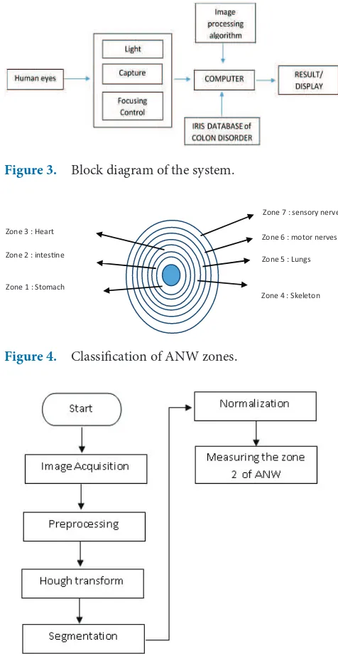
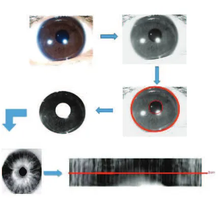
he Autonomic Nervous Wreath (ANW) is a major landmark feature of iris topography for practitioner of iridology to analysis the iris eye [4]. his major land mark describes the condition of the digestive ANW resulting in autonomic nervous. ANW illustrates the large intestine (colon). he ANW can be the basis to determine the con-dition of a person’s colon. According to Figure 4, Circle ANW for bowel problems is shown in between zones 2 and 3.
2.2 Iris Recognition
In biometric systems, iris recognition is becoming a most widely used to verify that the human iris patterns is uniqueness. he iris recognition is an important step in iridology, to extract the iris code, some steps should be done. Flowchart to extract iris in this paper is shown in Figure 5.
According to ANW, the colon is placing in zone 2 (Figure 4), from the expert iridology, the maximum wide zone 2 from the pupil is 3 cm, if the iris pattern from the human eyes shown more than 3 cm means the person had problem with the colon.
2.2.1 Hough Transform
his method is a standard computer vision algorithm to determine the parameters in image for a simple geomet-ric objects, such as lines and circles [5]. his method uses edge information of the image for deining a mapping
Figure 1. A front view of human eye.
Figure 2. Iris map of human.
Figure 3. Block diagram of the system.
Figure 4. Classiication of ANW zones.
Zone 7 : sensory nerves
Zone 6 : motor nerves
Zone 5 : Lungs
Zone 4 : Skeleton Zone 3 : Heart
Zone 2 : intesne
Zone 1 : Stomach
Development of Iridology System Database for Colon Disorders Identiication using Image Processing
Indian Journal of Bioinformatics and Biotechnology | Print ISSN 2319-6580 | Online ISSN 2319-6599 102 Vol 2(6) | June 2013 | www.ijbb.informaticspublishing.com
from the orientation of and edge point to reference point of the shape.
2.2.2 Segmentation
Iris segmentation is very important for an iris recognition system [6, 7]. Segmenting iris sometimes is diicult task due to complex structure and the boundary of the pupil as it is not circle on many cases. If the iris regions were not correctly segmented, there would possibly exist four kinds of noises in segmented iris regions such as eyelashes, eyelids, reflections and pupil, which will result in poor rec-ognition performance[8]. here are many methods had been used to eliminate the noises such as edge detection, region growing, statistical approach, mathematical mor-phology and Active contour based method or snake [9]. In this research, the edge detection is applied due to simplify.
2.2.3 Normalization
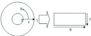
his transformation is done in order to make it easier to extract the value of the iris image. Activity image trans-formation can be likened to the process of normalization from image transformation operations that take advan-tage of the geometry of the image. Normalization here is done with re-map every point on the iris area (polar coordinates) into Cartesian coordinates. For polar to Cartesian coordinate transformation, this transformation can be illustrated in Figure 6.
his can be modeled as:
(x, y) are the original Cartesian coordinates,
(r, θ) are the corresponding normalized polar coordi-nates,
he normalization is done by taking a reference point from the center of the pupil and radial vectors pass via iris area.
2.2.4 Matching
Matching is the process where the organ matched to iri-dology maps to ind the colon organ.
2.3 Colon Disorder
he large intestine is the last part of the digestive tract that has the following functions:
a. Place to collect letover food then will be excreted through the anus
b. Place for absorbing water and some minerals
c. Place of bacterial growth; may form some vitamins such as vitamins B and K
he shape and size of the ANW is analogous to the large intestine (colon). If ANW seen tending to the let side, then there is a problem in the colon. According to medical science, there is also a gut thing down (descend-ing) in the form of accumulation of dirt, so that this section of the looned. he bowel conditions of human shown in Table 1.
3. Preliminary Experiment
Experiment was conducted at the Laboratory Industrial Automation (Figure 7), the Subjects to verify the database were a random take from student volunteers. he process in database to measure the colon disorder is shown in Figure 8. Ater the system suggests a result, the subject is sent to hospital for a medical check-up to validate the result from system. he percentage error of the iris data-base for colon disorder was 8%.
4. Conclusion
We proposed an iris database for helping medical doc-tors in detecting colon disorder using image processing, however this database still need to be improved since the percentage error was 8%. More databases on pattern of the iris are needed to gain better results.
5. Acknowledgment
his work was supported by University of Sriwijaya under BPOTN 2013 grant number: 144a/UN9.3.1/PL/2013.
Rossi Passarella, Erwin, M. Fachrurrozi and Sutarno
Indian Journal of Bioinformatics and Biotechnology | Print ISSN 2319-6580 | Online ISSN 2319-6599 103 Vol 2(6) | June 2013 | www.ijbb.informaticspublishing.com
6. References
1. Daugman J (2002). How iris recognition works, Proceedings of 2002 International Conference on Image Processing, vol 1, I-33–I-36.
2. Hauser H, Karl J et al. (2000). Information from Structure and Colour, Iridology 1. Heimsheim: Felke Institut, 134–243. 3. Jackson P (2004). Practical iridology, Working with the
chart, Carrol and Brown Publisher Limited, 76–78. 4. Li Y, Wang K et al. (2007). Extracting the autonomic
nerve wreath of iris based on an improved snake approach, Neurocomputing, vol 70(4), 743–748.
5. Ballard H D (1981). Generalizing the Hough transform to detect arbitrary shapes, Pattern Recognition, vol 13(2), 111–122.
6. Cui J, Wang Y et al. (2004). A fast and robust iris localiza-tion method based on texture segmentalocaliza-tion, Proceedings SPIE, Biometric Technology for Human Identiication, vol 5404, doi:10.1117/12.541921.
7. Huang J, Wang Y et al. (2004). A new iris segmenta-tion method for recognisegmenta-tion, Proceedings of the 17th International Conference on Pattern Recognition, ICPR 2004, vol. 3, 554–557.
8. He Z, Tan T et al. (2009). Toward accurate and fast iris segmentation for iris biometrics, IEEE Transactions on Pattern Analysis and Machine Intelligence, vol 31(9), 1670–1684.
9. Othman Z, and Prabuwono A S (2010). Preliminary study on iris recognition system: tissues of body organs in iridol-ogy, IEEE EMBS conference on Biomedical Engineering and Sciences (IECBES), 115–119.
Table 1. Bowel Conditions
No Type of condition Indication
1. Colon Normal Visible iber fabric evenly and tightly around the radial slices. his shows the body of the owner of the iris has a strong endurance, able to cope with diseases and is able to develop mental, emotional status better.
2. Ballooned Sigmoid Swelling (balloon) around the sigmoid indicates constipation. In the iris it is visible aswidened ANW. 3. Prolapse Transverse colon falls down and pressing other organs. In such iris case, ANW is at the center and
below the pupil.
4. Pocket Bowel associated with symptoms: Irritable Bowel Syndrome (IBS).
5. Stricture ANW on iris looks rather sharply to the pupil. Downsizing the colon caused food poisoning or administration of anti-diarrhealdrugs.
6. Spasm Reduction occurs in the colon. It disrupts the process of sewage. ANW portraits like wave. 7. Radii Solaris When poison spreads throughout the body, it becomes diicult to determine the defective part.
Disruption of hormones and emotions.
Figure 7. Experiment conducted.